. 2013 Dec;34(38):9853-62.
doi: 10.1016/j.biomaterials.2013.08.015. Epub 2013 Sep 21.

The promotion of mandibular defect healing by the targeting of S1P receptors and the recruitment of alternatively activated macrophages

Affiliations

The promotion of mandibular defect healing by the targeting of S1P receptors and the recruitment of alternatively activated macrophages

Anusuya Das et al. Biomaterials. 2013 Dec.

Abstract

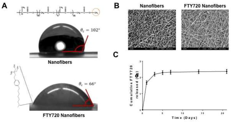
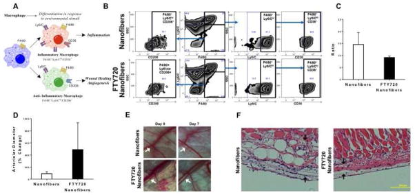
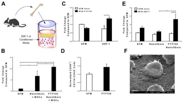
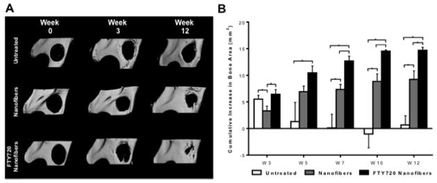
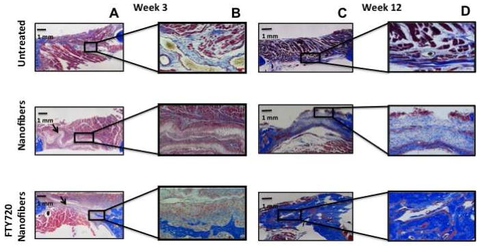
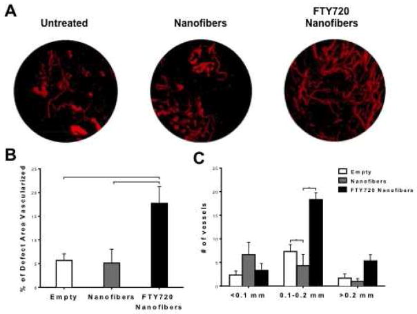
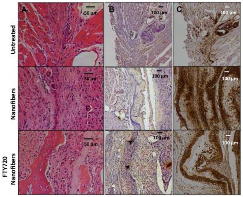
Endogenous signals originating at the site of injury are involved in the paracrine recruitment, proliferation, and differentiation of circulating progenitor and diverse inflammatory cell types. Here, we investigate a strategy to exploit endogenous cell recruitment mechanisms to regenerate injured bone by local targeting and activation of sphingosine-1-phosphate (S1P) receptors. A mandibular defect model was selected for evaluating regeneration of bone following trauma or congenital disease. The particular challenges of mandibular reconstruction are inherent in the complex anatomy and function of the bone given that the area is highly vascularized and in close proximity to muscle. Nanofibers composed of poly(DL-lactide-co-glycolide) (PLAGA) and polycaprolactone (PCL) were used to delivery FTY720, a targeted agonist of S1P receptors 1 and 3. In vitro culture of bone progenitor cells on drug-loaded constructs significantly enhanced SDF1α mediated chemotaxis of bone marrow mononuclear cells. In vivo results show that local delivery of FTY720 from composite nanofibers enhanced blood vessel ingrowth and increased recruitment of M2 alternatively activated macrophages, leading to significant osseous tissue ingrowth into critical sized defects after 12 weeks of treatment. These results demonstrate that local activation of S1P receptors is a regenerative cue resulting in recruitment of wound healing or anti-inflammatory macrophages and bone healing. Use of such small molecule therapy can provide an alternative to biological factors for the clinical treatment of critical size craniofacial defects.

Keywords: Bone healing; Craniofacial reconstruction; FTY720; Neovascularization; S1P.


Figures

Fig 1
Fig 1
Characterization of FTY720 loading in PCL/PLAGA nanofibers at a drug:polymer ratio of 1:200 by weight. (A) Glycerine drop contact angle analysis demonstrates that unloaded nanofibers are more hydrophobic than FTY720 loaded nanofibers. (B) Scanning electron micrographs of unloaded and FTY720 loaded nanofibers (original magnification x2000) demonstrates similar morphologies of randomly aligned nanofibers. (C) Cumulative release of FTY720 from nanofibers using HPLC-MS. Approximately 2.4 μg of FTY720 was released by 21 days.
Fig 2
Fig 2
FTY720 modulates inflammatory response after scaffold implantation by (A) changing macrophage phenotype surrounding the implant. (B) Flow cytometric analysis of digested mouse dorsal skinfold tissue treated with unloaded or FTY720 loaded nanofibers. Gating strategy for F4/80 and Ly6/C. (C) FTY720 nanofibers decrease the ratio of inflammatory Ly6Chi/CD36+ to Ly6Clo/CD206+ tissue macrophages (F4/80+). (D) FTY720 nanofibers increase the arteriolar diameter of blood vessels surrounding the implant. (E) Intravital images of dorsal skinfold window chamber microvasculature showing the arteriolar diameter increase. H&E staining of (F) unloaded nanofibers or FTY720 loaded nanofibers in dorsal skinfold tissue (arrows show fibrous tissue surrounding implant).
Fig 3
Fig 3
FTY720 loaded nanofibers interact with MSCs to direct their secretory profile. (A, B) CM generated from fat vascular stromal fraction derived MSCs cultured on unloaded or FTY720 loaded nanofibers induces migration of bone marrow cells towards SDF-1. (C) FTY720 pre-treatment increases the migration of bone marrow stromal cells and (D) bone marrow-derived progenitor cells towards SDF-1. (E) Conditioned media (CM) generated from FTY720 loaded nanofibers enhances migration of bone marrow cells towards SDF-1. (*p<0.01). Scanning electron micrographs demonstrate that MSCs adhere to and interact with (F) FTY720 nanofibers (original magnification x1500).
Fig 4
Fig 4
Assessment of new bone formation in rat critical size mandibular defects. (A) Three-dimensional representative microCT images of the mandibular ramus on the day of surgery and 3 or 12 weeks post-surgery. Defects were left untreated or treated with unloaded nanofibers or FTY720 loaded nanofibers. (B) Two-dimensional quantification of new bone formation in the defect region was calculated using ImageJ image analysis software. FTY720 promotes more osseous ingrowth than unloaded nanofibers or no treatment. Bars indicate significance between groups (p<0.1 or *p<0.01).
Fig 5
Fig 5
Assessment of new bone formation in rat critical size mandibular defects by Masson’s trichrome staining. Defects were left untreated or treated with unloaded nanofibers or FTY720-loaded nanofibers. (A) Representative images of mandibular defect 3 weeks post-surgery (original magnification x10). (B) High magnification image of a region with new bone formation 3 weeks post surgery (original magnification x40). (C) Representative images of mandibular defect 12 weeks post-surgery (original magnification x10). (D) High magnification image of a region with new bone formation 12 weeks post surgery (original magnification x40).
Fig 6
Fig 6
Neovascularization in the defect area visualized with MICROFIL® enhanced microCT. (A) Representative images at week 12 demonstrate vessel perfusion with MICROFIL within the defect area. (B) Two-dimensional quantification of blood vessel area in the defect region was calculated using ImageJ image analysis software. (C) Number of vessels by vessel diameter within the defect area for the three treatment groups. (p<0.05 or **p<0.001).
Fig 7
Fig 7
FTY720 treatment decreases the recruitment of M1 macrophages to the defect site. Mandibular defect tissue stained 3 weeks after treatment for (A) H&E (B) CD163 (M2 marker, black arrowheads) and (C) CCR7 (M1 marker) to qualitatively evaluate inflammatory response following treatment with unloaded or FTY720-loaded nanofibers.

Similar articles

Cited by

References

    1. Maurer P, Eckert AW, Kriwalsky MS, Schubert J. Scope and limitations of methods of mandibular reconstruction: a long-term follow-up. Brit J Oral Maxill. 2010;48(2):100–4. - PubMed
    1. Mariani PB, Kowalski LP, Magrin J. Reconstruction of large defects postmandibulectomy for oral cancer using plate and myocutaneous flaps: a long-term follow-up. Int J Oral Maxillofac Surg. 2006;35(5):427–32. - PubMed
    1. Schliephake H, Weich HA, Dullin C, Gruber R, Frahse S. Mandibular bone repair by implantation of rhBMP-2 in a slow release carrier of polylactic acid--an experimental study in rats. Biomaterials. 2008;29(1):103–10. - PubMed
    1. Herford AS, Lu M, Buxton AN, Kim J, Henkin J, Boyne PJ, Caruso JM, Rungcharassaeng K, Hong J. Recombinant human bone morphogenetic protein 2 combined with an osteoconductive bulking agent for mandibular continuity defects in nonhuman primates. J Oral Maxillofac Surg. 2012;70(3):703–16. - PubMed
    1. Zétola A, Ferreira FM, Larson R, Shibli JA. Recombinant human bone morphogenetic protein-2 (rhBMP-2) in the treatment of mandibular sequelae after tumor resection. Oral Maxillofac Surg. 2011;15(3):169–74. - PubMed

Publication types

LinkOut - more resources