Clinical Trial
. 2013 Nov 8;288(45):32172-32183.
doi: 10.1074/jbc.M113.502955. Epub 2013 Sep 24.

Binding of complement inhibitor C4b-binding protein to a highly virulent Streptococcus pyogenes M1 strain is mediated by protein H and enhances adhesion to and invasion of endothelial cells

Affiliations
Clinical Trial

Binding of complement inhibitor C4b-binding protein to a highly virulent Streptococcus pyogenes M1 strain is mediated by protein H and enhances adhesion to and invasion of endothelial cells

David Ermert et al. J Biol Chem. .

Abstract

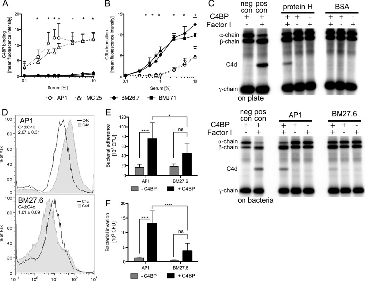
Streptococcus pyogenes AP1, a strain of the highly virulent M1 serotype, uses exclusively protein H to bind the complement inhibitor C4b-binding protein (C4BP). We found a strong correlation between the ability of AP1 and its isogenic mutants lacking protein H to inhibit opsonization with complement C3b and binding of C4BP. C4BP bound to immobilized protein H or AP1 bacteria retained its cofactor activity for degradation of (125)I-C4b. Furthermore, C4b deposited from serum onto AP1 bacterial surfaces was processed into C4c/C4d fragments, which did not occur on strains unable to bind C4BP. Recombinant C4BP mutants, which (i) lack certain CCP domains or (ii) have mutations in single aa as well as (iii) mutants with additional aa between different CCP domains were used to determine that the binding is mainly mediated by a patch of positively charged amino acid residues at the interface of domains CCP1 and CCP2. Using recombinant protein H fragments, we narrowed down the binding site to the N-terminal domain A. With a peptide microarray, we identified one single 18-amino acid-long peptide comprising residues 92-109, which specifically bound C4BP. Biacore was used to determine KD = 6 × 10(-7) M between protein H and a single subunit of C4BP. C4BP binding also correlated with elevated levels of adhesion and invasion to endothelial cells. Taken together, we identified the molecular basis of C4BP-protein H interaction and found that it is not only important for decreased opsonization but also for invasion of endothelial cells by S. pyogenes.

Keywords: Complement; Host-pathogen Interactions; Peptide Arrays; Protein-protein Interactions; Streptococcus pyogenes; Virulence Factors.


Figures

FIGURE 1.
FIGURE 1.
Protein H binds C4BP thus degrading C4b and reducing C3b deposition on S. pyogenes as well as increasing adherence to and invasion of endothelial cells. For analysis of complement activation and opsonization S. pyogenes strains AP1, MC25, BM27.6, and BMJ71 were incubated in increasing concentrations of NHS for 1 h at 37 °C. Subsequently, bacteria were stained with the respective antibodies and subjected to flow cytometry analysis to asses C4BP binding (A) and C3b deposition on the bacterial surface (B). C4BP bound to protein H serves as factor I cofactor in degradation of C4b (C). Immobilized protein H was incubated with 20 μg C4BP, washed thoroughly, and then incubated with Factor I and 125I-C4b as indicated. As control, soluble purified proteins were used. Subsequently, samples were separated by SDS-PAGE. Because BSA cannot bind C4BP, even in the presence of factor I, C4b degradation could not be observed (C, top). S. pyogenes AP1 and BM27.6 were incubated with 100 μg of C4BP, washed thoroughly, and then incubated with factor I and 125I-C4b as indicated. Only AP1 binding C4BP was able to support degradation of C4b (C, lower panel) as indicated by the appearance of the C4d band. BM27.6 unable to bind C4BP could nearly not degrade C4b. S. pyogenes AP1 and BM27.6 were preincubated with 10% NHS and subsequently stained with antibodies for C4d and C4d. Flow cytometry analysis revealed that AP1 had decreased levels C4c compared with C4d, indicating that C4c was released by cleavage. BM27.6 had comparable values of C4c and C4d judged by mean fluorescence intensities (D) confirming that cleavage did not occur. S. pyogenes AP1 and BM27.6 were preincubated with 25 μg of C4BP for 30 min and surplus C4BP was removed. Bacteria were then incubated with HUVECs at an multiplicity of infection of 25 at 37 °C for 3 h to let S. pyogenes adhere. After removing all unbound S. pyogenes, remaining bacteria were plated on blood agar plates and counted (E). To assess invasion, prior to plating, extracellular bacteria were killed using gentamicin and penicillin (F). S. pyogenes strains featuring protein H show strong C4BP binding but decreased deposition of C3b as well as increased adherence to and invasion of endothelial cells. Results from at least three independent experiments are shown. In C and D, representative experiments are shown. Error bars indicate S.D. values. Statistical significance is shown: *, p < 0.05 or ****, p < 0.0001 by two-way analysis of variance. ns, not significant; neg con, negative control; pos con, positive control.
FIGURE 2.
FIGURE 2.
Protein H binds C4BP in a partially ionic interaction. Increasing amounts of 125I-labeled C4BP were incubated with immobilized protein H (3 μg/ml), M1 protein (3 μg/ml), or BSA (10 μg/ml). C4BP specifically bound to protein H but barely to M1 protein and not to BSA (A). As expected, immobilized C4BP (1 μg/ml) also bound 125I-labeled protein H in a dose-dependent manner (B). Binding of 125I-labeled C4BP (50 kcpm) to immobilized protein H was analyzed under increasing amounts of either NaCl (C) or ethylene glycol (D), revealing a partly ionic and hydrophobic interaction. Using Biacore, binding of C4BP to immobilized C4met (E) or protein H (F) was analyzed. The binding is illustrated as response units obtained at plateau of sensorgrams (Req) plotted against concentrations of injected protein and as the sensorgrams obtained for several concentrations of injected C4BP (inserted graph). Results from three independent experiments performed in duplicates are shown in A–D. Error bars indicate S.D. values. RU, resonance units.
FIGURE 3.
FIGURE 3.
Protein H binding to C4BP can be inhibited with C4met or mAb against CCP1 but not with heparin. Schematic representation of C4BP and the involved CCP domains in binding of C4met, heparin, and different monoclonal antibodies on the C4BP α-chain (A). 125I-labeled C4BP (50 kcpm) was incubated for 30 min at 37 °C with increasing amounts of indicated antibodies prior to binding on immobilized protein H (3 μg/ml) (B). Exclusively, MK104, binding to C4BP CCP1, blocked the interaction. Binding of 125I-labeled C4BP with immobilized protein H could be competed out by addition of C4BP and C4met to the fluid phase (C), but not with BSA or heparin (D). Results from three independent experiments are shown. Error bars indicate S.D. values. Statistical significance is shown as: *, p < 0.05 or ****, p < 0.0001 by two-way analysis of variance.
FIGURE 4.
FIGURE 4.
Protein H binds specifically to CCP1-CCP2 domain on C4BP. 125I-labeled C4BP (25 kcpm) was incubated with increasing amounts of indicated C4BP mutants on immobilized protein H (0.5 μg/ml). A, a schematic representation of C4BP mutants used to compete for C4BP-protein H binding. Increasing amounts of C4BP mutants lacking different CCP domains were tested to compete out binding of 125I-C4BP to immobilized protein H (B and C). C4BP ΔCCP1 could not, and ΔCCP2 could only partially inhibit the binding. All other mutants were able to block binding of C4BP and protein H. Ala-Ala insertion between CCP1 and CCP2 abrogated C4BP binding completely, whereas insertions between CCP2-CCP3 and CCP3-CCP4 only had little or no influence at all, respectively (D). Results from three independent experiments performed in duplicates are shown. Error bars indicate S.D. values. rec. wt, recombinant wild type.
FIGURE 5.
FIGURE 5.
Amino acids at the interface of C4BP CCP1 and CCP2 form the binding site for protein H binding. To compete for the binding of protein H on C4BP, increasing amounts of indicated C4BP mutants were incubated together with 125I-labeled C4BP (25 kcpm) on immobilized protein H (0.5 μg/ml). Some single amino acid mutants could not interfere with the binding of 125I-C4BP to protein H (A). Point mutations in the vicinity of the putative binding site exhibited only a minor blocking effect on the binding (B), whereas the rest of the mutations inhibited the interaction as efficient as the recombinant wild type C4BP (C and D). In the solution structure (E, lower 180° turn around the y axis; Protein Data Bank code 2A55 (34)) of C4BP CCP1–2 aa pivotal for the binding of protein H are marked in red, whereas orange indicates residues influencing the binding to a minor extent. Green mutations do not interfere with binding or even enhance the interaction. Results from at least three independent experiments are shown. Error bars indicate S.D. values. Statistical significance of all curves versus recombinant WT C4BP tested by two-way analysis of variance with Bonferronis post test (*, p < 0.05; +, p < 0.001; and #, p < 0.0001 for all curves of indicated point). rec, recombinant.
FIGURE 6.
FIGURE 6.
The COOH-terminal part of protein H domain A is responsible for the interaction with C4BP. Five different truncations of protein H, as indicated in A were expressed in E. coli and subsequently purified. The purity and size of full-length protein H as well as the corresponding fragments were analyzed by SDS-PAGE and subsequent Coomassie staining (B). Increasing amounts of 125I-labeled C4BP were incubated with different immobilized protein H fragments (each molar equivalent to 3 μg/ml full-length protein H) (C), showing that the binding site is located within domain A. A peptide microarray comprising 14 consecutive 18-aa-long peptides (each with a 13-aa overlap with the previous peptide) from protein H domain A (aa 41–122, depicted in D) was incubated with 125I-labeled C4BP (10,000 kcpm) to identify the binding motif on protein H (E). C4BP binding was located in peptide 11 at the COOH-terminal part of domain A. In C, results from three independent experiments performed in technical duplicates are shown. Error bars indicate S.D. values.

Similar articles

Cited by

References

    1. Carapetis J. R., Steer A. C., Mulholland E. K., Weber M. (2005) The global burden of group A streptococcal diseases. Lancet Infect Dis. 5, 685–694 - PubMed
    1. Nowak R. (1994) Flesh-eating bacteria: not new, but still worrisome. Science 264, 1665. - PubMed
    1. Bisno A. L., Stevens D. L. (1996) Streptococcal infections of skin and soft tissues. N. Engl. J. Med. 334, 240–245 - PubMed
    1. Steer A. C., Law I., Matatolu L., Beall B. W., Carapetis J. R. (2009) Global emm type distribution of group A streptococci: systematic review and implications for vaccine development. Lancet Infect. Dis. 9, 611–616 - PubMed
    1. Horstmann R. D., Sievertsen H. J., Knobloch J., Fischetti V. A. (1988) Antiphagocytic activity of streptococcal M protein: selective binding of complement control protein factor H. Proc. Natl. Acad. Sci. U.S.A. 85, 1657–1661 - PMC - PubMed

Publication types

MeSH terms

LinkOut - more resources