. 2013 Nov 8;288(45):32585-32598.
doi: 10.1074/jbc.M113.508838. Epub 2013 Sep 24.

Characterization of a membrane-active peptide from the Bordetella pertussis CyaA toxin

Affiliations

Characterization of a membrane-active peptide from the Bordetella pertussis CyaA toxin

Orso Subrini et al. J Biol Chem. .

Abstract

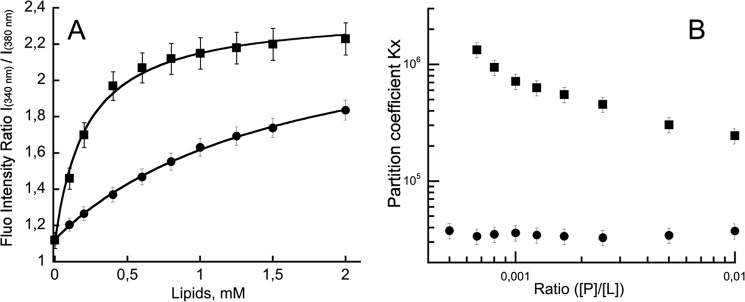
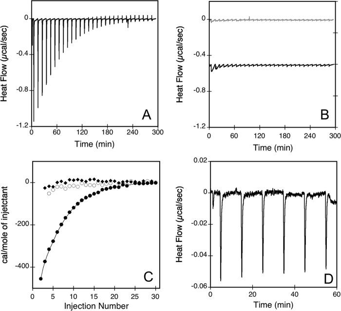
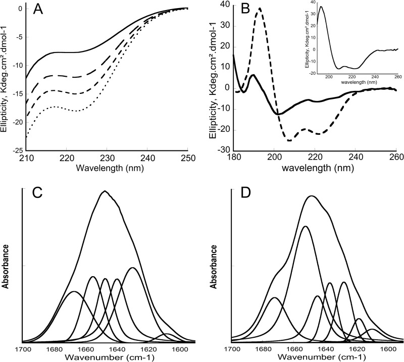
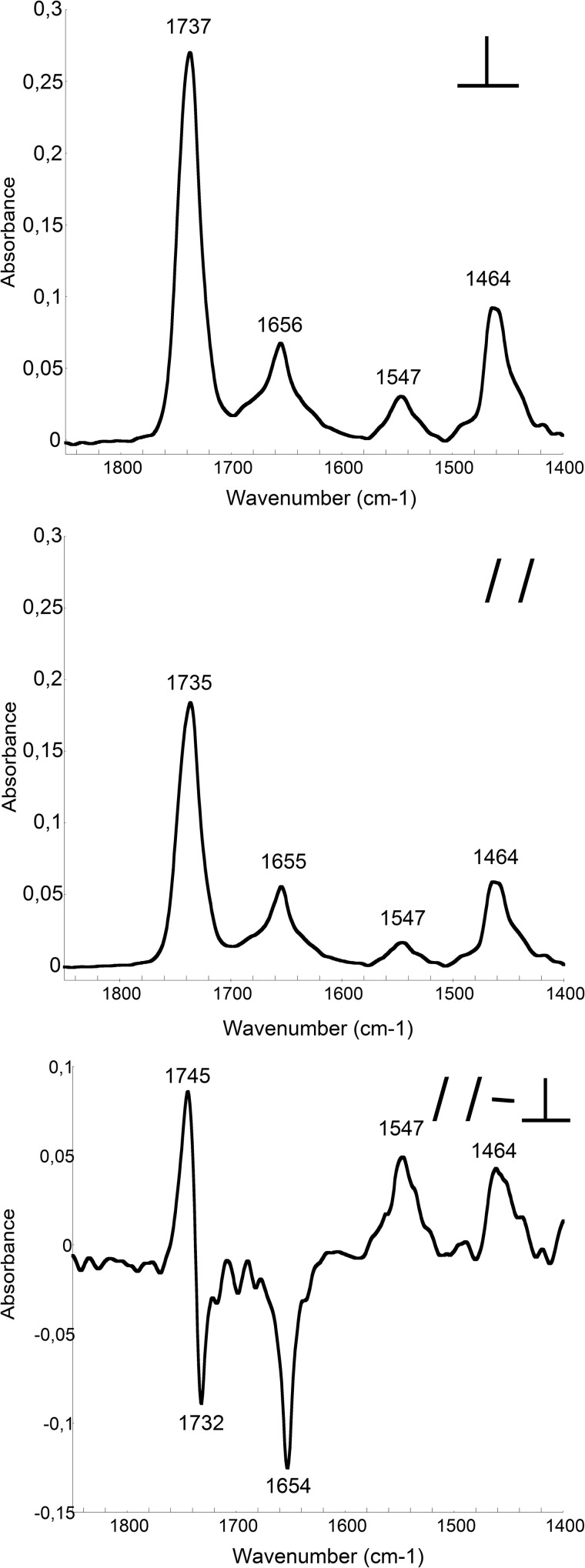
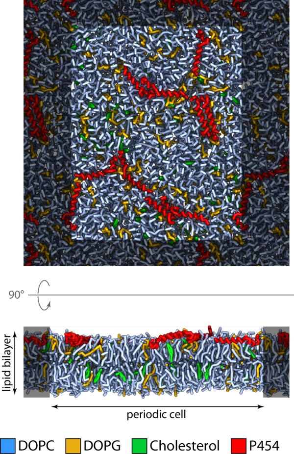
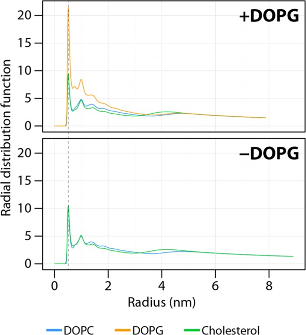
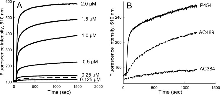
Bordetella pertussis, the pathogenic bacteria responsible for whooping cough, secretes several virulence factors, among which is the adenylate cyclase toxin (CyaA) that plays a crucial role in the early stages of human respiratory tract colonization. CyaA invades target cells by translocating its catalytic domain directly across the plasma membrane and overproduces cAMP, leading to cell death. The molecular process leading to the translocation of the catalytic domain remains largely unknown. We have previously shown that the catalytic domain per se, AC384, encompassing residues 1-384 of CyaA, did not interact with lipid bilayer, whereas a longer polypeptide, AC489, spanning residues 1-489, binds to membranes and permeabilizes vesicles. Moreover, deletion of residues 375-485 within CyaA abrogated the translocation of the catalytic domain into target cells. Here, we further identified within this region a peptidic segment that exhibits membrane interaction properties. A synthetic peptide, P454, corresponding to this sequence (residues 454-485 of CyaA) was characterized by various biophysical approaches. We found that P454 (i) binds to membranes containing anionic lipids, (ii) adopts an α-helical structure oriented in plane with respect to the lipid bilayer, and (iii) permeabilizes vesicles. We propose that the region encompassing the helix 454-485 of CyaA may insert into target cell membrane and induce a local destabilization of the lipid bilayer, thus favoring the translocation of the catalytic domain across the plasma membrane.

Keywords: Adenylate Cyclase (Adenylyl Cyclase); Bacterial Toxins; Circular Dichroism (CD); Infrared Spectroscopy; Membrane; Molecular Dynamics; Peptide Conformation; Phospholipid Vesicle.


Figures

FIGURE 1.
FIGURE 1.
P454 membrane partition followed by fluorescence. A, ratio of fluorescence intensity at 340 and 380 nm as a function of lipid concentration. The negatively charged small unilamellar vesicles (squares) or large unilamellar vesicles (circles) were made of DOPC/DOPE/DOPG/Chol (4:3:2:1). The P454 fluorescence intensity ratio values were fitted as described under “Materials and Methods,” providing partition coefficient KX and the Hill number (KX = 2.9 × 10⁵, nH = 1.35 for SUVs; KX = 3.9 × 10⁴, nH = 1.04 for LUVs). B, partition coefficient KX as a function of peptide/lipid ratio. Thermodynamic parameters are reported in supplemental Table S1. Error bars correspond to S.D.
FIGURE 2.
FIGURE 2.
Isothermal titration calorimetry of peptide membrane partition. The heat flow traces show the calorimetric titrations of P454 (A) and P414 (B, gray traces) by consecutive injections of negatively charged SUVs (DOPC/DOPE/DOPG/Chol, 4:3:2:1). B also shows the heat flow produced by injections of neutral SUVs (DOPC/DOPE/Chol, 6:3:1) into P454 (black traces; offset −0.5 μcal/s in the figure for a better visualization). Each peak corresponds to the injection of 5 μl of 20 mm SUVs into the ITC reaction cell containing 1.41 ml of 42 μm peptide in buffer B at 25 °C. C, integrated binding isotherm of P454 with negative SUVs (filled circles) or with neutral SUVs (empty circles) and P414 with negative SUVs (filled squares). D, heat flow trace of anionic SUVs titrated by a solution of P454. Each peak corresponds to the injection of 5 μl of 85 μm P454 into the ITC reaction cell containing 1.41 ml of 5 mm anionic SUVs in buffer B at 25 °C. Thermodynamic parameters are reported in supplemental Table S2.
FIGURE 3.
FIGURE 3.
Circular dichroism and FTIR spectra of P454. A, far-UV CD spectra of P454 (40 μm) in the absence (solid line) or in the presence (dashed lines) of increasing concentrations (0.5, 1, and 2 mm from top to bottom) of anionic SUVs made of DOPC/DOPE/DOPG/cholesterol (4:3:2:1) in buffer B at 25 °C. B, synchrotron radiation CD spectra of P454 (200 μm) in the absence (solid line) or in the presence (dashed line) of anionic SUVs (10 mm) in buffer B at 25 °C. C and D, transmission FTIR spectra and band deconvolutions of P454 (150 μm) in D2O buffer B (C) or in the presence (D) of anionic SUVs (5 mm lipids) in D2O buffer B at 25 °C. The S.D. value is within 5%. Secondary structure content analysis is reported in supplemental Table S3.
FIGURE 4.
FIGURE 4.
ATR-FTIR spectra of P454. The ATR-FTIR spectra of P454 mixed with lipids (DOPC/DOPE/DOPG/cholesterol, 4:3:2:1) were recorded with perpendicular (top) or parallel (middle) polarization of the infrared light. The resulting dichroic spectrum of P454 in membranes (bottom) is multiplied by a scaling factor of 8 for convenience (see “Materials and Methods”). The S.D. value is within 5%. Secondary structure content analysis is reported in supplemental Table S4.
FIGURE 5.
FIGURE 5.
Molecular dynamics of P454 inserted into an anionic lipid bilayer. Snapshot of the system after 1 μs of molecular dynamics simulation at constant temperature (323.15 K) and pressure (1 bar). Red, P454 peptides; light blue, DOPC; green, cholesterol; yellow-orange, DOPG. For clarity, water is not depicted.
FIGURE 6.
FIGURE 6.
Radial distribution function of the lipids around the center of mass of P454 in the presence or absence of DOPG in the lipid bilayer. The radial distribution function was calculated using the phosphate beads for DOPC and DOPG or the hydroxyl beads for cholesterol. The dashed line indicates the first shell of lipids around the P454 mass center. The small and wide peak at ∼4 nm corresponds to lipids on the opposite side of the bilayer.
FIGURE 7.
FIGURE 7.
Membrane permeabilization by P454. A, time course of ANTS and DPX efflux from negatively charged LUVs (0.45 mm lipids) made of DOPC/DOPE/DOPG/Chol (4:3:2:1 lipid ratio) in the presence of the indicated concentrations of P454 (solid lines). Dashed line, permeabilization of neutral LUVs (DOPC/DOPE/Chol at a 6:3:1 ratio) in the presence of 1 μm P454. B, time course of ANTS and DPX efflux from negatively charged LUVs (0.45 mm lipids) made of DOPC/DOPE/DOPG/Chol (4:3:2:1) in the presence of 0.3 μm P454, AC384, or AC489.

References

    1. Ladant D., Brezin C., Alonso J. M., Crenon I., Guiso N. (1986) Bordetella pertussis adenylate cyclase. Purification, characterization, and radioimmunoassay. J. Biol. Chem. 261, 16264–16269 - PubMed
    1. Glaser P., Ladant D., Sezer O., Pichot F., Ullmann A., Danchin A. (1988) The calmodulin-sensitive adenylate cyclase of Bordetella pertussis. Cloning and expression in Escherichia coli. Mol. Microbiol. 2, 19–30 - PubMed
    1. Ladant D., Ullmann A. (1999) Bordatella pertussis adenylate cyclase. A toxin with multiple talents. Trends Microbiol. 7, 172–176 - PubMed
    1. Guo Q., Shen Y., Lee Y. S., Gibbs C. S., Mrksich M., Tang W. J. (2005) Structural basis for the interaction of Bordetella pertussis adenylyl cyclase toxin with calmodulin. EMBO J. 24, 3190–3201 - PMC - PubMed
    1. Guo Q., Jureller J. E., Warren J. T., Solomaha E., Florián J., Tang W. J. (2008) Protein-protein docking and analysis reveal that two homologous bacterial adenylyl cyclase toxins interact with calmodulin differently. J. Biol. Chem. 283, 23836–23845 - PMC - PubMed

Publication types