. 2014 Jan;42(1):137-52.
doi: 10.1093/nar/gkt855. Epub 2013 Sep 24.

Differential involvement of E2A-corepressor interactions in distinct leukemogenic pathways

Affiliations

Differential involvement of E2A-corepressor interactions in distinct leukemogenic pathways

Chien-Hung Gow et al. Nucleic Acids Res. 2014 Jan.

Abstract

E2A is a member of the E-protein family of transcription factors. Previous studies have reported context-dependent regulation of E2A-dependent transcription. For example, whereas the E2A portion of the E2A-Pbx1 leukemia fusion protein mediates robust transcriptional activation in t(1;19) acute lymphoblastic leukemia, the transcriptional activity of wild-type E2A is silenced by high levels of corepressors, such as the AML1-ETO fusion protein in t(8;21) acute myeloid leukemia and ETO-2 in hematopoietic cells. Here, we show that, unlike the HEB E-protein, the activation domain 1 (AD1) of E2A has specifically reduced corepressor interaction due to E2A-specific amino acid changes in the p300/CBP and ETO target motif. Replacing E2A-AD1 with HEB-AD1 abolished the ability of E2A-Pbx1 to activate target genes and to induce cell transformation. On the other hand, the weak E2A-AD1-corepressor interaction imposes a critical importance on another ETO-interacting domain, downstream ETO-interacting sequence (DES), for corepressor-mediated repression. Deletion of DES abrogates silencing of E2A activity by AML1-ETO in t(8;21) leukemia cells or by ETO-2 in normal hematopoietic cells. Our results reveal an E2A-specific mechanism important for its context-dependent activation and repression function, and provide the first evidence for the differential involvement of E2A-corepressor interactions in distinct leukemogenic pathways.


Figures
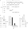
Figure 1.
Figure 1.
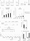
E2A-AD1 is a weak ETO-binding domain and requires DES for strong corepressor binding. (A) Schematic representations of E2A and its derivatives used in the experiment, indicating the regions of AD1, DES and AD2. (B and C) GST pull-down assays using the indicated GST fusion proteins and the in vitro translated and 35S labeled E2A derivatives. These derivatives were expressed as fusion proteins with the Gal4-DNA-binding domain. The right panel of (B) shows quantification of the relative binding of each E2A fragment to GST-ETO as the percentage of total input.
Figure 2.
Figure 2.
Mapping E2A-AD1 residues responsible for its reduced binding to the ETO corepressor. (A, C and F) GST pull-down assays comparing the interactions of wild-type and mutant E2A-AD1 and HEB-AD1 (A, C, F), or full-length HEB and E2A (A) with GST-ETO (A, C, F), GST-TAFH (A) or GST-KIX (C). In the right panel of (A), HEB and E2A were premixed before GST pull-down. (B) Luciferase reporter assays and mammalian two-hybrid assays comparing both the sensitivity of Gal4-E2A-AD1 and Gal4-HEB-AD1 to ETO-mediated repression, and their interaction with ETO. ‘Fold Repression’ (i.e. ETO-mediated repression) was calculated as the ratio of luciferase activities observed in the absence of ETO versus in the presence of ETO. ‘Fold Activation’ (i.e. ETO interaction) was calculated as the ratio of the luciferase activities observed in the presence of VP16-ETO versus in the presence of ETO. P-values were calculated from two-tailed t-test. *P < 0.05. (D) The quantification of the relative binding of E2A-AD1 and HEB-AD1 to GST-KIX and GST-ETO as shown in (C). The bindings of E2A-AD1 and HEB-AD1 to GST-KIX were similar and set to 1. (E) Comparison of E2A and HEB PCET sequences with E2A-specific residues at the C-terminal region underlined. (G) The quantification of the relative binding of wild-type and mutant E2A-AD1 and HEB-AD1 to GST-ETO as shown in (F).
Figure 3.
Figure 3.
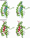
Modeling of TAFH interactions with HEB-PCET and E2A-PCET reveals direct involvement of HEB-specific C-terminal residues of PCET in complex formation with TAFH. Shown are stereo views of the modeled structures of HEB-PCET-TAFH (top) and E2A-PCET-TAFH (bottom). The final HEB (cyan) and E2A (red) docking structures from results involving semiflexible refinement of the PCET structure reveal differential intermolecular interactions of TAFH with HEB versus E2A in two areas: one area involving R151 and its interactions with S28 as well as the base of the HEB-PCET helix, and another area involving D19 and D22 of the HEB-PCET and their interactions with K98. In comparison, E2A only shows the interaction among the top portion of the helix involving D18. This, in turn, may explain the difference in binding affinities between HEB and E2A. Hydrogen-bond interactions are depicted by thin red lines.
Figure 4.
Figure 4.
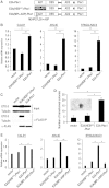
The weak corepressor binding of E2A-AD1 allows E2A-Pbx1 to bypass corepressor-mediated repression to facilitate its gene activation and oncogenic activities. (A) Schematic representation of wild-type E2A-Pbx1, E2A(HEBAD1)-Pbx1 that has E2A-AD1 replaced by HEB-AD1, and E2A(MPL-ASP)-Pbx1 that has residues M24, P27 and L28 mutated to A, S and P, respectively. (B) RT-qPCR showing that replacing E2A-AD1 by HEB-AD1 abolished the ability of E2A-Pbx1 to activate its endogenous target genes in transduced Namalwa cells. (C) Co-IP assays showing enhanced ETO-2 corepressor binding by E2A(HEBAD1)-Pbx1 in transduced Namalwa cells. (D) Colony formation assays performed in NIH3T3 cells showing dramatically reduced transforming capacity of E2A(HEBAD1)-Pbx1. The inset shows a typical transformed colony. (E) Effect of converting E2A-specific PCET residues to those of HEB on the ability of E2A-Pbx1 to activate target genes in transduced Namalwa cells. P values were calculated from two-tailed t-test. *P < 0.05. **P < 0.01.
Figure 5.
Figure 5.
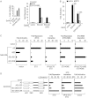
Both corepressor-binding domains (AD1 and DES) are important for the corepressor sensitivity of E2A. (A and B) Reporter assays showing that both DES (A) and AD1 (B) were required for the optimal E2A sensitivity to ETO-mediated repression. ‘Fold Activation’ was calculated as the ratio of the luciferase activity observed with each E2A derivative to the luciferase activity observed with Gal4-DBD. ‘Fold Repression’ was calculated as the ratio of the luciferase activities observed with each E2A derivative in the absence versus presence of ETO. (C) Mammalian two-hybrid assays showing that AD1 and DES cooperatively regulated the binding of E2A to ETO and the repression of E2A-dependent transcription. ‘ETO/ETO-K98E interaction’ was calculated as the ratio of the luciferase activities observed with each E2A derivative in the presence of VP16-ETO (or VP16-ETO-K98E) versus in the presence of ETO (or ETO-K98E). (D) Similar assays as in (C) showing that AD2 and DES opposingly regulated the corepressor sensitivity of E2A.
Figure 6.
Figure 6.
In vivo importance of DES for corepressor-mediated silencing of the transcriptional activity of E2A. (A) Co-IP showing that DES was critical for E2A to form complex with endogenous AML1-ETO in Kasumi-1 cells. (B) GST pull-down of Kasumi-1 whole cell lysate showing that E2A-AD1 had a much weaker interaction with AML1-ETO, but acquired increased binding to coactivators as compared with HEB-AD1. (C) Knockdown of AML1-ETO in Kasumi-1 cells increased the expression of SLA as assayed by RT-PCR. The 18S rRNA was used as the internal control. (D) FLAG-tagged E2A and E2AΔDES showed similar binding to an enhancer element of SLA in transduced Kasumi-1 cells. The anti-FLAG antibody was used. (E) DES was required for the silencing of E2A transcriptional activity by AML1-ETO in Kasumi-1 cells. (Left) RT-qPCR showing that ectopic expression of E2AΔDES, but not E2A, activated SLA expression. (Middle and Right) ChIP assays showing that ectopic expression of E2AΔDES, but not E2A, reduced the occupancy of AML1-ETO and enhanced GCN5 occupancy on SLA enhancer. (F) RT-qPCR assays showing that DES was required for ETO-2 to repress E2A-dependent activation or to potentiate E2A-dependent repression of distinct target genes in transduced Jurkat cells. P-values were calculated from two-tailed t-test. *P < 0.05. **P < 0.01. n.s., not significant.
Figure 7.
Figure 7.
A mechanistic model depicting the role of E2A domains in the assembly of corepressor and coactivator complexes, and their importance for the transcriptional activity of E2A-Pbx1 and AML1-ETO leukemia fusion proteins. (A) We propose that the formation of stable E2A complexes with coactivators (CoAs) or corepressors (CoRs) requires cooperative interactions with two E2A domains. TAFH and NHR2 are the conserved domains of ETO family of CoRs that interact with AD1 and DES, respectively. At low CoR levels, due to the defective E2A-AD1-CoR interaction (shown by the yellow asterisk), the balance of CoR and CoA interactions favors the assembly of an E2A-CoA complex, explaining the high level of gene activation by E2A-Pbx1. At high CoR levels, CoRs such as AML1-ETO and ETO-2 drive the formation of their complexes with E2A through a stepwise mechanism that involves the formation of an intermediate complex containing both CoRs and CoAs. The formation of the intermediate complex before the formation of the stable E2A-CoR complex is facilitated by DES-dependent interactions with CoRs, explaining the importance of DES in CoR-mediated repression of E2A. The dashed lines denote competitive AD1 interactions with CoA and CoR, which dictate the formation of stable CoA or CoR complexes. (B) The E2A-specific changes in the PCET motif destabilize CoR binding, which is required for the ability of E2A-Pbx1 to drive gene activation in t(1;19) leukemia. (C) The DES domain is required for the repression of AML1-ETO/E2A target genes in AML1-ETO-dependent t(8;21) leukemia.

Similar articles

Cited by

References

    1. Murre C, Bain G, van Dijk MA, Engel I, Furnari BA, Massari ME, Matthews JR, Quong MW, Rivera RR, Stuiver MH. Structure and function of helix-loop-helix proteins. Biochim. Biophys. Acta. 1994;1218:129–135. - PubMed
    1. Massari ME, Murre C. Helix-loop-helix proteins: regulators of transcription in eucaryotic organisms. Mol. Cell. Biol. 2000;20:429–440. - PMC - PubMed
    1. Kee BL, Quong MW, Murre C. E2A proteins: essential regulators at multiple stages of B-cell development. Immunol. Rev. 2000;175:138–149. - PubMed
    1. Bain G, Maandag EC, Izon DJ, Amsen D, Kruisbeek AM, Weintraub BC, Krop I, Schlissel MS, Feeney AJ, van Roon M, et al. E2A proteins are required for proper B cell development and initiation of immunoglobulin gene rearrangements. Cell. 1994;79:885–892. - PubMed
    1. Zhuang Y, Soriano P, Weintraub H. The helix-loop-helix gene E2A is required for B cell formation. Cell. 1994;79:875–884. - PubMed

Publication types

MeSH terms

Substances