. 2014 Feb 15;23(4):843-54.
doi: 10.1093/hmg/ddt468. Epub 2013 Sep 24.

Neuronal accumulation of glucosylceramide in a mouse model of neuronopathic Gaucher disease leads to neurodegeneration

Affiliations

Neuronal accumulation of glucosylceramide in a mouse model of neuronopathic Gaucher disease leads to neurodegeneration

Tamar Farfel-Becker et al. Hum Mol Genet. .

Abstract

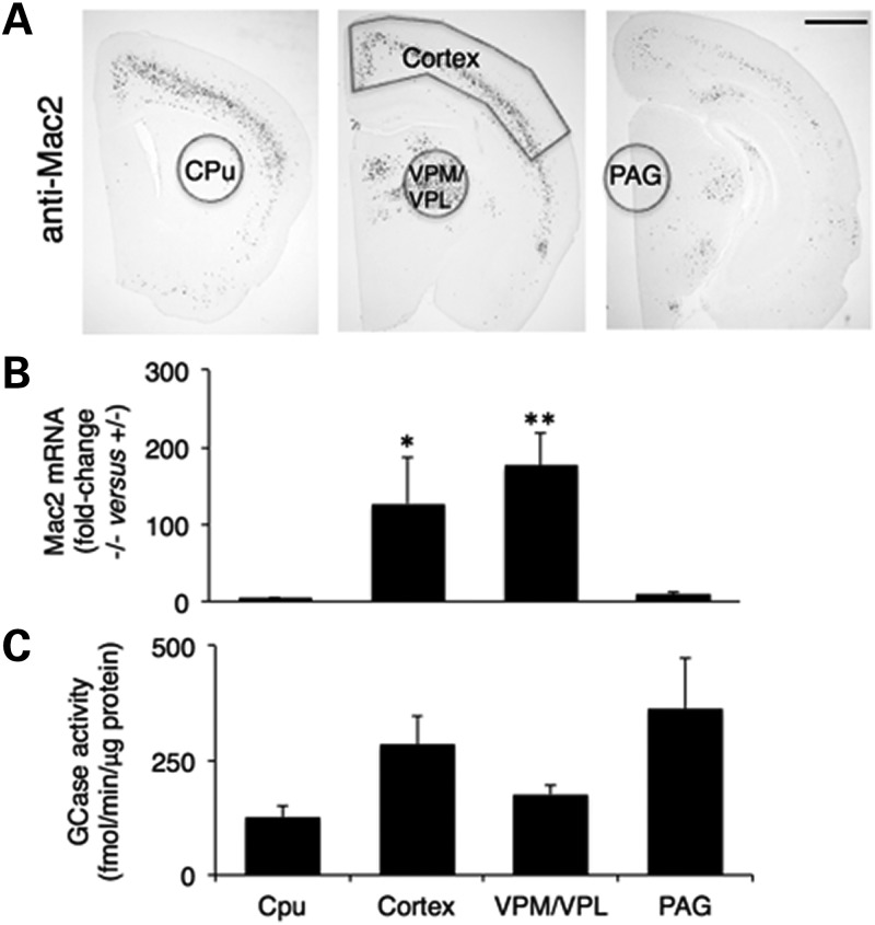
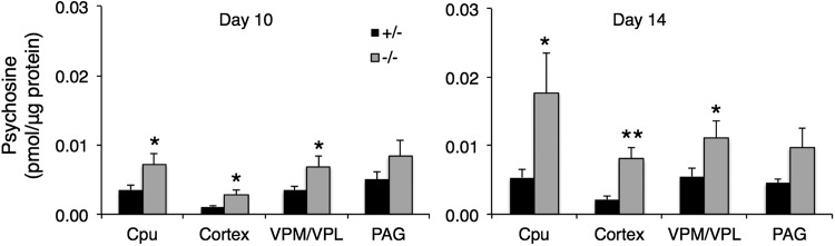
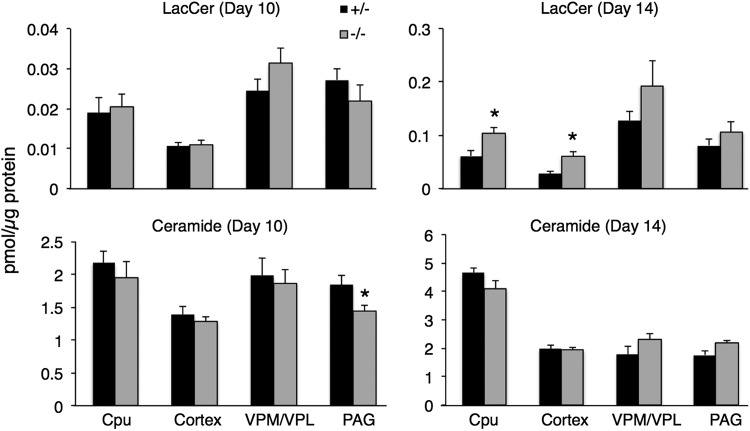
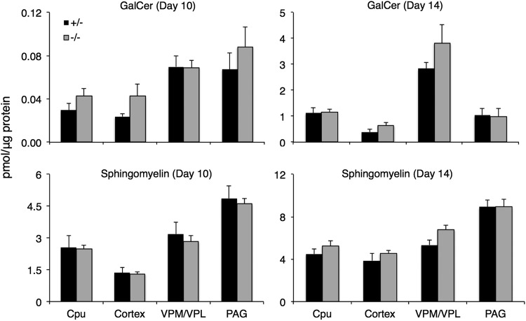
Gaucher disease has recently received wide attention due to the unexpected discovery that it is a genetic risk factor for Parkinson's disease. Gaucher disease is caused by the defective activity of the lysosomal enzyme, glucocerebrosidase (GCase; GBA1), resulting in intracellular accumulation of the glycosphingolipids, glucosylceramide and psychosine. The rare neuronopathic forms of GD (nGD) are characterized by profound neurological impairment and neuronal cell death. We have previously described the progression of neuropathological changes in a mouse model of nGD. We now examine the relationship between glycosphingolipid accumulation and initiation of pathology at two pre-symptomatic stages of the disease in four different brain areas which display differential degrees of susceptibility to GCase deficiency. Liquid chromatography electrospray ionization tandem mass spectrometry demonstrated glucosylceramide and psychosine accumulation in nGD brains prior to the appearance of neuroinflammation, although only glucosylceramide accumulation correlated with neuroinflammation and neuron loss. Levels of other sphingolipids, including the pro-apoptotic lipid, ceramide, were mostly unaltered. Transmission electron microscopy revealed that glucosylceramide accumulation occurs in neurons, mostly in the form of membrane-delimited pseudo-tubules located near the nucleus. Highly disrupted glucosylceramide-storing cells, which are likely degenerating neurons containing massive inclusions, numerous autophagosomes and unique ultrastructural features, were also observed. Together, our results indicate that a certain level of neuronal glucosylceramide storage is required to trigger neuropathological changes in affected brain areas, while other brain areas containing similar glucosylceramide levels are unaltered, presumably because of intrinsic differences in neuronal properties, or in the neuronal environment, between various brain regions.


Figures

Figure 1.
Figure 1.
Characterization of brain areas used in this study. (A) Immunohistochemical labeling for Mac2 on coronal brain sections from 16-day-old −/− mice. Brain areas that were subsequently used for biochemical analysis are indicated. No staining was observed in control brains. Scale bar = 1 mm. (B) Quantitative PCR analysis of Mac2 mRNA in different brain areas of 14-day-old mice. Values are means ± s.e.m, n = 5. *P < 0.05, **P < 0.01. (C) GCase activity in nGD brains. Values are means ± s.e.m., n = 3.
Figure 2.
Figure 2.
GlcCer levels and N-acyl chain species in nGD brains. Total GlcCer levels are shown in the upper panels and N-acyl chain length distribution in the bottom panels. Values are means ± s.e.m., n = 4–5. *P < 0.05, **P < 0.01.
Figure 3.
Figure 3.
Psychosine levels in nGD brains at 10 and 14 days. Values are means ± s.e.m., n = 4–5. *P < 0.05, **P < 0.01.
Figure 4.
Figure 4.
Lactosylceramide and ceramide levels in nGD brains at 10 and 14 days. Values are means ± s.e.m., n = 4–5.*P < 0.05.
Figure 5.
Figure 5.
Galactosylceramide and sphingomyelin levels in nGD brains at 10 and 14 days. Values are means ± s.e.m., n = 4–5.
Figure 6.
Figure 6.
Levels of long chain bases in nGD brains at 10 and 14 days. n = 4–5 ± s.e.m. Values are means ± s.e.m., n = 4–5. *P < 0.05.
Figure 7.
Figure 7.
Ultrastructure of GlcCer storage in nGD neurons. Neurons were from the cerebral cortex of −/− mice at 14 (AF) and 21 (G) days of age. (A–D) A prominent vacuolar inclusion (arrows in A) in the perikaryoplasm of a large neuron to the left of the nucleus, containing dilute arrays of fibrils and pseudotubules, most of which are longitudinal but sometimes transverse as in the upper vacuole (arrowhead in A), which is probably a continuation of the same membrane-bound compartment indicated by the arrows. (B–D) Higher magnifications of the inclusions so as to resolve the encompassing membrane and the tubular appearance of its contents (C and D) seen in cross-section in (C) (arrowheads). An unrelated inclusion body (arrow) is also present in (B). (E and F) A large neuron with an inclusion body (arrows) (∼9 µm in length) showing more densely packed pseudotubules. A rounded vacuole in (F) includes dense fibrils (arrowhead) that might correspond to flattened GlcCer bilayer stacks (32). (G) A neuron with pseudotubules (arrows) running into a process which is not clearly membrane-delimited (further magnified in the inset). Scale bars are all 2 µm except for (C) and (D) which are 0.5 µm.
Figure 8.
Figure 8.
Distention of ER cisternae in nGD neurons. Sample arrays of rough ER in cerebral cortex neurons of (A) +/− and (B) −/− mice at 14 days of age. Scale bars are 2 µm.
Figure 9.
Figure 9.
Neurons showing progression of storage and additional pathology. Samples were from the cerebral cortex of nGD mice at 18 days of age (AF) and from the VPM/VPL at 21 days of age (G). (A) A cell with a modestly heterochromatic nucleus (arrow) and an eccentric nucleolus showing numerous pseudotubular inclusions (arrowhead), or amassed with other inclusions (lower left-half). (B) A magnification of the lower-left corner of (A) shows some inclusions that are continuous (arrowhead sequence) with the plasma membrane and extracellular space (electron-lucent area in lower left). (C) A magnification of the upper right region of the cell shown in (A) shows cytoplasmic extensions (dots mark the cell perimeter). Note the presence of long, relatively-narrow ER cisternae (indicated by arrowheads). (D–F) Enlargement of portions from the lower part of the cell shown in (A) shows morphologically diverse constituents (D and F), separate inclusion bodies tethered together through narrow extensions (arrowheads in D and E), and bodies containing densely packed arrays of pseudotubules at various orientations (arrows in D and F). (G) A cell with a somewhat heterochromatic nucleus (arrow) and huge inclusion (arrowheads) of up to ∼12 µm in length, containing densely packed pseudotubules, many of which are oriented longitudinally. Scale bars: (A) 4 µm; (B) 1 µm; (C and G) 2 µm; (D–F) 0.5 µm.
Figure 10.
Figure 10.
Additional ultrastructural features of highly disrupted neurons. Samples were from the cerebral cortex of nGD mice at 18 days of age. (A) A cell with a large mass of pseudotubules (arrow) and one very large (∼7 µm) membrane-bound, pleiomorphic inclusion (curved arrow). The cell nucleus, cut off at the upper right, is shown fully in the inset. The large, independent pseudotubule mass shows an extension (thin arrow) leading to another small inclusion. The path of another longer extension suggests continuity with the plasma membrane and extracellular space (arrowheads). The cell has several autophagosomes (on the right of the asterisks). (B and C) Magnifications of two autophagosomes from the cell shown in (A), with both showing a double membrane and one (B) in the process of fusing with the pleiomorphic body on its right. (D) This cell also shows a large pseudotubule-containing body (arrow), a pleiomorphic membrane-bounded mass (curved arrow) (up to 8 µm), several short cytoplasmic extensions (upper and lower right) and numerous autophagosomes (thin arrows). The cell displays two heterochromatic nuclear profiles (arrowheads). Scale bars: (A), (A) inset, (D) 2 µm; (B and C) 0.5 µm.

References

    1. Farfel-Becker T., Futerman A.H. Cellular pathogenesis in sphingolipid storage disorders: the quest for new therapeutic approaches. Clin. Lipidol. 2010;5:255–265.
    1. Vitner E.B., Platt F.M., Futerman A.H. Common and uncommon pathogenic cascades in lysosomal storage diseases. J. Biol. Chem. 2010;285:20423–20427. - PMC - PubMed
    1. Beutler E., Grabowski G.A. Gaucher Disease. In: Scriver C., Beaudet A.L., Valle D., Sly W.S., Childs B., editors. The Metabolic and Molecular Bases of Inherited Disease. Vol. 3. New-York: McGraw-Hill; 2001. pp. 3635–3668.
    1. Mignot C., Doummar D., Maire I., De Villemeur T.B. French Type 2 Gaucher Disease Study Group. Type 2 Gaucher disease: 15 new cases and review of the literature. Brain Dev. 2006;28:39–48. - PubMed
    1. Gupta N., Oppenheim I.M., Kauvar E.F., Tayebi N., Sidransky E. Type 2 Gaucher disease: phenotypic variation and genotypic heterogeneity. Blood Cells Mol. Dis. 2011;46:75–84. - PMC - PubMed

Publication types