Review
. 2013 Dec;154(12):4450-68.
doi: 10.1210/en.2013-1652. Epub 2013 Sep 24.

A role for glucocorticoids in stress-impaired reproduction: beyond the hypothalamus and pituitary

Affiliations
Review

A role for glucocorticoids in stress-impaired reproduction: beyond the hypothalamus and pituitary

Shannon Whirledge et al. Endocrinology. 2013 Dec.

Abstract

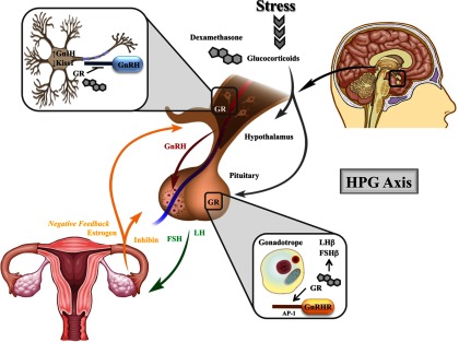
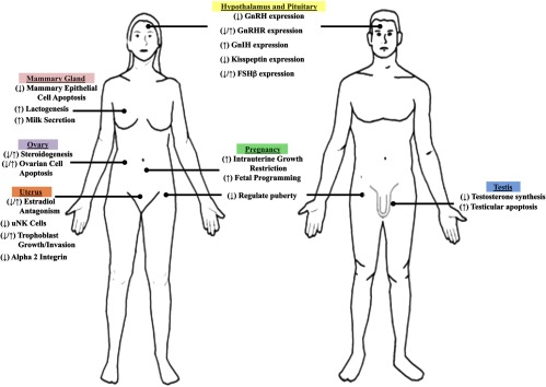
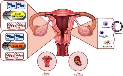
In addition to the well-characterized role of the sex steroid receptors in regulating fertility and reproduction, reproductive events are also mediated by the hypothalamic-pituitary-adrenal axis in response to an individual's environment. Glucocorticoid secretion in response to stress contributes to the well-characterized suppression of the hypothalamic-pituitary-gonadal axis through central actions in the hypothalamus and pituitary. However, both animal and in vitro studies indicate that other components of the reproductive system are also regulated by glucocorticoids. Furthermore, in the absence of stress, it appears that homeostatic glucocorticoid signaling plays a significant role in reproduction and fertility in all tissues comprising the hypothalamic-pituitary-gonadal axis. Indeed, as central regulators of the immune response, glucocorticoids are uniquely poised to integrate an individual's infectious, inflammatory, stress, nutritional, and metabolic status through glucocorticoid receptor signaling in target tissues. Endocrine signaling between tissues regulating the immune and stress response and those determining reproductive status provides an evolutionary advantage, facilitating the trade-off between reproductive investment and offspring fitness. This review focuses on the actions of glucocorticoids in tissues important for fertility and reproduction, highlighting recent studies that show glucocorticoid signaling plays a significant role throughout the hypothalamic-pituitary-gonadal axis and characterizing these effects as permissive or inhibitory in terms of facilitating reproductive success.


Figures

Figure 1.
Figure 1.
Organization of and modifications to the human GR gene contribute to signaling diversity A, The human GR gene (NR3C1) is contained in one locus on chromosome 5 (5q31). Nine exons comprise the human GR primary transcript. Exon 1 forms the 5′-untranslated region, whereas exons 2–9 form the protein-coding region. Exon 2 encodes most of the NTD (N-terminal domain), exons 3 and 4 encode the DBD (DNA-binding domain), and exons 5–9 encode the hinge (H) region and LBD (ligand-binding domain). Alternative splicing of the primary transcript results in the α- and β-transcriptional human GR isoforms. B, Translational initiation at 8 different AUG start codons in a single human GR mRNA produces 8 receptor isoforms with progressively shorter NTDs (isoforms A–D3). Alternative posttranslational modifications also contribute to the diversity of human GR. Suggested and validated sites of phosphorylation (P), sumoylation (S), and acetylation (A) are indicated. C, The GR signals as a ligand-dependent transcription factor, where unliganded GR resides primarily in the cytoplasm of cells as part of a multiprotein complex. Cortisol, the most abundant natural glucocorticoid in humans, is converted from inactive cortisone by 11β-HSD I. As a mechanism for local regulation of glucocorticoid action, the biologically active form cortisol can be converted to the inactive cortisone by 11β-HSD II. Upon binding glucocorticoids, GR undergoes a conformational change, dissociates from the heterocomplex, and translocates into the nucleus. Ligand binding is also responsible for a hyperphosphorylated state of the GR protein. Once within the nucleus, GR regulates transcription of target genes by direct binding to GREs (5′-AGAACAnnnTGTTCT-3′, where “n” is any nucleotide) or nGREs (5′-CTCCnGGAGA-3′ nGRE 1, nGRE identified with variable number of spacer nucleotides 0–2), in a composite manner bound to a GRE and interacting with adjacent transcription factor binding sites (STAT5), or tethered to other transcription factors without direct DNA binding (NFκB). 11β-HSB, 11-β hydroxysteroid dehydrogenase; HSP: heat shock protein; NPC: nuclear pore complex; nGRE: negative GRE; p65: nuclear factor NF-κ-B p65 subunit; p50: nuclear factor NF-κ-B p50 subunit.
Figure 2.
Figure 2.
Neuroendocrine regulation of the HPG axis by glugocorticoids. As part of the HPG axis, GnRH is a central driver of the reproductive axis, responsible for the synthesis and secretion of the gonadotropins LH and FSH from the pituitary. In the female reproductive tract, LH and FSH stimulate the ovaries to produce estrogen and are required for ovulation. As part of a classical feedback loop, hormones produced by the ovaries (estrogen and inhibin) inhibit the production of GnRH and the gonadotropins. Stress-induced increases in circulating glucocorticoids or exogenous administration of the synthetic glucocorticoid dexamethasone are largely inhibitory to the reproductive functions of the hypothalamus and pituitary. GR signaling in the hypothalamus is believed to be directly involved in repressing transcription of GnRH. Furthermore, glucocorticoids are also believed to regulate the number of GnIH and kisspeptin (Kiss1) expressing neurons. In the pituitary, both the natural ligand and dexamethasone regulate gonadotropin subunit expression, although both inhibitory and stimulatory effects have been described. In vitro studies suggest dexamethasone directly induces GnRHR expression. These findings indicate glucocorticoids may influence the neuroendocrine functions of the HPG axis both positively and negatively, which provides a mechanism by which glucocorticoids can respond to periods of extreme stress, as well as homeostatic conditions. AP-1, activator protein 1.
Figure 3.
Figure 3.
The role of glucocorticoids in tissues of the reproductive tract. The schematic represents several highlighted roles of glucocorticoids (stress-induced or exogenous exposure) in major organs of the reproductive tract. Common central functions in the hypothalamus and pituitary are indicated for the male and female. Sex-specific organs are also indicated. Glucocorticoids induce both repressive and enhancing effects, indicated by right arrow and left arrow, respectively.
Figure 4.
Figure 4.
Impact of glucocorticoid signaling in the uterus. Glucocorticoids regulate many of the signal transduction and biological processes that are important for uterine function and successful reproduction. In a human uterine leiomyoma cell line and human uterine endometrial cell line, glucocorticoids regulate many unique gene targets and also target many estradiol-regulated genes. In endometrial cells, estradiol antagonizes the transcription of common immune-modulatory GR-target genes, GILZ, MAPK phosphatase 1 (MKP-1), and SGK. Altered GR and ERα recruitment to the promoter of GILZ is likely part of the mechanism by which GILZ expression is antagonized. It is not yet known what mechanisms are involved in the coregulation of common glucocorticoid and estradiol target genes. As potent regulators of the immune system, glucocorticoids inhibit aspects of the inflammatory-like response to estradiol. The immunosuppressant actions of glucocorticoids may play a role in preventing immune rejection of the implanting blastocyst. Clinical studies have indicated that glucocorticoids can influence uterine NK cells, enhancing pregnancy outcomes in some women. Macrophages and dendritic cells play a role in the immune control of endometrial receptivity to embryo implantation and are immune cell types regulated by glucocorticoids. Glucocorticoids can act in both early and late pregnancy to affect the growth and development of the fetus. Both prenatal stress and maternal exposure to exogenous glucocorticoids can lead to fetal programming of the HPA axis. MKP-1: MAPK phosphatase 1; SGK: serum- and glucocorticoid-induced protein kinase.

References

    1. Stearns SC. Life-history tactics: a review of the ideas. Q Rev Biol. 1976;51(1):3–47 - PubMed
    1. Wingfield JC, Sapolsky RM. Reproduction and resistance to stress: when and how. J Neuroendocrinol. 2003;15(8):711–724 - PubMed
    1. Sayers G. The adrenal cortex and homoestasis. Physiol Rev. 1950;30(3):241–320 - PubMed
    1. Welt ID, Stetten D, Jr, Ingle DJ, Morley EH. Effect of cortisone upon rates of glucose production and oxidation in the rat. J Biol Chem. 1952;197(1):57–66 - PubMed
    1. Galosy RA, Clarke LK, Vasko MR, Crawford IL. Neurophysiology and neuropharmacology of cardiovascular regulation and stress. Neurosci Biobehav Rev. 1981;5(1): 137–175 - PubMed

Publication types

Substances