

Revealing the properties of plant defensins through dynamics
- PMID: 24064452
- PMCID: PMC6270066
- DOI: 10.3390/molecules180911311
Revealing the properties of plant defensins through dynamics
Abstract
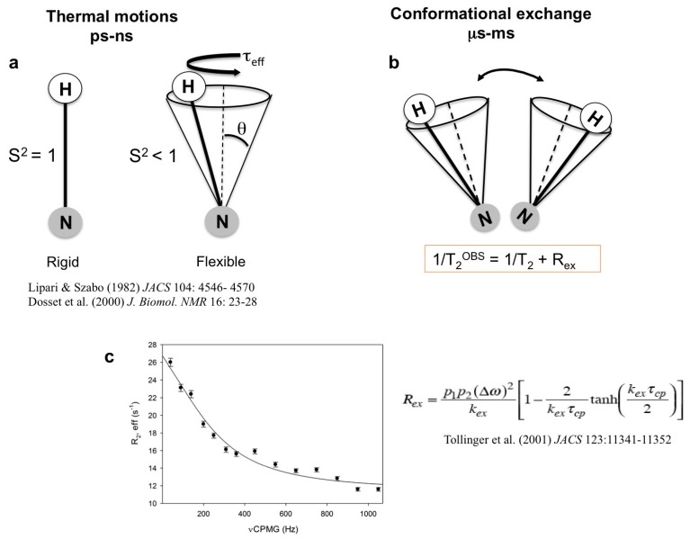
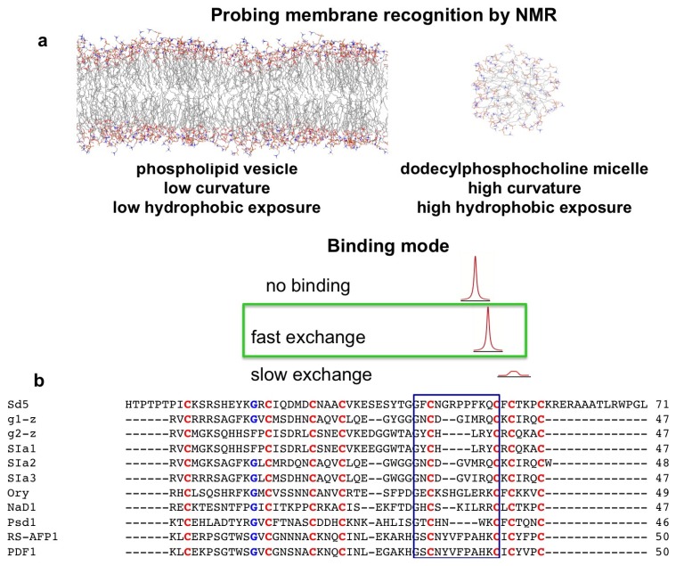
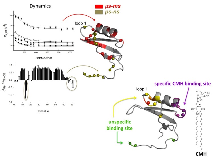
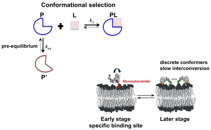
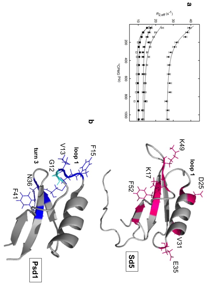
Defensins are potent, ancient natural antibiotics that are present in organisms ranging from lower organisms to humans. Although the structures of several defensins have been well characterized, the dynamics of only a few have been studied. This review discusses the diverse dynamics of two plant defensins for which the structure and dynamics have been characterized, both in the free state and in the presence of target membranes. Multiple motions are observed in loops and in secondary structure elements and may be related to twisting or breathing of the α-helix and β-sheet. This complex behavior is altered in the presence of an interface and is responsive to the presence of the putative target. The stages of membrane recognition and disruption can be mapped over a large time scale range, demonstrating that defensins in solution exist as an ensemble of different conformations, a subset of which is selected upon membrane binding. Therefore, studies on the dynamics have revealed that defensins interact with membranes through a mechanism of conformational selection.
Figures
Similar articles
-
Portrayal of complex dynamic properties of sugarcane defensin 5 by NMR: multiple motions associated with membrane interaction.Structure. 2011 Jan 12;19(1):26-36. doi: 10.1016/j.str.2010.11.011. Structure. 2011. PMID: 21220113
-
Antifungal plant defensins: mechanisms of action and production.Molecules. 2014 Aug 14;19(8):12280-303. doi: 10.3390/molecules190812280. Molecules. 2014. PMID: 25153857 Free PMC article. Review.
-
X-ray structure of a carpet-like antimicrobial defensin-phospholipid membrane disruption complex.Nat Commun. 2018 May 17;9(1):1962. doi: 10.1038/s41467-018-04434-y. Nat Commun. 2018. PMID: 29773800 Free PMC article.
-
Plant defensins--prospects for the biological functions and biotechnological properties.Peptides. 2009 May;30(5):1007-20. doi: 10.1016/j.peptides.2009.01.018. Epub 2009 Feb 7. Peptides. 2009. PMID: 19428780 Review.
-
Structural characterization of plant defensin protein superfamily.Mol Biol Rep. 2012 Apr;39(4):4461-9. doi: 10.1007/s11033-011-1235-y. Epub 2011 Sep 25. Mol Biol Rep. 2012. PMID: 21947884
Cited by
-
In Vivo Applicability of Neosartorya fischeri Antifungal Protein 2 (NFAP2) in Treatment of Vulvovaginal Candidiasis.Antimicrob Agents Chemother. 2019 Jan 29;63(2):e01777-18. doi: 10.1128/AAC.01777-18. Print 2019 Feb. Antimicrob Agents Chemother. 2019. PMID: 30478163 Free PMC article.
-
Antifungal defensins and their role in plant defense.Front Microbiol. 2014 Apr 2;5:116. doi: 10.3389/fmicb.2014.00116. eCollection 2014. Front Microbiol. 2014. PMID: 24765086 Free PMC article. Review.
-
Overexpression of a defensin enhances resistance to a fruit-specific anthracnose fungus in pepper.PLoS One. 2014 May 21;9(5):e97936. doi: 10.1371/journal.pone.0097936. eCollection 2014. PLoS One. 2014. PMID: 24848280 Free PMC article.
-
Plant Defensins from a Structural Perspective.Int J Mol Sci. 2020 Jul 26;21(15):5307. doi: 10.3390/ijms21155307. Int J Mol Sci. 2020. PMID: 32722628 Free PMC article. Review.
-
Structural and Dynamic Insights of the Interaction between Tritrpticin and Micelles: An NMR Study.Biophys J. 2016 Dec 20;111(12):2676-2688. doi: 10.1016/j.bpj.2016.10.034. Biophys J. 2016. PMID: 28002744 Free PMC article.
References
-
- Fisher E. Eifluss der configuration auf die wirkung der enzyme. Ber. Dtsch. Chem. Ges. 1894;27:2984–2993.
Publication types
MeSH terms
Substances
LinkOut - more resources
Full Text Sources
Other Literature Sources
