Review
. 2013 Nov;14(11):713-26.
doi: 10.1038/nrm3667. Epub 2013 Sep 25.

Functions and mechanics of dynein motor proteins

Affiliations
Review

Functions and mechanics of dynein motor proteins

Anthony J Roberts et al. Nat Rev Mol Cell Biol. 2013 Nov.

Abstract

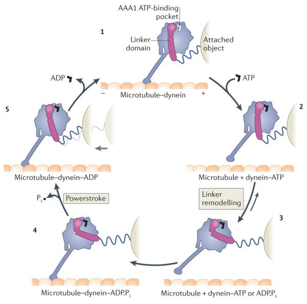
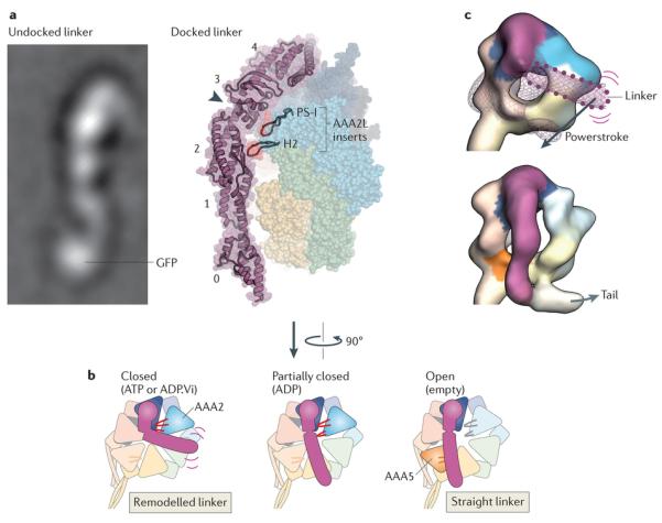
Fuelled by ATP hydrolysis, dyneins generate force and movement on microtubules in a wealth of biological processes, including ciliary beating, cell division and intracellular transport. The large mass and complexity of dynein motors have made elucidating their mechanisms a sizable task. Yet, through a combination of approaches, including X-ray crystallography, cryo-electron microscopy, single-molecule assays and biochemical experiments, important progress has been made towards understanding how these giant motor proteins work. From these studies, a model for the mechanochemical cycle of dynein is emerging, in which nucleotide-driven flexing motions within the AAA+ ring of dynein alter the affinity of its microtubule-binding stalk and reshape its mechanical element to generate movement.


Figures

Figure 1
Figure 1. Sites of dynein action in the cell
a | Example functions of cytoplasmic dynein (green) are shown in an interphase cell (left) and a dividing cell (right). The polarity of microtubules is indicated by plus signs. The arrow depicts the direction of dynein movement towards the microtubule minus end. Note that in some cell types and regions, such as the dendritic arbors of neurons, the microtubule network can have mixed polarity. b | Dynein functions in cilia. Intraflagellar transport (IFT) dynein (pink) performs retrograde IFT, whereas axonemal dyneins (cyan) power the beating of motile cilia.
Figure 2
Figure 2. Overview of dynein composition
a | Linear representation of domains within the dynein heavy chain. The amino-terminal tail domain is involved in dynein oligomerization, cargo-binding and regulation, but is not part of the minimal motor domain capable of producing movement in vitro. The motor domain comprises the linker domain, six AAA+ modules (1–6), the coiled-coil stalk and strut, and the carboxy-terminal region. The motor domain of Dictyostelium discoideum dynein has a molecular mass of ~380 kDa. In many fungal dyneins, the C-terminal region is shorter and the motor domain is ~330 kDa. Each of the AAA+ modules is composed of a large N-terminal subdomain and a smaller C-terminal subdomain (see inset and Box 2). b | The cytoplasmic dynein complex contains a pair of identical heavy chains. Within each heavy chain, the six AAA+ modules fold into a ring. The stalk protrudes as an extension from the small subdomain of AAA4. The tail is connected to AAA1 by the linker domain, which arches over the AAA+ ring. In Chlamydomonas reinhardtii inner arm dynein-c, a sharp ~90° kink exists between the linker and the tail, , . Although not yet visualized in cytoplasmic dynein, a similar kink might exist, as it would prevent a steric clash between the tail and the microtubule. In dynein-c, this neck region of the tail is a natural site of flexibility in the molecule, allowing the angle of the tail to vary with respect to the motor domain, , . The cytoplasmic dynein heavy chains assemble with up to five types of associated subunit, which are also dimers. The associated subunits comprise the intermediate chain, the light-intermediate chain and three classes of light chain: TCTEX, LC8 and Roadblock. Dashed lines indicate reported interactions of the associated subunits with each other and with the tail. The three-dimensional (3D) arrangement of the associated subunits with respect to the tail is unknown. c | A 3D model of the cytoplasmic dynein motor domain bound to the microtubule (the associated subunits are not shown). As no high-resolution structure currently exists for the entire motor domain bound to a tubulin dimer, this model is based on a 2.8 Å crystal structure of the D. discoideum dynein motor domain lacking the microtubule-binding domain (Protein Data Bank ID: 3VKG), joined to a cryo-electron microscopy-derived model of the mouse microtubule-binding domain bound to an α-tubulin–β-tubulin dimer (Protein Data Bank ID: 3J1T). Subdomains are shown in surface representation, with the two long α-helices in the stalk rendered separately to emphasize their coiled-coil arrangement. The six AAA+ modules are numerically labelled.
Figure 3
Figure 3. Model of the mechanochemical cycle of a cytoplasmic dynein motor domain
Model for the Dictyostelium discoideum cytoplasmic dynein motor domain, , , , , . Plus and minus signs indicate microtubule polarity. The dynein motor domain moves towards the minus end of the microtubule. Force exerted on an attached object is shown schematically by the stretching of a spring, which does not represent a part of the dynein structure. Conceptually, the attached object could represent a partnering motor domain in a cytoplasmic dynein dimer or the cargo microtubule of an axonemal dynein. With no nucleotide bound at the AAA1 ATPase site, the dynein motor domain is tightly bound to the microtubule (1). ATP binding induces rapid dissociation from the microtubule (2) (Fig. 4). The dissociation rate is ~460 s–1 for the D . discoideum dynein motor domain. A slower ATP-driven change (~200 s–1) is the remodelling of the linker domain, which is displaced across the AAA+ ring (3) (Fig. 5). Remodelling of the linker extends the search range of the microtubule-binding domain along the microtubule. After hydrolysis of ATP to ADP and inorganic phosphate (Pi), the motor domain is thought to engage a new binding site on the microtubule, initially via a weak interaction (4). Strong binding to the microtubule accelerates the release of Pi from AAA1, inducing the linker to revert to its straight form. This transition is speculated to represent the powerstroke: the main step in which force (indicated by the light grey arrow) is transmitted to the attached object (5). Finally, ADP is released from AAA1 and the cycle restarts. The occupancy of other nucleotide-binding AAA+ modules of dynein, AAA2, AAA3 and AAA4, during the cycle, is unknown. Although the cycle is shown starting with dynein having no nucleotide bound at AAA1, it is not meant to imply that this is the longest-lived state. Indeed, in vivo, where the ATP concentration is in the millimolar range, ATP would rapidly bind at AAA1 once ADP is released, and thus this state would be populated only transiently.
Figure 4
Figure 4. Cyclic microtubule binding
A cartoon of the dynein motor domain is shown, with regions of interest enlarged in parts a–c. a | Crystal structure of the microtubule-binding domain from mouse cytoplasmic dynein in its weak binding state (Protein Data Bank (PDB) code: 3ERR), shown above a surface representation of a tubulin dimer. b | Model of the mouse microtubule-binding domain strongly bound to the tubulin dimer, based on a ~10 Å cryo-electron microscopy (cryo-EM) map and steered molecular dynamics (PDB code: 3J1T). The binding site of dynein between α-tubulin and β-tubulin overlaps with that of kinesin. Although the positioning of atoms in the cryo-EM derived model of the microtubule-binding domain is less certain than in the crystal structure, it is clear that helix H1 undergoes a large displacement on forming a strong interface with the microtubule. Movement of H1 is thought to be associated with changes in the relative alignment or registry of the α-helices (CC1 and CC2) of the stalk, thereby allowing the microtubule-binding domain and the AAA+ ring to communicate through the stalk. This communication is vital for dynein movement, as it allows the microtubule-binding domain to sequentially bind and release its track during the ATPase cycle (see Fig. 3). A marker on CC1 (purple) and CC2 (orange) is coloured in each panel to highlight the registry change between the models. c | Communication between the AAA+ ring and the microtubule-binding domain depends critically on interactions between the stalk (an outgrowth of AAA4) and the strut (an outgrowth of AAA5). These stalk–strut interactions are expected to change during the ATPase cycle but, thus far, they have only been visualized in the ADP-bound state of the Dictyostelium discoideum dynein motor, in which the stalk adopts the α-registry. Four highly conserved residues on the strut (Glu3831, Leu3835, Leu3838 and Leu3846; green) and a highly conserved triad of residues on CC2 of the stalk (Glu3570, Arg3573 and Trp3574; purple) might be important for the structure and dynamics of the stalk–strut interface.
Figure 5
Figure 5. Linker domain structure and remodelling
Rearrangements in the linker domain’s structure are crucial for dynein motility. a | Electron microscopy image of the linker domain from Dictyostelium discoideum dynein when it is undocked from the AAA+ ring, showing that the linker is a stable structural entity (left). A GFP tag on the amino-terminal end of the linker is labelled. Linker undocking has, thus far, been observed by electron microscopy only in purified dyneins, , , and it is not known whether it can occur under physiological settings. View of the linker arching over the AAA+ ring in the D. discoideum dynein crystal structure in the ADP-bound state (right). The five subdomains in the linker are numbered (0–4). The cleft between subdomains 2 and 3, which is spanned by a single α-helix, is indicated by the arrowhead. Two β-hairpin inserts (PS-I and H2) within the large subdomain of AAA2 (AAA2L) that contact the linker near the cleft are highlighted. In addition to the cleft, note that there is a tenuous connection between linker subdomains 0 and 1 that could potentially deform, for example, under strain. b | Model of linker remodelling based on available structural data, , , , , . When ATP or ADP.Vi (ADP plus vanadate) is bound in AAA1, the linker seems to be mobile (indicated by purple lines) but bent towards AAA2 in most molecules, according to cryo-EM studies of Chlamydomonas reinhardtii inner arm dynein-c and D. discoideum cytoplasmic dynein (left). The PS-I and H2 inserts in AAA2 (shown in red) are strong candidates to mediate this remodelling of the linker, . Following inorganic phosphate (Pi) release, the linker is thought to undergo a powerstroke, in which it straightens to lie over AAA4, based on the D. discoideum dynein crystal structure (middle). AAA1 is partially closed around ADP, and the PS-I and H2 inserts form a limited interaction with the linker. Finally, the linker docks at AAA5, as has been observed for Saccharomyces cerevisiae cytoplasmic dynein and C. reinhardtii inner arm dynein-c in the absence of nucleotide (right). This step is proposed to fully open AAA1 and thus eject ADP. It is unknown if the inserts within the core folds of AAA3 and AAA4 (not shown) can also interact with the linker. c | Cryo-electron microscopy maps of C. reinhardtii inner arm dynein-c in the ADP.Vi-bound state (upper) and in the absence of nucleotide (lower) (Electron Microscopy Data Bank codes: 2156 and 2155, respectively). The maps are coloured as in part b, with AAA+ modules interacting with the linker shown in saturated colours and other modules in pale colours. In the ADP.Vi map, density for the distal linker is missing, owing to variability in its position. The position of the distal linker suggested by a variance map (purple wiremesh) and GFP-based tagging is shown with a dotted outline. Straightening of the linker is thought to represent the powerstroke of dynein (Fig. 3). Electron microscopy image in part a courtesy of B. Malkova, Biomolecular Research Laboratory, Paul Scherrer Institute, Switzerland.

References

    1. Moughamian AJ, Holzbaur E. G protein-related fold. In: King SM, editor. Dyneins: Structure, Biology and Disease. Elsevier Inc.; 2011. pp. 584–601.
    1. Fliegauf M, Benzing T, Omran H. When cilia go bad: cilia defects and ciliopathies. Nat. Rev. Mol. Cell Biol. 2007;8:880–893. - PubMed
    1. Gibbons IR. Studies on the protein components of cilia from Tetrahymena pyriformis. Proc. Natl Acad. Sci. USA. 50 - PMC - PubMed
    1. Gibbons I, Rowe A. Dynein: a protein with adenosine triphosphatase activity from cilia. Science. 1965;149:424–426. [References 3 and 4 mark the discovery of dynein motor proteins.] - PubMed
    1. Paschal BM, Shpetner HS, Vallee RB. MAP 1C is a microtubule-activated ATPase which translocates microtubules in vitro and has dynein-like properties. J. Cell Biol. 1987;105:1273–1282. [Describes the isolation and characterization of cytoplasmic dynein.] - PMC - PubMed

Publication types