. 2013 Dec;43(6):1817-23.
doi: 10.3892/ijo.2013.2109. Epub 2013 Sep 23.

P53 regulates the migration of mesenchymal stromal cells in response to the tumor microenvironment through both CXCL12-dependent and -independent mechanisms

Affiliations

P53 regulates the migration of mesenchymal stromal cells in response to the tumor microenvironment through both CXCL12-dependent and -independent mechanisms

Siang-Yo Lin et al. Int J Oncol. 2013 Dec.

Abstract

Mesenchymal stromal cells (MSCs) are multipotent fibroblast-like cells located in the bone marrow that localize to areas of tissue damage including wounds and solid tumors. Within the tumor microenvironment, MSCs adopt the phenotype of carcinoma-associated fibroblasts (CAFs) and stimulate tumor growth. Production of the chemokine CXCL12, also known as stromal cell-derived factor 1 (SDF-1), by MSCs is required for their in vitro migration in response to tumor cells and has also been implicated in stimulation of tumor growth. The tumor suppressor p53 regulates cellular migration, CXCL12 production and the promotion of tumor growth by carcinoma-associated fibroblasts (CAFs). We investigated the role of p53 in MSC migration to tumors. P53 inhibits the migration of MSCs in response to tumor cells in conjunction with a decrease in CXCL12 transcription. Conversely, decreased p53 activity leads to enhanced MSC migration. Interestingly, increased p53 activity inhibits MSC migration even in the context of high concentrations of exogenous CXCL12. These data show that stromal p53 status impacts the recruitment of MSCs to solid tumors through both regulation of CXCL12 production as well as other mechanisms. Stromal p53 may influence other important aspects of tumor biology such as tumor growth and metastasis through mechanisms distinct from CXCL12.


Figures
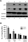
Figure 1.
Figure 1.
p53 regulates migration of MSCs in response to tumor cells. Human MSCs were treated with 25 μM Nutlin-3 or vehicle (DMSO). (A) Nutlin-3 treatment led to increased levels of p53 protein as well as the p53 target p21 as assessed by western blotting. (B) Nutlin-3 treatment decreased hMSC migration in response to MDA-MB-231 cells (*P<0.001) and caused a non-significant decrease in hMSC migration in response to tumor cell conditioned medium. Nutlin-3 treatment did not change the migration of hMSCs in response to 10 ng/ml recombinant IL-8 (a known chemotactic stimulus for MSCs) or control medium. (C) Targeted siRNA was used to knockdown expression of p53 in hMSCs as demonstrated by western blot analysis of cell lysate. (D) Decreased levels of p53 led to increased MSC migration in response to tumor cells as well as to control medium (*P<0.0037 compared to si-RNA control).
Figure 1.
Figure 1.
p53 regulates migration of MSCs in response to tumor cells. Human MSCs were treated with 25 μM Nutlin-3 or vehicle (DMSO). (A) Nutlin-3 treatment led to increased levels of p53 protein as well as the p53 target p21 as assessed by western blotting. (B) Nutlin-3 treatment decreased hMSC migration in response to MDA-MB-231 cells (*P<0.001) and caused a non-significant decrease in hMSC migration in response to tumor cell conditioned medium. Nutlin-3 treatment did not change the migration of hMSCs in response to 10 ng/ml recombinant IL-8 (a known chemotactic stimulus for MSCs) or control medium. (C) Targeted siRNA was used to knockdown expression of p53 in hMSCs as demonstrated by western blot analysis of cell lysate. (D) Decreased levels of p53 led to increased MSC migration in response to tumor cells as well as to control medium (*P<0.0037 compared to si-RNA control).
Figure 2.
Figure 2.
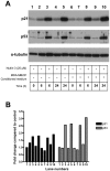
Treatment with tumor conditioned medium does not affect MSC p53 levels. hMSCs were treated with MDA-MB-231 conditioned medium or control medium with or without 25 μM Nutlin-3. A, Western blot analysis of protein extracts collected over 24 h revealed that tumor conditioned medium alone did not influence MSC p53 or p21 levels. B, Treatment with Nutlin-3 led to increased p53 activity in the presence of both control medium and tumor-conditioned medium.
Figure 3.
Figure 3.
Nutlin-3 decreases CXCL12 mRNA levels in hMSCs. Human MSCs were treated with control medium or tumor conditioned medium with or without 25 μM Nutlin-3. Q-RT-PCR of the extracted RNA after 24 h of Nutlin-3 treatment revealed decreased CXCL12 mRNA levels in MSCs cultured in control medium. Decreased CXCL12 mRNA levels were seen in hMSCs cultured in tumor-conditioned medium at both 6 and 24 h (*P<0.04).
Figure 4.
Figure 4.
Enhanced MSC motility due to decreased p53 level is dependent on CXCL12. Human MSCs with siRNA-mediated p53 knockdown demonstrated enhanced migration in response to MDA-MB-231 cells. Treatment with 25 μM Nutlin-3 did not cause a decrease in migration. Knockdown of CXCL12 production using siRNA decreased the migration of p53−/− MSCs suggesting that decreased p53 activity leads to increased MSC mobility through increased CXCL12 transcription. (n=4) (*P= 0.007).
Figure 5.
Figure 5.
Decreased MSC motility in response to increased p53 activity is not reversed by CXCL12. MSCs were treated with 25 μM Nutlin-3. Recombinant CXCL12 was added to both the upper and lower chambers of the Boyden chamber and migration of MSCs in response to MDA-MB-231 cell conditioned media was then allowed to proceed overnight. Exogenous CXCL12 did not reverse the decreased migration of MSCs observed with Nutlin-3 treatment. **Migration of MSCs in response to Tumor CM was significantly decreased in the presence of Nutlin-3 (P<0.005).
Figure 6.
Figure 6.
MSCs with p53 knock-down localize more efficiently to tumors in vivo than MSCs with wild-type p53. Murine MSCs were isolated from C57BL/6J p53−/− mice. In order to generate cells with functional p53, murine p53−/− cells were transfected with a plasmid encoding wild-type murine p53. (A) Western blot analysis of protein isolated from p53−/− murine cells and p53-transfected murine cells shows expression of human p53. (B) MSCs expressing wt p53 and p53−/− MSCs were differentially labeled using green (CFSE) and red (CM-DiI) fluorescent dyes, respectively. The cells were combined in a ratio of 1:1 and injected subcutaneously 5 mm from established MDA-MB-231 tumors in nude mice. Three days after injection the animals were sacrificed and tumors were harvested. Single cell suspensions were made from the tumors and the percentages of both p53 knockdown and wild-type MSCs were determined using flow cytometry. An increased percentage of labeled p53 knockdown MSCs were present in the tumors compared to wild-type MSCs. These data indicate that decreased p53 levels lead to increased chemokinesis of MSCs in response to tumors.
Figure 6.
Figure 6.
MSCs with p53 knock-down localize more efficiently to tumors in vivo than MSCs with wild-type p53. Murine MSCs were isolated from C57BL/6J p53−/− mice. In order to generate cells with functional p53, murine p53−/− cells were transfected with a plasmid encoding wild-type murine p53. (A) Western blot analysis of protein isolated from p53−/− murine cells and p53-transfected murine cells shows expression of human p53. (B) MSCs expressing wt p53 and p53−/− MSCs were differentially labeled using green (CFSE) and red (CM-DiI) fluorescent dyes, respectively. The cells were combined in a ratio of 1:1 and injected subcutaneously 5 mm from established MDA-MB-231 tumors in nude mice. Three days after injection the animals were sacrificed and tumors were harvested. Single cell suspensions were made from the tumors and the percentages of both p53 knockdown and wild-type MSCs were determined using flow cytometry. An increased percentage of labeled p53 knockdown MSCs were present in the tumors compared to wild-type MSCs. These data indicate that decreased p53 levels lead to increased chemokinesis of MSCs in response to tumors.

Similar articles

Cited by

References

    1. Studeny M, Marini FC, Dembinski JL, Zompetta C, Cabreira-Hansen M, Bekele BN, Champlin RE, Andreeff M. Mesenchymal stem cells: potential precursors for tumor stroma and targeted-delivery vehicles for anticancer agents. J Natl Cancer Inst. 2004;96:1593–1603. - PubMed
    1. Studeny M, Marini FC, Champlin RE, Zompetta C, Fidler IJ, Andreeff M. Bone marrow-derived mesenchymal stem cells as vehicles for interferon-beta delivery into tumors. Cancer Res. 2002;62:3603–3608. - PubMed
    1. Shen FH, Visger JM, Balian G, Hurwitz SR, Diduch DR. Systemically administered mesenchymal stromal cells transduced with insulin-like growth factor-I localize to a fracture site and potentiate healing. J Orthop Trauma. 2002;16:651–659. - PubMed
    1. Rochefort GY, Delorme B, Lopez A, Herault O, Bonnet P, Charbord P, Eder V, Domenech J. Multipotential mesenchymal stem cells are mobilized into peripheral blood by hypoxia. Stem Cells. 2006;24:2202–2208. - PubMed
    1. Pittenger MF, Mackay AM, Beck SC, et al. Multilineage potential of adult human mesenchymal stem cells. Science. 1999;284:143–147. - PubMed

Publication types

MeSH terms