

Oleic acid increases synthesis and secretion of VEGF in rat vascular smooth muscle cells: role of oxidative stress and impairment in obesity
- PMID: 24065093
- PMCID: PMC3794811
- DOI: 10.3390/ijms140918861
Oleic acid increases synthesis and secretion of VEGF in rat vascular smooth muscle cells: role of oxidative stress and impairment in obesity
Abstract
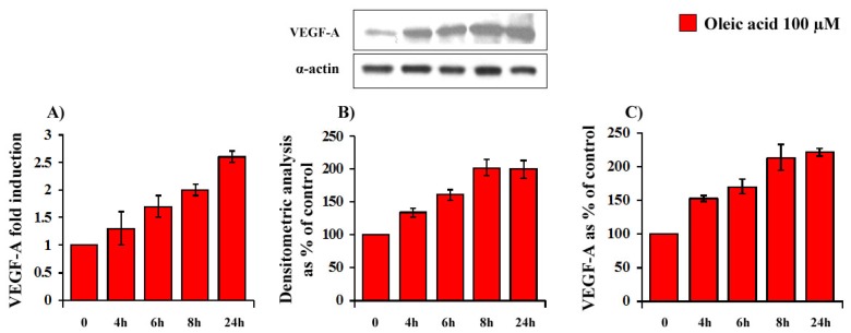
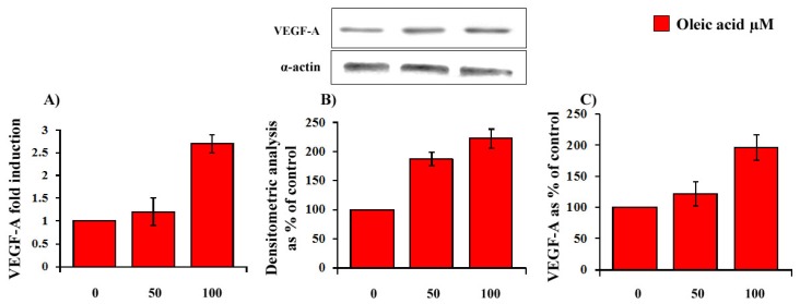
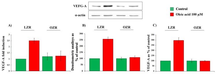
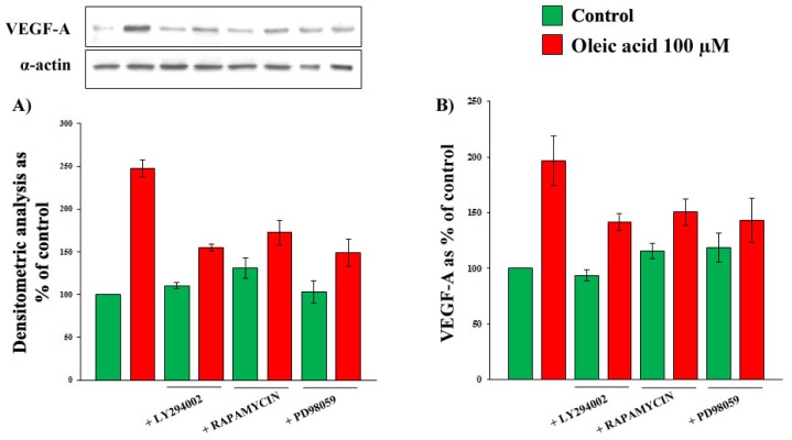
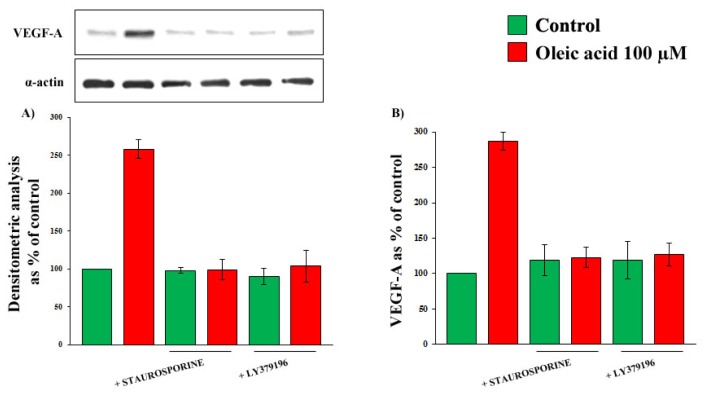
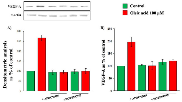
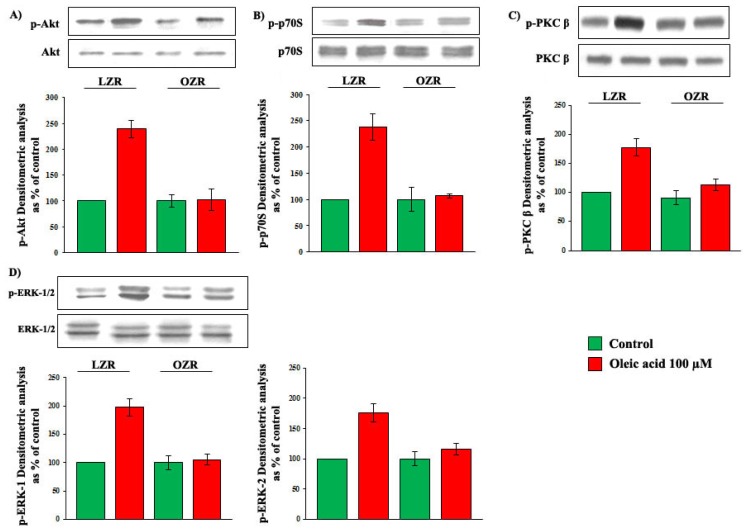
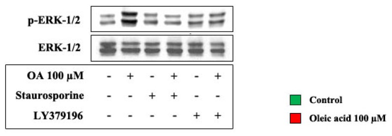
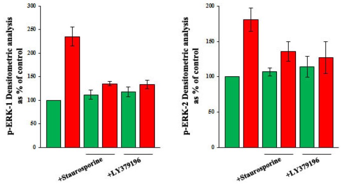
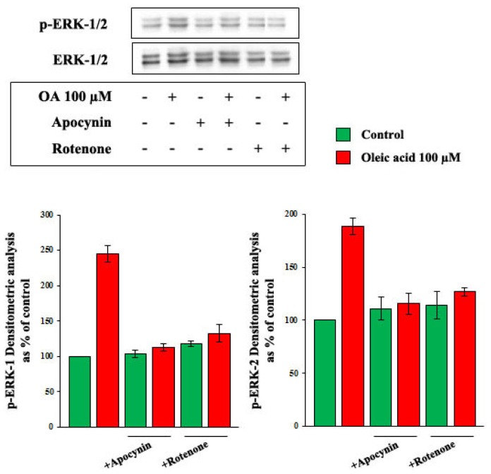
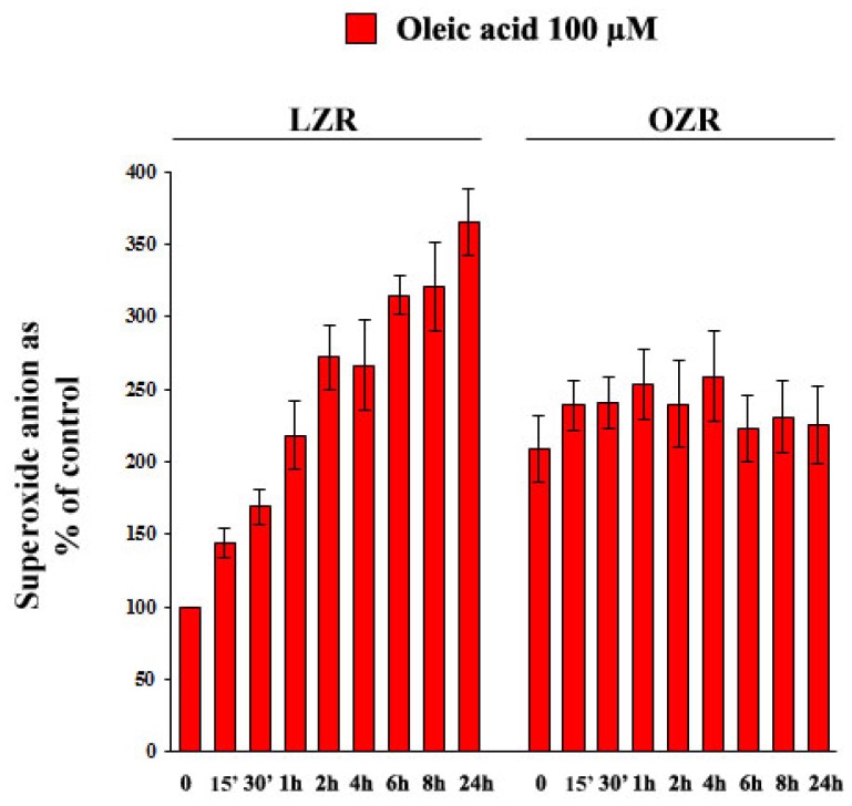
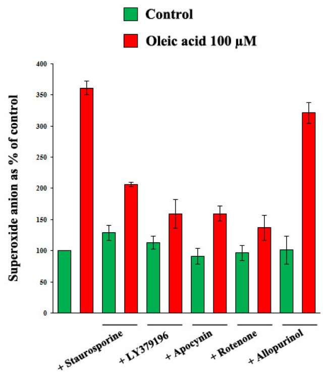
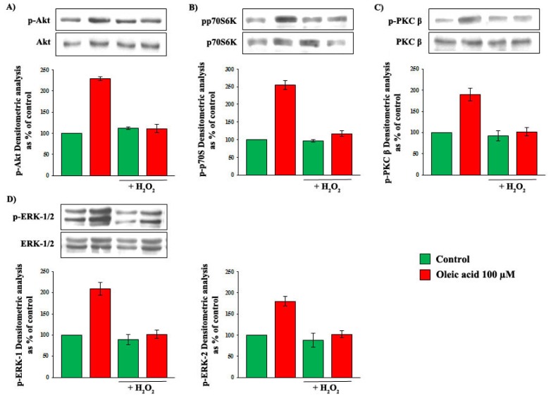
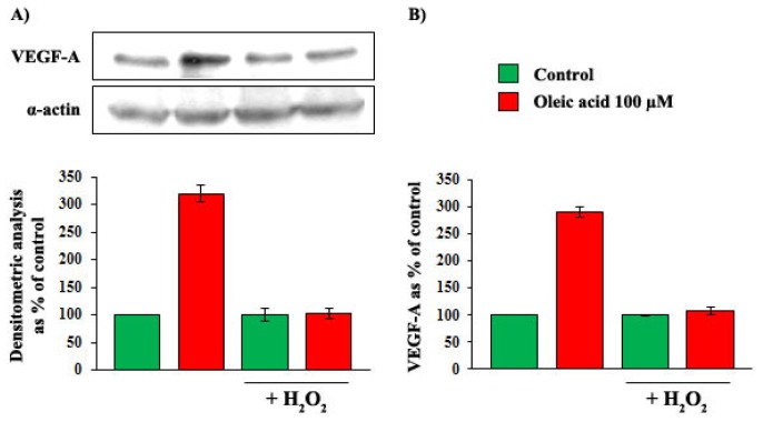
Obesity is characterized by poor collateral vessel formation, a process involving vascular endothelial growth factor (VEGF) action on vascular smooth muscle cells (VSMC). Free fatty acids are involved in the pathogenesis of obesity vascular complications, and we have aimed to clarify whether oleic acid (OA) enhances VEGF synthesis/secretion in VSMC, and whether this effect is impaired in obesity. In cultured aortic VSMC from lean and obese Zucker rats (LZR and OZR, respectively) we measured the influence of OA on VEGF-A synthesis/secretion, signaling molecules and reactive oxygen species (ROS). In VSMC from LZR we found the following: (a) OA increases VEGF-A synthesis/secretion by a mechanism blunted by inhibitors of Akt, mTOR, ERK-1/2, PKC-beta, NADPH-oxidase and mitochondrial electron transport chain complex; (b) OA activates the above mentioned signaling pathways and increases ROS; (c) OA-induced activation of PKC-beta enhances oxidative stress, which activates signaling pathways responsible for the increased VEGF synthesis/secretion. In VSMC from OZR, which present enhanced baseline oxidative stress, the above mentioned actions of OA on VEGF-A, signaling pathways and ROS are impaired: this impairment is reproduced in VSMC from LZR by incubation with hydrogen peroxide. Thus, in OZR chronically elevated oxidative stress causes a resistance to the action on VEGF that OA exerts in LZR by increasing ROS.
Figures
Similar articles
-
Nitric oxide activates PI3-K and MAPK signalling pathways in human and rat vascular smooth muscle cells: influence of insulin resistance and oxidative stress.Atherosclerosis. 2011 May;216(1):44-53. doi: 10.1016/j.atherosclerosis.2011.01.019. Epub 2011 Jan 21. Atherosclerosis. 2011. PMID: 21316056
-
Effects of high glucose on vascular endothelial growth factor synthesis and secretion in aortic vascular smooth muscle cells from obese and lean Zucker rats.Int J Mol Sci. 2012;13(8):9478-9488. doi: 10.3390/ijms13089478. Epub 2012 Jul 26. Int J Mol Sci. 2012. PMID: 22949809 Free PMC article.
-
Adipokines enhance oleic acid-induced proliferation of vascular smooth muscle cells by inducing CD36 expression.Arch Physiol Biochem. 2015;121(3):81-7. doi: 10.3109/13813455.2015.1045520. Epub 2015 Jul 1. Arch Physiol Biochem. 2015. PMID: 26135380
-
Modulation of protein kinase activity and gene expression by reactive oxygen species and their role in vascular physiology and pathophysiology.Arterioscler Thromb Vasc Biol. 2000 Oct;20(10):2175-83. doi: 10.1161/01.atv.20.10.2175. Arterioscler Thromb Vasc Biol. 2000. PMID: 11031201 Review.
-
Synchronous activation of ERK 1/2, p38mapk and PKB/Akt signaling by H2O2 in vascular smooth muscle cells: potential involvement in vascular disease (review).Int J Mol Med. 2003 Feb;11(2):229-34. Int J Mol Med. 2003. PMID: 12525883 Review.
Cited by
-
Distinguish fatty acids impact survival, differentiation and cellular function of periodontal ligament fibroblasts.Sci Rep. 2020 Sep 24;10(1):15706. doi: 10.1038/s41598-020-72736-7. Sci Rep. 2020. PMID: 32973207 Free PMC article.
-
Oleic Acid, Cholesterol, and Linoleic Acid as Angiogenesis Initiators.ACS Omega. 2020 Aug 10;5(32):20575-20585. doi: 10.1021/acsomega.0c02850. eCollection 2020 Aug 18. ACS Omega. 2020. PMID: 32832811 Free PMC article.
-
Oxidative stress in cardiovascular disease.Int J Mol Sci. 2014 Apr 9;15(4):6002-8. doi: 10.3390/ijms15046002. Int J Mol Sci. 2014. PMID: 24722571 Free PMC article.
-
Stearoyl-CoA Desaturase Regulates Angiogenesis and Energy Metabolism in Ischemic Cardiomyocytes.Int J Mol Sci. 2022 Sep 9;23(18):10459. doi: 10.3390/ijms231810459. Int J Mol Sci. 2022. PMID: 36142371 Free PMC article.
-
Role of Dietary Nutritional Treatment on Hepatic and Intestinal Damage in Transplantation with Steatotic and Non-Steatotic Liver Grafts from Brain Dead Donors.Nutrients. 2021 Jul 26;13(8):2554. doi: 10.3390/nu13082554. Nutrients. 2021. PMID: 34444713 Free PMC article.
References
-
- Hubert H.B., Feinleib M., McNamara P.M., Castelli W.P. Obesity as an independent risk factor for cardiovascular disease: A 26-year follow-up in the Framingham Heart Study. Circulation. 1983;67:968–977. - PubMed
-
- Lakka H.M., Lakka T.A., Tuomiletho J., Salonen J.T. Abdominal obesity is associated with increased risk of acute coronary events in men. Eur. Heart J. 2002;23:706–713. - PubMed
-
- Hu F.B., Willett W.C., Li T., Stampfer M.J., Colditz G.A., Manson J.E. Adiposity as compared with physical activity in predicting mortality among women. N. Engl. J. Med. 2004;351:2694–2703. - PubMed
-
- Reaven G., Abbasi F., McLaughlin T. Obesity, insulin resistance, and cardiovascular disease. Recent Prog. Horm. Res. 2004;59:207–223. - PubMed
-
- Anfossi G., Russo I., Doronzo G., Trovati M. Contribution of insulin resistance to vascular dysfunction. Arch. Physiol. Biochem. 2009;115:199–217. - PubMed
Publication types
MeSH terms
Substances
LinkOut - more resources
Full Text Sources
Other Literature Sources
Miscellaneous
