. 2014 Apr;22(4):564-7.
doi: 10.1038/ejhg.2013.215. Epub 2013 Sep 25.

Germ-line deletion in DICER1 revealed by a novel MLPA assay using synthetic oligonucleotides

Affiliations

Germ-line deletion in DICER1 revealed by a novel MLPA assay using synthetic oligonucleotides

Nelly Sabbaghian et al. Eur J Hum Genet. 2014 Apr.

Abstract

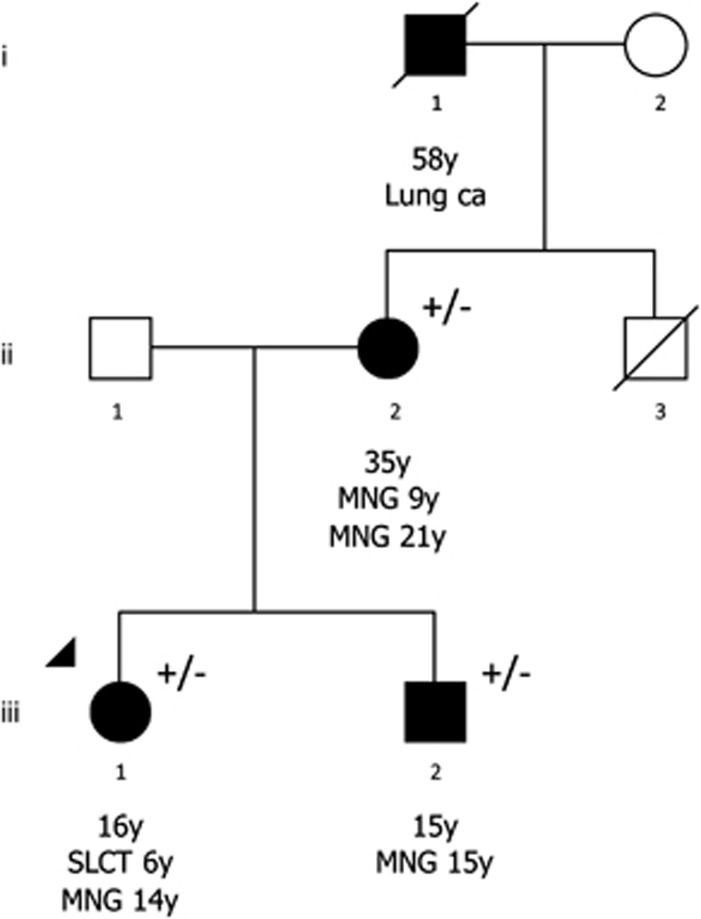
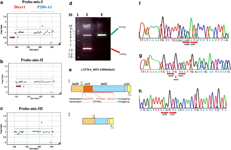
DICER1 is an endoribonuclease responsible for the production of mature microRNAs which are small, single-stranded RNA molecules that regulate gene expression post-transcriptionally by binding to mRNA and repressing the expression of target genes. Germ-line mutations in DICER1 are responsible for a rare cancer syndrome, including tumors that can co-occur with multinodular goiter (MNG). Using Sanger sequencing, we screened all DICER1 exons and intron boundaries in 20 suspected mutation carriers: nine with ovarian sex cord-stromal tumors (including Sertoli-Leydig cell tumors (SLCTs)), five with pleuropulmonary blastoma, one with cystic nephroma, one with nasal chondromesenchymal hamartoma and four with more than one manifestation suggestive of a germ-line DICER1 mutation. All were negative for any apparently deleterious variants. We developed a Multiplex Ligation-based Probe Amplification assay for DICER1 to screen for large deletions or duplications. Synthetic oligonucleotides were designed to cover all exons in three probe-mixes. In a child with a SLCT and MNG, and in her mother and brother (both diagnosed with MNG), we identified a heterozygous germ-line deletion of approximately 3 kilobases that eliminates exon 21 of DICER1 and two-thirds of intron 21, accompanied by an insertion of a G nucleotide at the 3' end of the deletion (c.3270-6_4051-1280delinsG). This allele is expressed in the patient's cDNA, creating an out-of-frame deletion predicted to result in a truncated protein (r.3270_4050del; p.Tyr1091Ser*28). Our novel finding of a disease-causing large deletion in DICER1 emphasizes the need to include assays that can detect rearrangements, duplications and deletions in any DICER1 screening protocol.


Figures

Figure 1
Figure 1
Pedigree of the proband with the exon 21 DICER1 deletion. Current age in years (y) is shown below the symbol (○: women; □: men; ▪/•: known diagnosis). The arrowhead indicates the proband. (+/−) indicates heterozygous status of the mutation. ii-2 required two separate operative removals of her MNG; at age 9 y, she had a partial left lobectomy of the thyroid, despite thyroid hormone replacement therapy, new lesions developed in both lobes were noted from age 13 y onwards and at age 21 y she underwent a total thyroidectomy. She was also diagnosed with a uterine leiomyoma at age 35 y.
Figure 2
Figure 2
DICER1 MLPA and deletion validation. (a, b and c) Representation of the MLPA results using probe-mixes I, II and III, respectively, for the proband. Panels (ac) were generated with GeneMarker v. 1.70 from SoftGenetics, LLC, (http://www.softgenetics.com/GeneMarker.html). The arrows point to the deletion. In a, the parentheses show the peak ratio values for the DICER1 probes and those for P200-A1 (control and reference probes from MRC-Holland). (d) 0.8% agarose gel showing in lane 1 the 2-log DNA ladder (NEB, Mississauga, Canada), lane 2 the two PCR fragments amplified in the proband to map the genomic deletion (e-1=4607 bp and e-2=1689 bp) and lane 3 is the PCR fragment of the control (e-1). (e) Graphic representation of the two PCR fragments shown in panel d. (e-1) shows the wild-type DICER1 sequence (large PCR fragment) from the beginning of the deleted sequence in intron 20, TTGCAG, until the breakpoint in intron 21, followed by an insertion of a ‘G' nucleotide; brown with white dots: intron 21; solid brown: exon 21; blue: intron 21; pink: exon 22 and yellow: intron 22. (e-2) The shorter fragment of the proband showing intron 20 next to intron 21. (f) Sequencing trace of the control sample (e-1) showing the junction between intron 20 and exon 21. (g) Sequencing trace of the shorter PCR fragment (e-2) using primers 21F and 22R (same primers used for the control sample). One arrow points at the ‘G' insertion and the two other arrows point in the direction of intron 20 and intron 21 showing the lack of exon 21. (h) Sequencing trace of the cDNA of the proband showing the junction between exon 20 and 22 (arrows) with the absence of exon 21 (r.3270_4050del).

Similar articles

Cited by

References

    1. Hill DA, Ivanovich J, Priest JR, et al. DICER1 mutations in familial pleuropulmonary blastoma. Science. 2009;325:965. - PMC - PubMed
    1. Priest JR, McDermott MB, Bhatia S, Watterson J, Manivel JC, Dehner LP. Pleuropulmonary blastoma: a clinicopathologic study of 50 cases. Cancer. 1997;80:147–161. - PubMed
    1. Bahubeshi A, Bal N, Frio TR, et al. Germ-line DICER1 mutations and familial cystic nephroma. J Med Genet. 2010;47:863–866. - PubMed
    1. Slade I, Bacchelli C, Davies H, et al. DICER1 syndrome: clarifying the diagnosis, clinical features and management implications of a pleiotropic tumour predisposition syndrome. J Med Genet. 2011;48:273–278. - PubMed
    1. Rio Frio T, Bahubeshi A, Kanellopoulou C, et al. DICER1 mutations in familial multinodular goiter with and without ovarian Sertoli–Leydig cell tumors. JAMA. 2011;305:68–77. - PMC - PubMed

Publication types

MeSH terms

LinkOut - more resources