. 2014 May 1;209(9):1331-42.
doi: 10.1093/infdis/jit504. Epub 2013 Sep 24.

Active replication of Middle East respiratory syndrome coronavirus and aberrant induction of inflammatory cytokines and chemokines in human macrophages: implications for pathogenesis

Affiliations

Active replication of Middle East respiratory syndrome coronavirus and aberrant induction of inflammatory cytokines and chemokines in human macrophages: implications for pathogenesis

Jie Zhou et al. J Infect Dis. .

Abstract

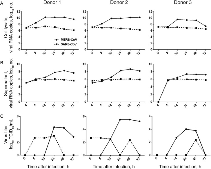
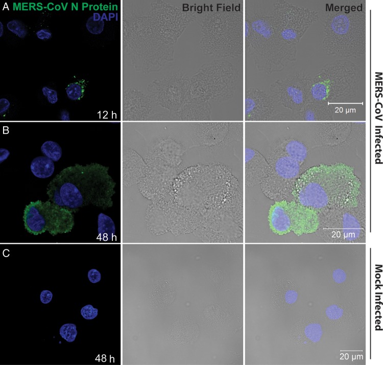
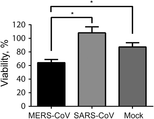
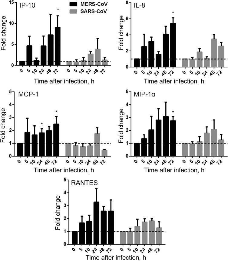
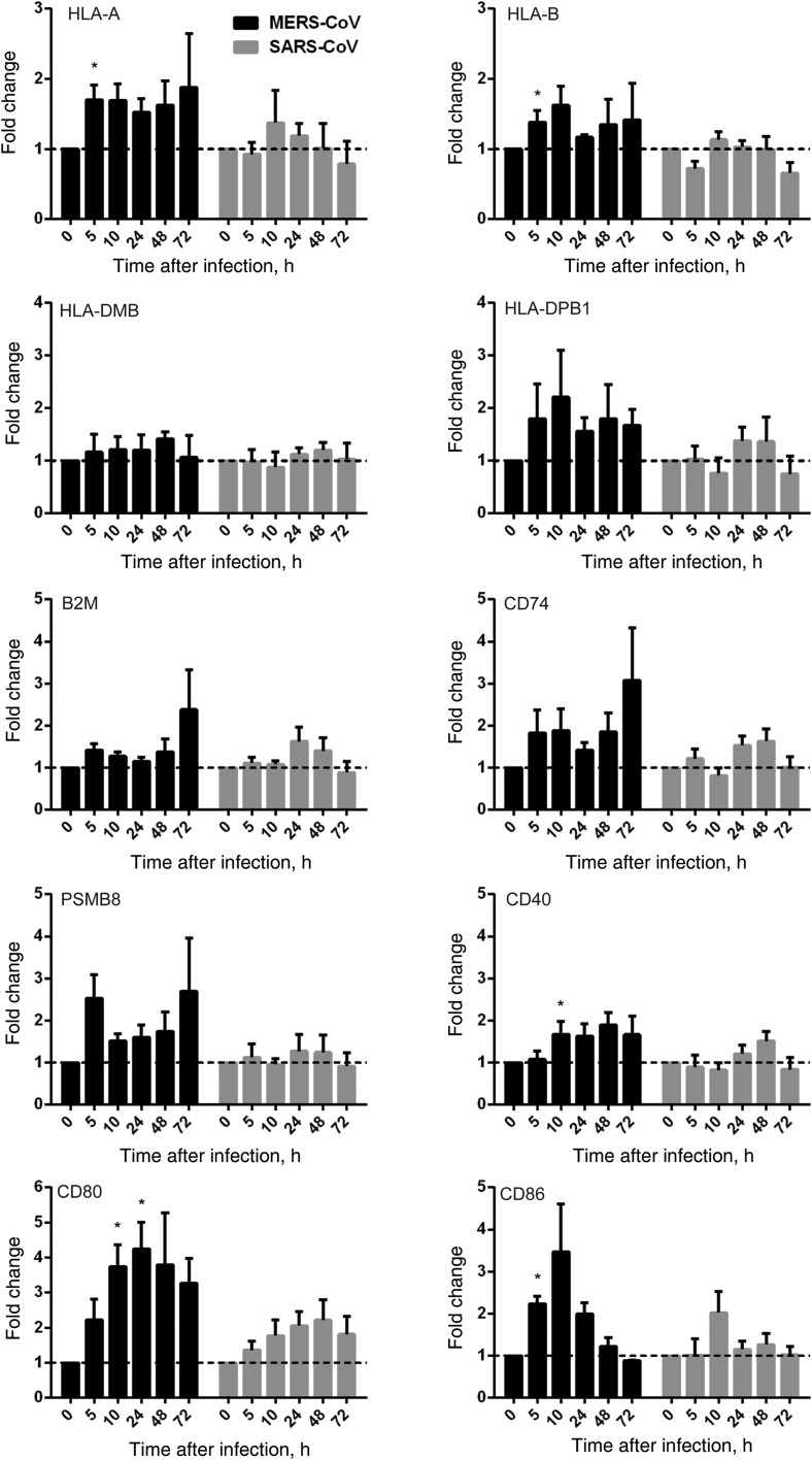
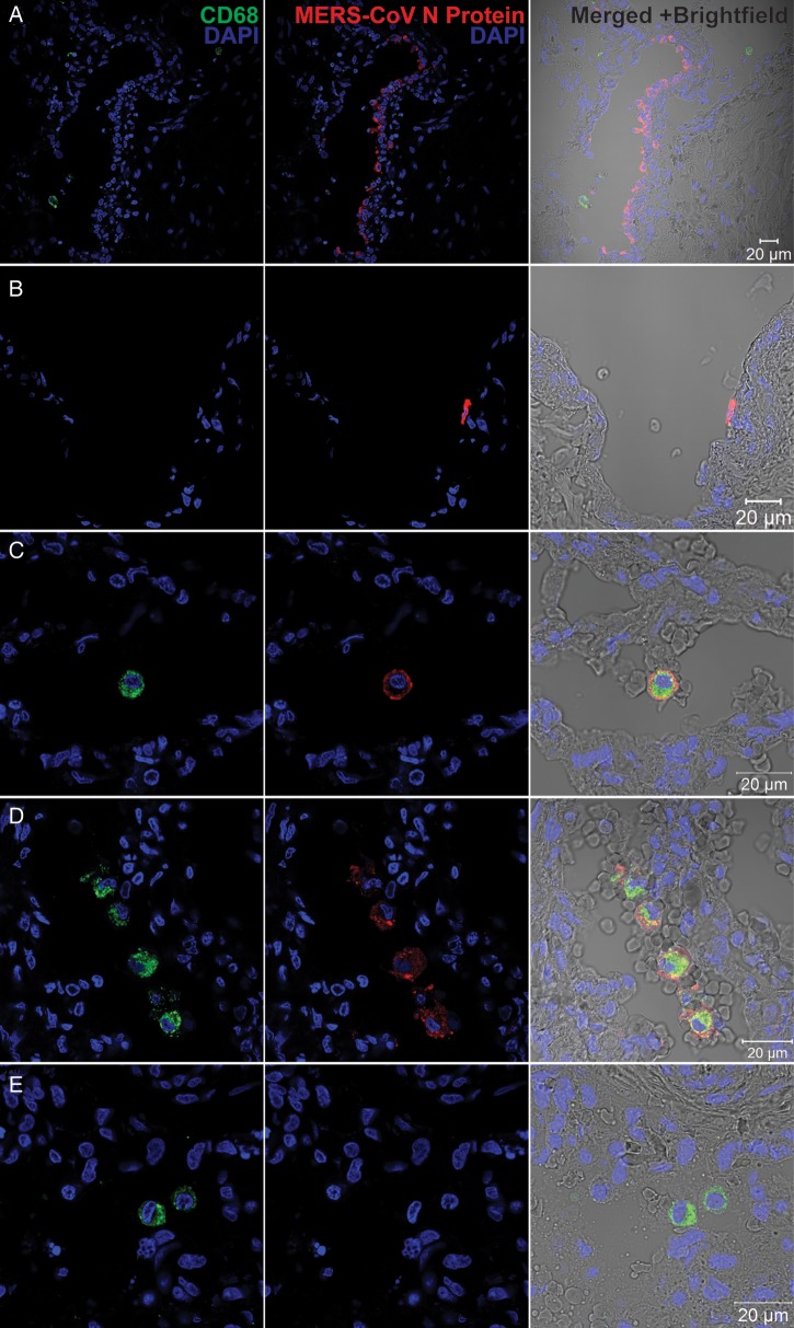
Middle East respiratory syndrome coronavirus (MERS-CoV) infection caused severe pneumonia and multiorgan dysfunction and had a higher crude fatality rate (around 50% vs. 10%) than SARS coronavirus (SARS-CoV) infection. To understand the pathogenesis, we studied viral replication, cytokine/chemokine response, and antigen presentation in MERS-CoV-infected human monocyte-derived macrophages (MDMs) versus SARS-CoV-infected MDMs. Only MERS-CoV can replicate in MDMs. Both viruses were unable to significantly stimulate the expression of antiviral cytokines (interferon α [IFN-α] and IFN-β) but induced comparable levels of tumor necrosis factor α and interleukin 6. Notably, MERS-CoV induced significantly higher expression levels of interleukin 12, IFN-γ, and chemokines (IP-10/CXCL-10, MCP-1/CCL-2, MIP-1α/CCL-3, RANTES/CCL-5, and interleukin 8) than SARS-CoV. The expression of major histocompatibility complex class I and costimulatory molecules were significantly higher in MERS-CoV-infected MDMs than in SARS-CoV-infected cells. MERS-CoV replication was validated by immunostaining of infected MDMs and ex vivo lung tissue. We conclusively showed that MERS-CoV can establish a productive infection in human macrophages. The aberrant induction of inflammatory cytokines/chemokines could be important in the disease pathogenesis.

Keywords: MERS-CoV; SARS-CoV; cytokine and chemokine response; pathogenesis; viral replication.


Figures

Figure 1.
Figure 1.
Middle East respiratory syndrome coronavirus (MERS-CoV) replication kinetics in human monocyte–derived macrophages (MDMs), compared with SARS coronavirus (SARS-CoV). MERS-CoV or SARS-CoV was inoculated onto MDMs at 2 tissue culture infective doses (TCID50) per cell. At the indicated hours after infection, cell lysate (A) and culture supernatant (B) were collected to detect the levels of positive-strand viral RNA by reverse-transcription quantitative polymerase chain reaction. Viral titration of the culture supernatants was performed with a TCID50 assay (C). The results of 3 representative donors are demonstrated.
Figure 2.
Figure 2.
Immunofluorescence staining of Middle East respiratory syndrome coronavirus (MERS-CoV) nucleocapsid protein (NP) in MERS-CoV–inoculated human monocyte–derived macrophages (MDMs). MDMs seeded on glass coverslips were inoculated with MERS-CoV at 2 tissue culture infective doses per cell or subjected to mock infection. Twelve hours (A) and 48 hours (B) after inoculation, cells were fixed, blocked, and permeabilized; incubated with anti-NP primary antibody for 1 hour; and stained with fluorescein isothiocyanate–conjugated secondary antibody for 1 hour. The slides were mounted with DAPI-containing mounting buffer and examined with a Carl Zeiss LSM 710 microscope. C, Findings for mock-inoculated MDMs that underwent the same procedure as that described for MERS-CoV–infected MDMs.
Figure 3.
Figure 3.
Cell viability of Middle East respiratory syndrome coronavirus (MERS-CoV)–infected and SARS coronavirus (SARS-CoV)–infected human monocyte–derived macrophages (MDMs). MDMs were seeded at 1.5 × 10⁵/well in a 24-well plate and inoculated with MERS-CoV or SARS-CoV at 2 median tissue culture infective doses per cell or subjected to mock infection. Before infection and 48 hours after infection, MDMs were trypsinized, stained with trypan blue, and counted in duplicate with disposable hemocytometers. Viability represents the ratio of viable cell number of inoculated MDMs 48 hours after infection, compared with that before infection. Results are the mean and standard error of the mean of at least 3 representative donors. *P < .05, by the Student t test.
Figure 4.
Figure 4.
Cytokine response in Middle East respiratory syndrome coronavirus (MERS-CoV)–infected and SARS coronavirus (SARS-CoV)–infected human monocyte–derived macrophages (MDMs). MDMs were inoculated with MERS-CoV or SARS-CoV at 2 median tissue culture infective doses per cell or subjected to mock infection. At the indicated hours after infection, cells were lysed for RNA extraction, and reverse-transcription quantitative polymerase chain reaction was performed to detect the messenger RNA expression levels for antiviral and proinflammatory cytokines. Results were normalized to those for GAPDH and are presented as the fold change in gene expression of virus-infected MDMs in relation to that of the mock-infected MDMs. Data displayed in this figure represent mean and standard error of the mean from at least 3 different donors. Statistical analyses between MERS-CoV– and SARS-CoV–inoculated MDMs at different time points were performed using the Student t test. *P < .05. Abbreviations: IFN-α, interferon α; IFN-β, interferon β; IFN-γ, interferon γ; IL-6, interleukin 6; IL-12, interleukin 12; TNF-α, tumor necrosis factor α.
Figure 5.
Figure 5.
Chemokine response in Middle East respiratory syndrome coronavirus (MERS-CoV)–infected and SARS coronavirus (SARS-CoV)–infected human monocyte–derived macrophages (MDMs). MDMs were inoculated with MERS-CoV or SARS-CoV at 2 median tissue culture infective doses per cell or subjected to mock infection. At the indicated hours after infection, cells were lysed for RNA extraction, and reverse-transcription quantitative polymerase chain reaction was performed to detect the messenger RNA expression levels for immunoregulatory chemokines. Results were normalized to those for GAPDH and are presented as the fold change in gene expression of virus-infected MDMs relative to that of the mock-infected MDMs. Statistical analyses between MERS-CoV– and SARS-CoV–infected MDMs at different time points were performed using the Student t test. *P < .05. Abbreviation: IL-8, interleukin 8.
Figure 6.
Figure 6.
Antigen presentation function of human monocyte–derived macrophages (MDMs) upon Middle East respiratory syndrome coronavirus (MERS-CoV) versus SARS coronavirus (SARS-CoV) infection. MDMs were inoculated with MERS-CoV or SARS-CoV at 2 median tissue culture infective doses per cell or subjected to mock infection. At the indicated hours after infection, cells lysates were collected for RNA extraction, and reverse-transcription quantitative polymerase chain reaction was performed to detect the messenger RNA expression levels for major histocompatibility complex (MHC) class I– and MHC class II–related molecules and costimulation molecules involved in antigen presentation. Results were normalized to those for GAPDH and are presented as the fold change in gene expression of virus-infected MDMs relative to that of the mock-infected MDMs.
Figure 7.
Figure 7.
Immunofluorescence double staining of Middle East respiratory syndrome coronavirus (MERS-CoV)–infected ex vivo lung tissue for MERS-CoV nucleocapsid protein (NP; red) and CD68 (green). Normal lung tissues were infected with MERS-CoV at 1 × 10⁷ tissue culture infective doses per milliliter or subjected to mock infection for 1 hour at 37°C. A total of 48 hours after infection, tissues were fixed, cryoprotected, and cryosectioned. Slides were sequentially stained with guinea pig anti-NP sera, Alexa 694 goat–anti-guinea pig immunoglobulin G (IgG), mouse anti-CD68 antibody and Alexa 488 goat–anti-mouse IgG. Cell nuclei were counterstained by DAPI in the mounting buffer. Images were captured with a Carl Zeiss LSM 710 microscope. Immunoreactivity to NP was detected in epithelial cells of terminal bronchiole (A) and vascular endothelial (B) cells. C and D, Colocalization of MERS-CoV NP and CD68. E, A representative image of mock-infected lung tissue.

Similar articles

Cited by

References

    1. Chan JF, Li KS, To KK, Cheng VC, Chen H, Yuen KY. Is the discovery of the novel human betacoronavirus 2c EMC/2012 (HCoV-EMC) the beginning of another SARS-like pandemic? J Infect. 2012;65:477–89. - PMC - PubMed
    1. Zaki AM, van Boheemen S, Bestebroer TM, Osterhaus AD, Fouchier RA. Isolation of a novel coronavirus from a man with pneumonia in Saudi Arabia. N Engl J Med. 2012;367:1814–20. - PubMed
    1. Chan JF, Lau S, Woo PC. The emerging novel Middle East respiratory syndrome coronavirus: the “knowns” and “unknowns”. J Formos Med Assoc. 2013;112:372–81. - PMC - PubMed
    1. World Health Organization. Middle East respiratory syndrome coronavirus (MERS-CoV)—update. 1 August 2013. http://www.who.int/csr/don/2013_08_01/en/index.html . Accessed 27 September 2013.
    1. Woo PC, Lau SK, Huang Y, Yuen KY. Coronavirus diversity, phylogeny and interspecies jumping. Exp Biol Med (Maywood) 2009;234:1117–27. - PubMed

Publication types

MeSH terms