. 2014 Mar;66(2):259-73.
doi: 10.1007/s10616-013-9565-3. Epub 2013 Sep 25.

Evaluation of YO-PRO-1 as an early marker of apoptosis following radiofrequency ablation of colon cancer liver metastases

Affiliations

Evaluation of YO-PRO-1 as an early marker of apoptosis following radiofrequency ablation of colon cancer liver metastases

Sho Fujisawa et al. Cytotechnology. 2014 Mar.

Abstract

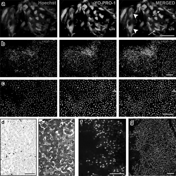
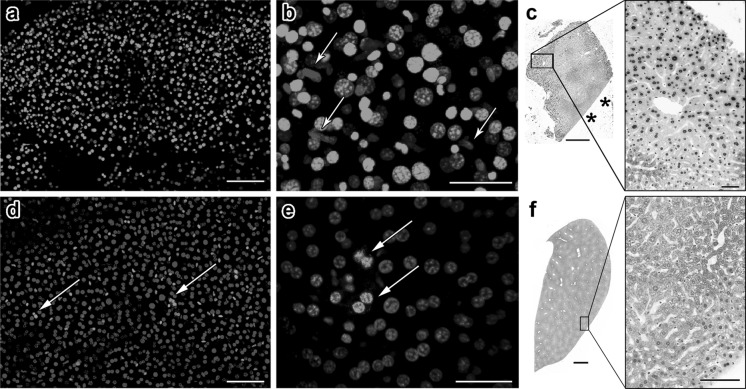
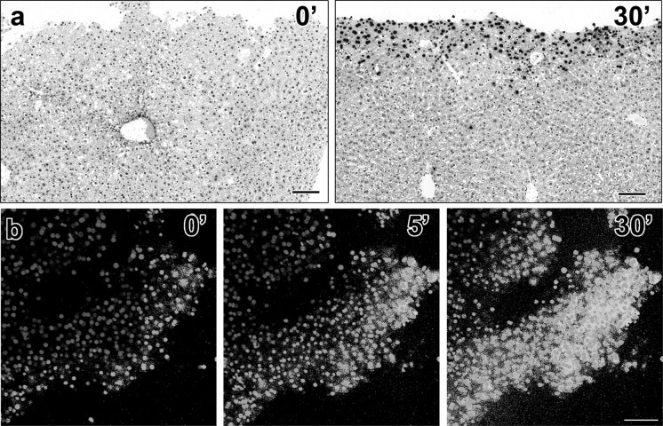
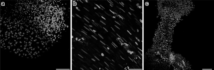
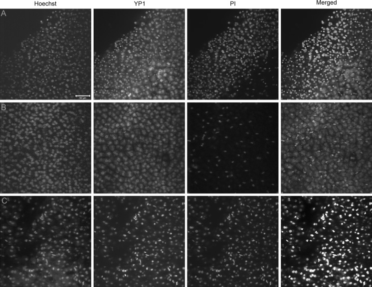
Radiofrequency (RF) ablation (RFA) is a minimally invasive treatment for colorectal-cancer liver metastases (CLM) in selected nonsurgical patients. Unlike surgical resection, RFA is not followed by routine pathological examination of the target tumor and the surrounding liver tissue. The aim of this study was the evaluation of apoptotic events after RFA. Specifically, we evaluated YO-PRO-1 (YP1), a green fluorescent DNA marker for cells with compromised plasma membrane, as a potential, early marker of cell death. YP1 was applied on liver tissue adherent on the RF electrode used for CLM ablation, as well as on biopsy samples from the center and the margin of the ablation zone as depicted by dynamic CT immediately after RFA. Normal pig and mouse liver tissues were used for comparison. The same samples were also immunostained for fragmented DNA (TUNEL assay) and for active mitochondria (anti-OxPhos antibody). YP1 was also used simultaneously with propidium iodine (PI) to stain mouse liver and samples from ablated CLM. Following RFA of human CLM, more than 90 % of cells were positive for YP1. In nonablated, dissected pig and mouse liver however, we found similar YP1 signals (93.1 % and 65 %, respectively). In samples of intact mouse liver parenchyma, there was a significantly smaller proportion of YP1 positive cells (22.7 %). YP1 and PI staining was similar for ablated CLM. However in dissected normal mouse liver there was initial YP1 positivity and complete absence of the PI signal and only later there was PI signal.

Conclusion: This is the first time that YP1 was applied in liver parenchymal tissue (rather than cell culture). The results suggest that YP1 is a very sensitive marker of early cellular events reflecting an early and widespread plasma membrane injury that allows YP1 penetration into the cells.


Figures

Fig. 1
Fig. 1
Human and pig RFA liver tissues exhibit ubiquitous YO-PRO-1 staining. Projection images were compiled from confocal z-stacks of human liver sections stained live with YO-PRO-1 (green, center panel) and Hoechst 33342 (blue, left panel). The right panel shows the merged images: a In tissues adherent to the RFA electrode, we observed a prominent heat artifact in which some nuclei were unnaturally elongated (arrows). Distinct nuclear and cytoplasmic regions are not well discernible, suggesting decompartmentalization of the cells (arrowhead). b In this sample from the center of an ablated region, morphologically intact hepatocytes are observed. A high proportion is YO-PRO-1 positive. c In this sample from the marginal region, an area in which we expected cell survival, we also observed high YO-PRO-1 positivity. d, e A subset of the margin sample was fixed and stained with TUNEL (d) and anti-OxPhos antibody (e). We observed significantly less TUNEL signal compared to YO-PRO-1, and abundant OxPhos signal compared to YO-PRO-1. These findings suggest that despite YO-PRO-1 positivity, the margin tissue contained metabolically active cells with intact DNA. f, g YO-PRO-1 staining of ablated (f) and nonablated (g) pig liver tissues shows that RFA does not influence the amount of YO-PRO-1 positivity. Scale bars: a = 25 μm, all others = 100 μm
Fig. 2
Fig. 2
YO-PRO-1 positivity in adult mouse liver tissue is influenced by the amount of physical damage inflicted during dissection. a A section of liver from an adult mouse was stained with YO-PRO-1 and Hoechst 33342. Exhibiting a general similarity to what was seen with human samples, a majority of the liver cells are YO-PRO-1-positive. b A higher magnification image reveals that nonhepatic cells with elongated nuclei seem to avoid YO-PRO-1 penetration (arrows). c TUNEL staining of mouse liver tissue shows that cells near sites of physical damage are particularly susceptible to apoptosis. We know that this is not an edge effect since TUNEL positivity is not observed near intact edge of the tissue (asterisks). d, e An entire lobe of liver was dissected gently and stained and imaged while intact. The proportion of YO-PRO-1-positive cells was significantly less and these cells formed small clusters (arrows). f TUNEL staining of intact liver lobe shows minimal apoptosis. Scale bars: a = 100 μm, b = 40 μm, c = 500 μm, 50 μm, d = 100 μm, e = 50 μm, f = 1 mm, 100 μm
Fig. 3
Fig. 3
A dissected liver tissue rapidly becomes positive for TUNEL and YO-PRO-1. When a dissected liver sample was fixed immediately following dissection, the tissue is void of TUNEL positivity (a, left panel). However when kept in the incubator for 30 min prior to fixation, the sample became positive for TUNEL staining near the cut edge (a, right panel). A time-lapse experiment in which YO-PRO-1 dye was added during imaging on confocal microscope showed that within 5 min the majority of cells became YO-PRO-1 positive b. Scale bars: a = 100 μm for both panels, b = 100 μm for all panels
Fig. 4
Fig. 4
Mouse non-liver tissues show distinct subpopulation of YO-PRO-1-positive cells that are largely exclusive from MitoTracker-positive cells. Tissues from lung a, intestinal walls b and seminiferous tubule c: each exhibit different proportions of apoptotic YO-PRO-1-positive cells. The results demonstrate the ability of YO-PRO-1 dye to specifically bind to metabolically inactive dying cells in tissues other than liver. Scale bars: a = 100 μm, b = 50 μm, c = 100 μm
Fig. 5
Fig. 5
The penetration of PI is slower than that of YP1 in healthy mouse liver, as opposed to RFA patient samples. a The edge of the healthy mouse liver sample shows a large number of both YP1 positive and PI positive nuclei with YP1 staining appearing earlier and covering more of the sample after 30 min of incubation. b Relatively intact healthy mouse liver shows significantly higher YP1 than PI signal, indicating that membrane pores allowing YP1 penetration can occur very early after any manipulation of the hepatocytes even in absence of mechanical damage to the tissue. c The human RFA patient sample shows ubiquitous YP1 and PI staining in all cells regardless of the size or location in the sample. This is most likely an indication of advanced injury and irreversible apoptosis due to the ablation treatment

References

    1. Abitabile P, Hartl U, Lange J, Maurer CA. Radiofrequency ablation permits an effective treatment for colorectal liver metastasis. Eur J Surg Oncol. 2007;33:67–71. doi: 10.1016/j.ejso.2006.10.040. - DOI - PubMed
    1. Amersi FF, McElrath-Garza A, Ahmad A, Zogakis T, Allegra DP, Krasne R, Bilchik AJ. Long-term survival after radiofrequency ablation of complex unresectable liver tumors. Arch Surg. 2006;141:581–587. doi: 10.1001/archsurg.141.6.581. - DOI - PubMed
    1. Boffa DJ, Waka J, Thomas D, Suh S, Curran K, Sharma VK, Besada M, Muthukumar T, Yang H, Suthanthiran M, Manova K. Measurement of apoptosis of intact human islets by confocal optical sectioning and stereologic analysis of YO-PRO-1-stained islets. Transplantation. 2005;79:842–845. doi: 10.1097/01.TP.0000155175.24802.73. - DOI - PubMed
    1. Castañeda F, Kinne RKH. Cytotoxicity of millimolar concentrations of ethanol on HepG2 human tumor cell line compared to normal rat hepatocytes in vitro. J Cancer Res Clin Oncol. 2000;126:503–510. doi: 10.1007/s004320000119. - DOI - PubMed
    1. Chagnon F, Fournier C, Charette PG, Moleski L, Payet MD, Dobbs LG, Lesur O. In vivo intravital endoscopic confocal fluorescence microscopy of normal and acutely injured rat lungs. Lab Invest J Tech Methods Pathol. 2010;90:824–834. doi: 10.1038/labinvest.2010.76. - DOI - PubMed

LinkOut - more resources