

Protocol to assess the neurophysiology associated with multi-segmental postural coordination
- PMID: 24065623
- PMCID: PMC3884551
- DOI: 10.1088/0967-3334/34/10/N97
Protocol to assess the neurophysiology associated with multi-segmental postural coordination
Abstract
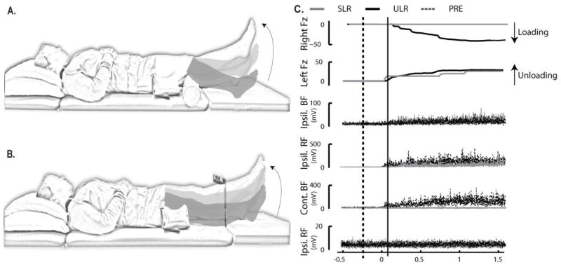
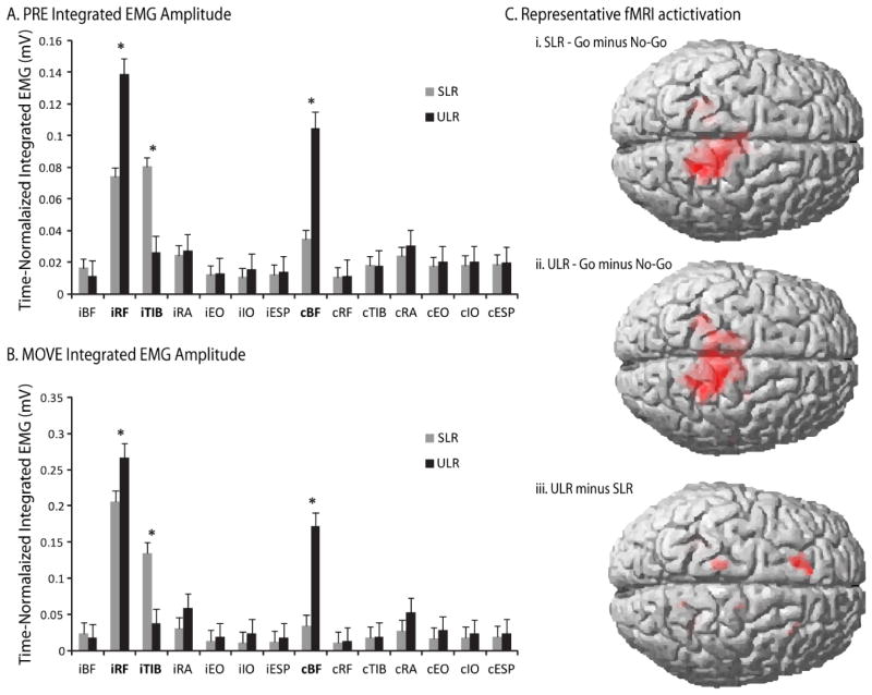
Anticipatory postural adjustments (APAs) stabilize potential disturbances to posture caused by movement. Impaired APAs are common with disease and injury. Brain functions associated with generating APAs remain uncertain due to a lack of paired tasks that require similar limb motion from similar postural orientations, but differ in eliciting an APA while also being compatible with brain imaging techniques (e.g., functional magnetic resonance imaging; fMRI). This study developed fMRI-compatible tasks differentiated by the presence or absence of APAs during leg movement. Eighteen healthy subjects performed two leg movement tasks, supported leg raise (SLR) and unsupported leg raise (ULR), to elicit isolated limb motion (no APA) versus multi-segmental coordination patterns (including APA), respectively. Ground reaction forces under the feet and electromyographic activation amplitudes were assessed to determine the coordination strategy elicited for each task. Results demonstrated that the ULR task elicited a multi-segmental coordination that was either minimized or absent in the SLR task, indicating that it would serve as an adequate control task for fMRI protocols. A pilot study with a single subject performing each task in an MRI scanner demonstrated minimal head movement in both tasks and brain activation patterns consistent with an isolated limb movement for the SLR task versus multi-segmental postural coordination for the ULR task.
Figures
References
-
- DRAKE JDM, CALLAGHAN JP. Elimination of electrocardiogram contamination from electromyogram signals: An evaluation of currently used removal techniques. Journal of Electromyography and Kinesiology. 2006;16:175–187. - PubMed
-
- FRISTON KJ, WILLIAMS S, HOWARD R, FRACKOWIAK RSJ, TURNER R. Movement-Related effects in fMRI time-series. Magnetic Resonance in Medicine. 1996;35:346–355. - PubMed
-
- HERMENS HJ, FRERIKS B, MERLETTI R, RAU G, DISSELHORST-KLUG C, STEGEMAN DF, HAGG GM. Sensor Locations. 2006 [Online]. Available: http://www.seniam.org 2011]
-
- HODGES PW, RICHARDSON CA. Relationship between limb movement speed and associated contraction of the trunk muscles. Ergonomics. 1997;40(11):1220–1230. - PubMed
-
- HODGES PW, RICHARDSON CA. Altered trunk muscle recruitment in people with low back pain with upper limb movement at different speeds. Archives of Physical Medical Rehabilitation. 1999;80:1005–12. - PubMed
Publication types
MeSH terms
Grants and funding
LinkOut - more resources
Full Text Sources
Other Literature Sources
Medical