. 2013 Nov;27(11):1796-807.
doi: 10.1210/me.2013-1095. Epub 2013 Sep 24.

Regulation of hypothalamic corticotropin-releasing hormone transcription by elevated glucocorticoids

Affiliations

Regulation of hypothalamic corticotropin-releasing hormone transcription by elevated glucocorticoids

Andrew N Evans et al. Mol Endocrinol. 2013 Nov.

Abstract

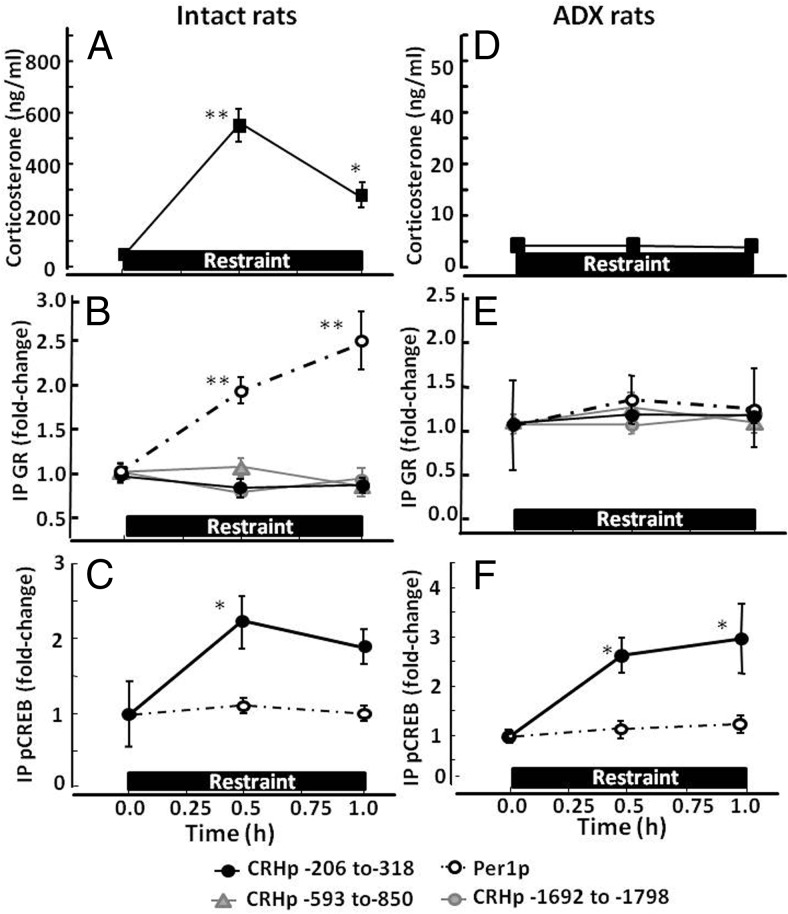
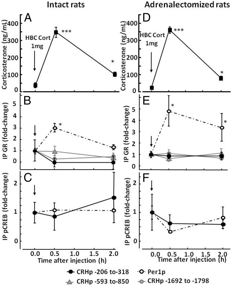
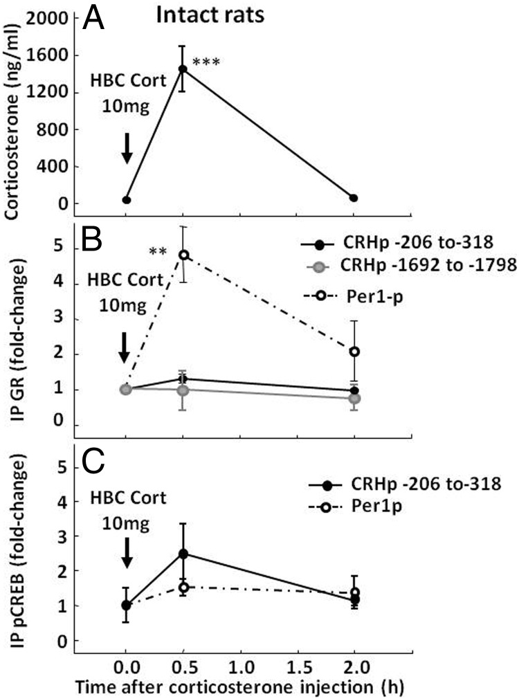
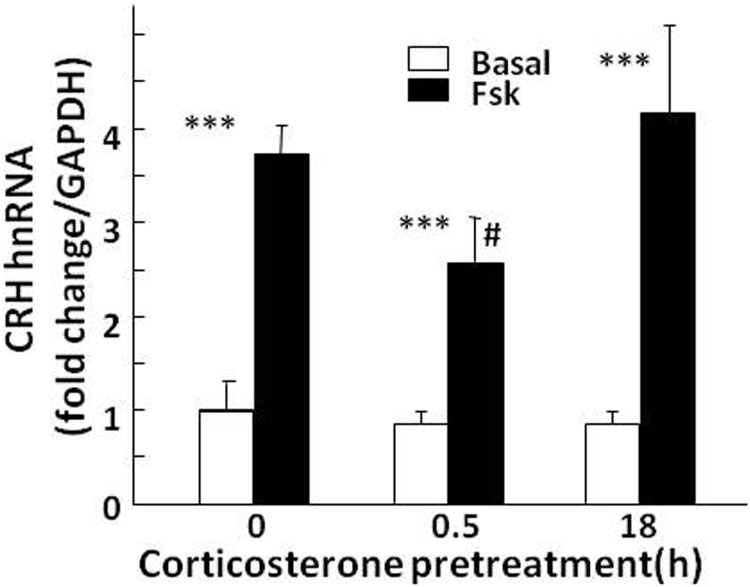
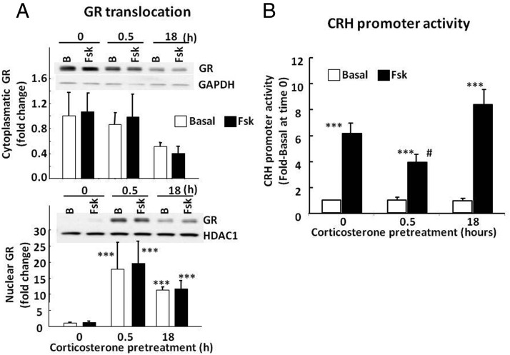
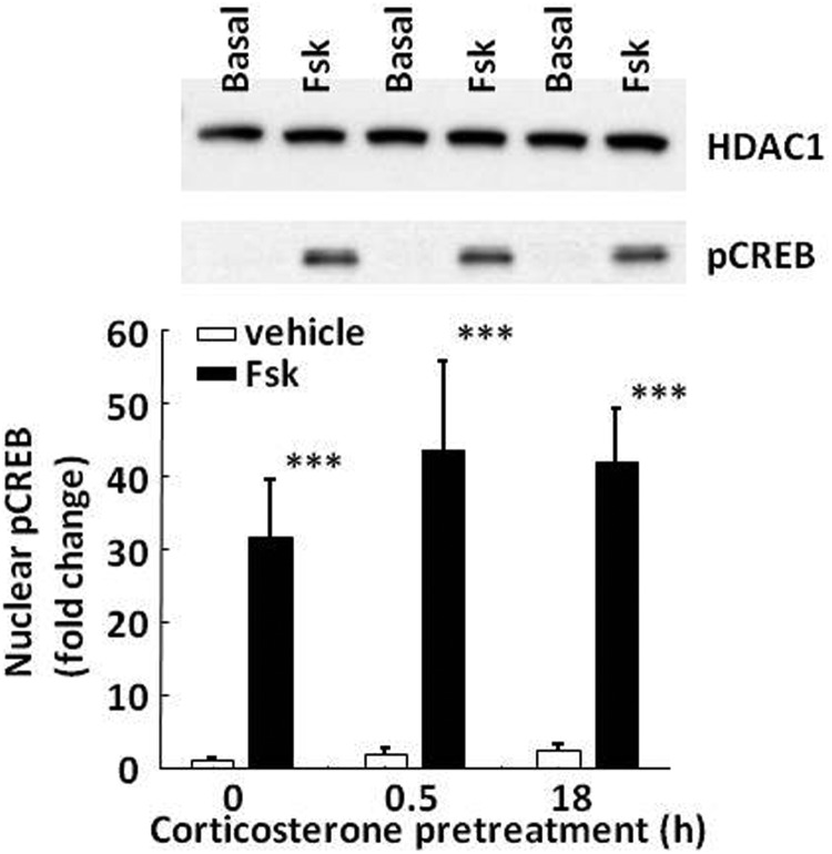
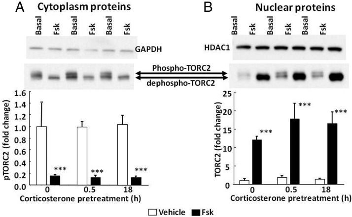
Negative glucocorticoid feedback is essential for preventing the deleterious effects of excessive hypothalamic pituitary adrenal axis axis activation, with an important target being CRH transcription in the hypothalamic paraventricular nucleus. The aim of these studies was to determine whether glucocorticoids repress CRH transcription directly in CRH neurons, by examining glucocorticoid effects on glucocorticoid receptor (GR)-CRH promoter interaction and the activation of proteins required for CRH transcription. Immunoprecipitation of hypothalamic chromatin from intact or adrenalectomized rats subjected to either stress or corticosterone injections showed minor association of the proximal CRH promoter with the GR compared with that with phospho-CREB (pCREB). In contrast, the Period-1 (Per1, a glucocorticoid-responsive gene) promoter markedly recruited GR. Stress increased pCREB recruitment by the CRH but not the Per1 promoter, irrespective of circulating glucocorticoids. In vitro, corticosterone pretreatment (30 minutes or 18 hours) only slightly inhibited basal and forskolin-stimulated CRH heteronuclear RNA in primary hypothalamic neuronal cultures and CRH promoter activity in hypothalamic 4B cells. In 4B cells, 30 minutes or 18 hours of corticosterone exposure had no effect on forskolin-induced nuclear accumulation of the recognized CRH transcriptional regulators, pCREB and transducer of regulated CREB activity 2. The data show that inhibition of CRH transcription by physiological glucocorticoids in vitro is minor and that direct interaction of GR with DNA in the proximal CRH promoter may not be a major mechanism of CRH gene repression. Although GR interaction with distal promoter elements may have a role, the data suggest that transcriptional repression of CRH by glucocorticoids involves protein-protein interactions and/or modulation of afferent inputs to the hypothalamic paraventricular nucleus.


Figures

Figure 1.
Figure 1.
Effect of restraint stress on GR and pCREB recruitment by the CRH and Per1 promoters in intact (A, B, and C) and adrenalectomized (Adx) (D, E, and F) rats. Plasma corticosterone measurements (A and D) and ChIP assays on hypothalamic chromatin for GR (B and E) and pCREB (C and F) were performed in basal conditions (time 0) and at 30 and 60 minutes during stress. Data points are the means and SE of the results of 4 and 3 experiments (using pooled hypothalamic tissue from 3 rats per experimental group) for intact and adrenalectomized rats, respectively. The dashed lines correspond to the Per1 promoter, and solid lines show different regions of the CRH promoter as indicated by the key at the bottom of the figure. The restraint stress period is shown by the horizontal boxes above the x-axis. **, P < .001 vs respective basal; *, P < .05 vs respective basal.
Figure 2.
Figure 2.
Effect of corticosterone injection on GR and pCREB recruitment by the CRH and Per1 promoters in intact (A, B, and C) and adrenalectomized (D, E, and F) rats. Intact and adrenalectomized rats received an ip injection of 1 mg of HBC-corticosterone (HBC-Cort) and were killed in basal conditions (time 0) and at 30 minutes and 2 hours after injection for measurement of plasma corticosterone (A and D) and ChIP assays on hypothalamic chromatin for GR (B and E) and pCREB (C and F). Data points are the means and SE of the results of 4 and 3 experiments (using pooled hypothalamic tissue from 3 rats per group in each experiment) for intact and adrenalectomized rats, respectively. The dashed lines correspond to the Per1 promoter, and solid lines show different regions of the CRH promoter as indicated by the key at the bottom of the figure. The time of injection is indicated by the arrow. ***, P < .001 vs respective basal; **, P < .01 vs respective basal; *, P < .05 vs respective basal.
Figure 3.
Figure 3.
Effect of a supraphysiological dose of corticosterone on GR and pCREB recruitment by the CRH and Per1 promoters in intact rats. Rats received an ip injection of 10 mg of HBC-corticosterone (HBC-Cort) and were killed either in basal conditions (time 0) or at 30 minutes and 2 hours after injection for measurement of plasma corticosterone (A) and ChIP assays on hypothalamic chromatin for GR (B) and pCREB (C). Data points are the means and SE of the results of 2 experiments using pooled hypothalamic tissue from 3 rats per group in each experiment. The dashed lines correspond to the Per1 promoter, and solid lines show 2 regions of the CRH promoter. Black circles, CRE/GRE region (−206 to 318); gray circles, −1692 to −1798. The time of injection is indicated by the arrow. ***, P < .001 vs respective basal; **, P < .01 vs respective basal.
Figure 4.
Figure 4.
Effect of corticosterone on forskolin-stimulated CRH hnRNA in primary cultures of hypothalamic neurons. At day 10 of culture, fetal rat hypothalamic neuronal cultures were exposed to 100 nM corticosterone for 18 hours or 30 minutes before addition of forskolin (Fsk) for an additional 45 minutes before RNA preparation. Data points are the means and SE of CRH hnRNA levels, normalized to GAPDH mRNA in 4 experiments. ***, P < .001 compared with basal; # P < .05 lower than forskolin at 0 minutes. Significance was calculated after log transformation of the data (only the effect of forskolin was significant without log transformation).
Figure 5.
Figure 5.
Effect of corticosterone on GR translocation (A) and CRH promoter activity (B) in 4B cells transfected with a CRH promoter-driven luciferase reporter gene Cells were incubated with 100 nM corticosterone for 18 hours or 30 minutes (0.5 hour) with and without forskolin (Fsk) for 30 minutes before protein extraction for Western blot or treatment with forskolin for 6 additional hours for luciferase assays. Western blot images are representative of 3 experiments. Data are expressed as fold change after normalization for GAPDH in the cytoplasm or HDAC in the nucleus. Data points are the means and SE of values obtained in 4 experiments. After log transformation of the data: ***, P < .001 vs basal at time 0; #, P < .05 compared with forskolin no corticosterone control (time 0) or 18-hour preincubation with corticosterone. Only the effect of forskolin was significant without log transformation of the data. B, basal.
Figure 6.
Figure 6.
Effect of corticosterone on nuclear accumulation of pCREB in 4B cells. Cells were exposed to 100 nM corticosterone for 30 minutes (0.5 hour) or 18 hours before treatment with forskolin (Fsk) for 30 additional minutes before protein extraction for Western blotting. The image is representative of 3 experiments. Bars represent the means and SE of data obtained from 3 experiments. Data are expressed as fold change after normalization for HDAC levels. ***, P < .001 compared with vehicle.
Figure 7.
Figure 7.
Effect of corticosterone on nuclear translocation of TORC2 in 4B cells. Cells were exposed to 100 nM corticosterone for 30 minutes (0.5 hour) or 18 hours before treatment with forskolin for 30 additional minutes before protein extraction for Western blotting. Images are representative of 3 experiments. The arrows indicate the 2 TORC2 bands corresponding to phospho-TORC (slower migrating band evident in the cytoplasm in the absence of forskolin) and dephospho-TORC. Bars represent the means and SE of data obtained from 3 experiments. Data are expressed as fold change after normalization for GAPDH in the cytoplasm or HDAC in the nucleus. ***, P < .001 compared with vehicle.

Similar articles

Cited by

References

    1. Aguilera G, Liu Y. The molecular physiology of CRH neurons. Front Neuroendocrinol. 2012;33:67–84 - PMC - PubMed
    1. Antoni FA. Hypothalamic control of adrenocorticotropin secretion: advances since the discovery of 41-residue corticotropin-releasing factor. Endocr Rev. 1986;7:351–378 - PubMed
    1. Claes S. Corticotropin-releasing hormone (CRH) in psychiatry: from stress to psychopathology. Ann Med (Helsinki). 2004;36:50–61 - PubMed
    1. McEwen BS. Protective and damaging effects of stress mediators. N Engl J Med. 1998;338:171–179 - PubMed
    1. Munck A, Náray-Fejes-Tóth A. Glucocorticoids and stress: permissive and suppressive actions. Ann NY Acad Sci. 1994;746:115–130; discussion 131–133 - PubMed

Publication types

MeSH terms

Substances