. 2013 Sep 15;27(18):2049-62.
doi: 10.1101/gad.222679.113.

De- and repolarization mechanism of flagellar morphogenesis during a bacterial cell cycle

Affiliations

De- and repolarization mechanism of flagellar morphogenesis during a bacterial cell cycle

Nicole J Davis et al. Genes Dev. .

Erratum in

  • Genes Dev. 2013 Oct 15;27(20):2292

Abstract

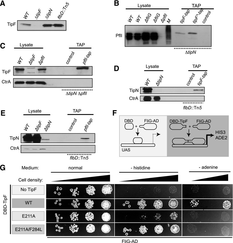
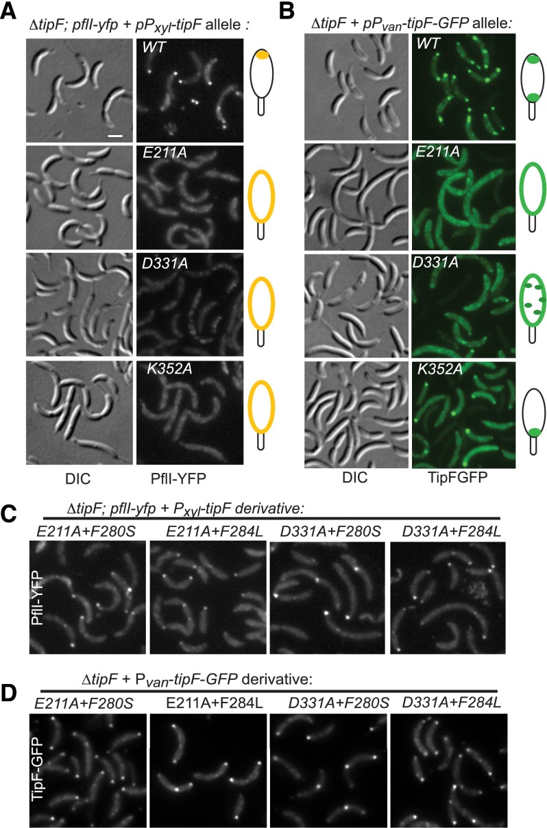
Eukaryotic morphogenesis is seeded with the establishment and subsequent amplification of polarity cues at key times during the cell cycle, often using (cyclic) nucleotide signals. We discovered that flagellum de- and repolarization in the model prokaryote Caulobacter crescentus is precisely orchestrated through at least three spatiotemporal mechanisms integrated at TipF. We show that TipF is a cell cycle-regulated receptor for the second messenger--bis-(3'-5')-cyclic dimeric guanosine monophosphate (c-di-GMP)--that perceives and transduces this signal through the degenerate c-di-GMP phosphodiesterase (EAL) domain to nucleate polar flagellum biogenesis. Once c-di-GMP levels rise at the G1 → S transition, TipF is activated, stabilized, and polarized, enabling the recruitment of downstream effectors, including flagellar switch proteins and the PflI positioning factor, at a preselected pole harboring the TipN landmark. These c-di-GMP-dependent events are coordinated with the onset of tipF transcription in early S phase and together enable the correct establishment and robust amplification of TipF-dependent polarization early in the cell cycle. Importantly, these mechanisms also govern the timely removal of TipF at cell division coincident with the drop in c-di-GMP levels, thereby resetting the flagellar polarization state in the next cell cycle after a preprogrammed period during which motility must be suspended.

Keywords: Caulobacter; asymmetric division; c-di-GMP; cell cycle; flagellum; morphogenesis; polarity.


Figures

Figure 1.
Figure 1.
Localization of TipN, TipF, and PflI to the flagellated pole. (A) Dynamic protein localization of TipN, TipF, and PflI and its coordination with other cell cycle events in C. crescentus. TipN (green) localized at the nonflagellated pole recruits newly synthesized TipF (red), which in turn recruits the PflI flagellar positioning protein (yellow). Thick blue arrows indicate the synthesis of TipF (by GcrA) and c-di-GMP. (B) Domain organization of TipN, TipF, and PflI. Shown are the predicted transmembrane domains (TM; gray), coiled-coil domains (CC; black), PDE domain (EAL; red), and proline-rich domain (PR; yellow). (C) Alignment of the EAL domains from TipF and the c-di-GMP PDEs PdeA of C. crescentus, VieA of Vibrio cholerae, RocR of Pseudomonas aeruginosa, and YkuI of Bacillus subtilis. Key residues that are predicted to be required for PDE activity (empty arrowheads) or required for c-di-GMP binding (filled black) are marked. Residues that bypass the requirement of E211 or D331 for motility (red triangles), the defining EAL motif, and the K332 residue of the conserved Asp–Asp loop (underlined) are indicated. (D) Motility assay of ΔtipF mutants harboring plasmids encoding wild-type (WT) or single- or double-mutant TipF. DTV and ADA denote the triple duplications of DTV or ADA residues at positions 121–124 or 128–130, respectively. Overnight cultures were spotted on PYE swarm agar plates and incubated for 60 h at 30°C. Compact swarms indicate the motility defect caused by mutations in the c-di-GMP-binding site, whereas the suppressive mutations yield diffuse and enlarged swarms.
Figure 2.
Figure 2.
TipF is enzymatically inactive but binds c-di-GMP. (A) c-di-GMP hydrolytic activity is not detected in recombinant (soluble) TipF but is in the control PDE YahA. (Left panel) Purified proteins (1 μM) were incubated with c-di-GMP for 5 min and then separated on a ResourceQ column to observe the cleavage product pGpG. No pGpG was detected after 1 h of incubation with wild-type (WT) or mutant (E211A or E211A/F284L) TipF with c-di-GMP. (B) ITC experiments showing that c-di-GMP binds to the soluble portion of wild-type TipF but not to the mutant derivatives E211A or E211A/F284L. The top panels show the raw ITC data curves collected at 25°C in binding buffer (50 mM Tris/HCl, 50 mM NaCl at pH 8.0). The bottom panels show the integrated titration peaks fitted to a one-site binding model (solid line). The average dissociation constant (KD) of TipF to c-di-GMP was estimated at 0.4 (±0.2 μM), the stoichiometry of binding (n) was estimated at 0.35 (±0.1), and the enthalpy of reaction was estimated at −2.1 (±0.3 kcal/mol).
Figure 3.
Figure 3.
TipF mediates the localization of flagellar proteins FliM, FliG, FliF, and PflI to the cell pole. (A) Schematic of the flagellum. The relative position of the MS ring (FliF), the switch complex (including FliG and FliM), the hook (FlgE), and the filament (flagellins) are indicated, as are the envelope layers: inner (cytoplasmic) membrane (IM), peptidoglycan layer (PG), outer membrane (OM), and S layer (SL). (B) ECT imaging reveals densities corresponding to the intact flagellum, including the MS ring and the switch complex in or near the inner membrane of the wild type (WT) that are absent in the ΔtipF mutant. The color-coding of the segmented structures corresponds to the color scheme in A. Flagellar structures (red) are shown in the left panel and in the segmentation of wild type. The black arrow points to the chemoreceptor array (orange) in ΔtipF cells that is also present in wild type. Bars, 200 nm. (C) The subcellular localization of FliM-GFP from the Pvan promoter on a medium copy number plasmid in a fliM mutant (fliM∷Tn5), a tipF mutant (ΔtipF; fliM∷Tn5), or a fliG mutant derivative (ΔfliG; fliM∷Tn5) analyzed by live-cell fluorescence microscopy. (D) Localization of PflI-YFP expressed from PpflI at the pflI locus in the presence (pflI-yfp) or absence of TipF (ΔtipF; pflI-yfp) or TipN (ΔtipN; pflI-yfp). (DIC) Differential interference contrast images. (E,F) Localization FliG-GFP (E) and FliF-GFP (F) expressed from Pxyl at the xylX locus in a ΔtipF mutant.
Figure 4.
Figure 4.
TipF directly interacts with FliG and forms a complex with TipN and PflI. (A) TipF steady-state levels as determined by immunoblotting using polyclonal antibodies to TipF in lysates from wild-type, ΔtipF, ΔtipN, and flbD∷Tn5. (B–E) TAP pull-down of TAP-tagged proteins expressed from Pvan on a medium copy plasmid followed by immunoblotting using polyclonal antibodies to TipF, PflI, TipN, and CtrA. The cell lysates derived from boiled cells shown in the lanes at the left of the panels provide negative and positive controls for the specificity for the antisera. CtrA immunoblots are shown as a control for loading. (B) TipF-TAP and TipF*-TAP (the E211A/F284L mutant is referred to as TipF*) were expressed from the Pvan promoter on a medium copy number plasmid in ΔtipN mutants. (M) Protein marker lane. (C) TipF is present in PflI-TAP purifications of ΔtipN ΔpflI double-mutant lysates. (D,E) TipN is present in pull-downs of TipF-TAP (D) or PfllI-TAP (E) from flbD∷Tn5 lysates. (F) Schematic showing the yeast two-hybrid (Y2H) assay using the C terminus of FliG (residues 237–340) as prey and the soluble part of TipF (residues 92–452) as bait to induce transcriptional activation of HIS3 and ADE2 (dark-gray box) as readout of the interaction. In the presence of DNA-binding domain (DBD)-TipF (TipF fused to the GAL4 DBD), transcriptional activation is seen, but not when DBD is used without a TipF fusion (light-gray box). (G) Growth of the yeast strains expressing the DBD-only (i.e., without TipF), wild-type, and mutant DBD-TipF variants along with FliG-AD (activation domain) on normal medium and selective medium lacking histidine (−histidine) or adenine (−adenine). The latter condition is more stringent. Gray scales from dark to light indicate the level of interaction corresponding to the growth readout on selective medium. Dark gray represents the strongest interaction as seen for wild-type DBD-TipF with FliG-AD.
Figure 5.
Figure 5.
Suppressor mutations render TipF and PflI localization c-di-GMP-independent. (A–D) Localization of PflI-YFP (A,C) or TipF-GFP (B,D) mutants in ΔtipF cells harboring plasmids encoding TipF loss-of-function mutants expressed from the Pxyl (A,C) or the Pvan (B,D) promoter a low copy number plasmid. (DIC) Differential interference contrast images. Note that TipF(K352A)-GFP can cluster at the stalked pole but not at the newborn pole and that the ectopic signals at the stalked pole of wild-type (WT) TipF-GFP are due to (constitutive) expression of TipF from Pvan on the low-copy plasmid compared with constructs expressing TipF-GFP or YFP from the chromosome (cf. B vs. Fig. 6B and Supplemental Fig. S3B).
Figure 6.
Figure 6.
Localization of TipF and PflI as well as expression of FlgE is c-di-GMP-dependent. (A–D) Localization of PflI-YFP (A), TipF-GFP (B), TipN-GFP (C), and YFP-tagged TipF suppressor mutants (D) under normal (PA5295-AAL, +cdG) or c-di-GMP-depleted (PA5295-WT, −cdG) conditions. PA5295 variants were expressed from the Pxyl promoter on a medium copy number plasmid in cells expressing the GFP fusion proteins from their respective promoters at the endogenous locus. In D, TipF-YFP variants were expressed from Pvan at the vanA locus in ΔtipF cells. (DIC) Differential interference contrast images. (E) Effects of c-di-GMP concentration on transcription of the flgE hook gene by β-galactosidase assays of cells (wild-type [WT], ΔtipF, or ΔtipF cells expressing mutant TipF from Pvan at the vanA locus) harboring a PflgE-lacZ transcriptional reporter fusion. (F) Effects of c-di-GMP depletion on FlgE steady-state levels in supernatants (Sup.) or cell lysates (Lys.), determined by immunoblotting using anti-FlgE antibody (α-FlgE). TipF mutants were expressed from the Pxyl promoter on a low copy number plasmid in ΔtipF cells.
Figure 7.
Figure 7.
c-di-GMP levels affect TipF protein stability. (A,B) Depletion of c-di-GMP by overexpression of a potent PDE reduces TipF steady-state levels. TipF, the c-di-GMP-binding mutant E211A, or the intragenic suppressor mutant E211A/F284L was expressed from Pxyl in ΔtipF cells grown in M2G containing xylose following a shift to M2G containing vanillate to induce the expression of the PDE PA5295 from P. aeruginosa or its active site mutant, PA5295-AAL, and repress Pxyl. Samples were taken every 20 min, and protein levels were quantified from the immunoblots and plotted as percentages of the highest value. (C) The dominant-negative clpXATP allele was expressed from plasmid in M2G containing xylose (M2GX) or repressed by growing the cells in M2G. Swarmer cells were isolated, and TipF was detected using a polyclonal anti-TipF antibody and ClpXATP*∷Flag using monoclonal anti-M2 antibodies. In the presence of wild-type (WT) ClpXP, TipF was not detectable, while inactivation of ClpXP by the dominant-negative allele encoding ClpXATP* led to stabilization of TipF. (D) The cell cycle abundance of TipF resembles that of GcrA. Synchronized wild-type or tipF-gfp (in which endogenous tipF is replaced by tipF-gfp) swarmer cells were released in fresh medium, and CtrA, TipF, TipF-GFP, and/or GcrA steady-state levels were determined by immunoblotting using antibodies to TipF (black asterisk), GFP (blue asterisk), CtrA, or GcrA at different times during cell cycle progression. (E) TipF and FtsZ (control) steady-state levels in wild-type and mutant cells, as determined by immunoblotting using antibodies to TipF and FtsZ. (F) GcrA binds the tipF promoter, as determined by qChIP experiments using polyclonal antibodies to GcrA. The abundance of the tipF and sciP promoters was quantified in the immunoprecipitates (left panel) and tipF (right panel) in wild-type and ΔgcrA; ΔgcrA2 double-mutant cells.

Similar articles

Cited by

References

    1. Abel S, Chien P, Wassmann P, Schirmer T, Kaever V, Laub MT, Baker TA, Jenal U 2011. Regulatory cohesion of cell cycle and cell differentiation through interlinked phosphorylation and second messenger networks. Mol Cell 43: 550–560 - PMC - PubMed
    1. Alley MR, Maddock JR, Shapiro L 1992. Polar localization of a bacterial chemoreceptor. Genes Dev 6: 825–836 - PubMed
    1. Amato S, Liu X, Zheng B, Cantley L, Rakic P, Man HY 2011. AMP-activated protein kinase regulates neuronal polarization by interfering with PI 3-kinase localization. Science 332: 247–251 - PMC - PubMed
    1. Balaban M, Joslin SN, Hendrixson DR 2009. FlhF and its GTPase activity are required for distinct processes in flagellar gene regulation and biosynthesis in Campylobacter jejuni. J Bacteriol 191: 6602–6611 - PMC - PubMed
    1. Barends TR, Hartmann E, Griese JJ, Beitlich T, Kirienko NV, Ryjenkov DA, Reinstein J, Shoeman RL, Gomelsky M, Schlichting I 2009. Structure and mechanism of a bacterial light-regulated cyclic nucleotide phosphodiesterase. Nature 459: 1015–1018 - PubMed

Publication types

LinkOut - more resources