

MicroRNA signature in wound healing following excimer laser ablation: role of miR-133b on TGFβ1, CTGF, SMA, and COL1A1 expression levels in rabbit corneal fibroblasts
- PMID: 24065814
- PMCID: PMC3808100
- DOI: 10.1167/iovs.13-12621
MicroRNA signature in wound healing following excimer laser ablation: role of miR-133b on TGFβ1, CTGF, SMA, and COL1A1 expression levels in rabbit corneal fibroblasts
Abstract
Purpose: The role of microRNA (miRNA) regulation in corneal wound healing and scar formation has yet to be elucidated. This study analyzed the miRNA expression pattern involved in corneal wound healing and focused on the effect of miR-133b on expression of several profibrotic genes.
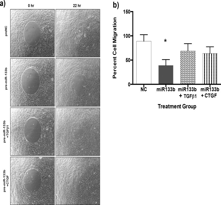
Methods: Laser-ablated mouse corneas were collected at 0 and 30 minutes and 2 days. Ribonucleic acid was collected from corneas and analyzed using cell differentiation and development miRNA PCR arrays. Luciferase assay was used to determine whether miR-133b targeted the 3' untranslated region (UTR) of transforming growth factor β1 (TGFβ1) and connective tissue growth factor (CTGF) in rabbit corneal fibroblasts (RbCF). Quantitative real-time PCR (qRT-PCR) and Western blots were used to determine the effect of miR-133b on CTGF, smooth muscle actin (SMA), and collagen (COL1A1) in RbCF. Migration assay was used to determine the effect of miR-133b on RbCF migration.
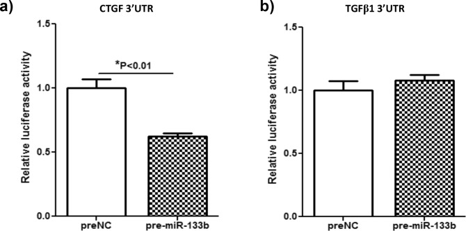
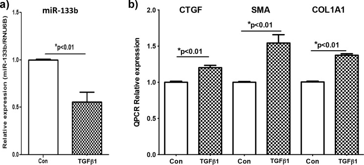
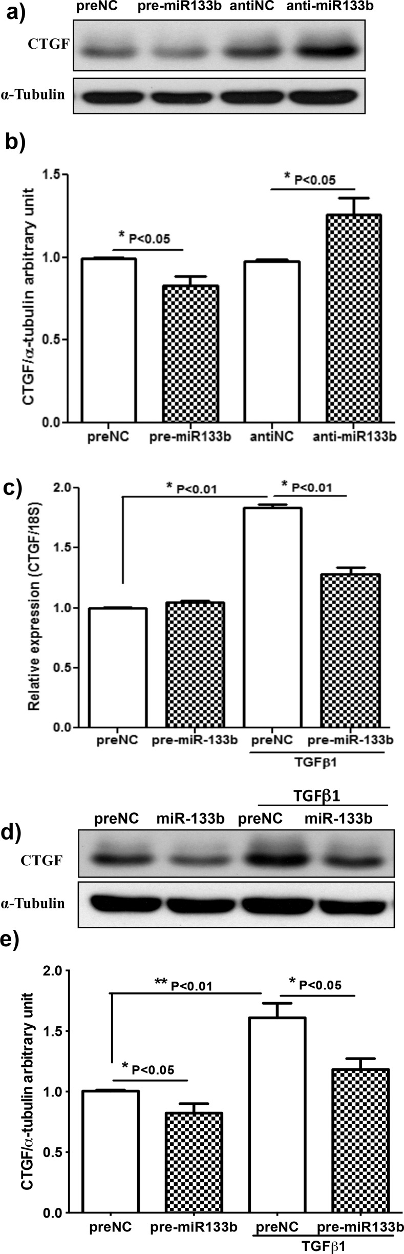
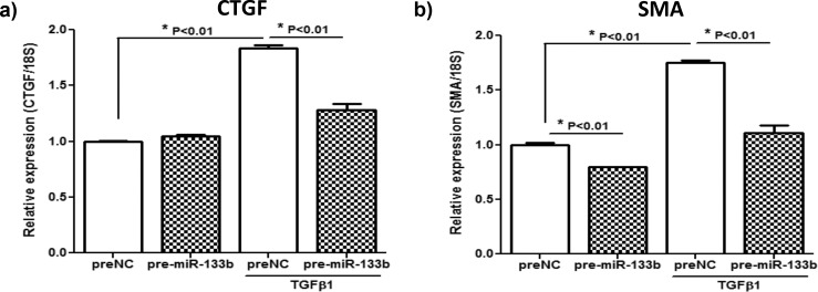
Results: At day 2, 37 of 86 miRNAs had substantial expression fold changes. miR-133b had the greatest fold decrease at -14.33. Pre-miR-133b targeted the 3' UTR of CTGF and caused a significant decrease of 38% (P < 0.01). Transforming growth factor β1-treated RbCF had a significant decrease of miR-133b of 49% (P < 0.01), whereas CTGF, SMA, and COL1A1 had significant increases of 20%, 54%, and 37% (P < 0.01), respectively. The RbCF treated with TGFβ1 and pre-miR133b showed significant decreases in expression of CTGF, SMA, and COL1A1 of 30%, 37%, and 28% (P < 0.01), respectively. Finally, there was significant decrease in migration of miR-133b-treated RbCF.
Conclusions: Significant changes occur in key miRNAs during early corneal wound healing, suggesting novel miRNA targets to reduce scar formation.
Keywords: CTGF; corneal wound healing; gene expression; microRNA.
Figures
References
-
- Mohan RR, Liang Q, Kim WJ, Helena MC, Baerveldt F, Wilson SE. Apoptosis in the cornea: further characterization of Fas/Fas ligand system. Exp Eye Res. 1997; 65: 575–589 - PubMed
-
- Mohan RR, Mohan RR, Kim WJ, Wilson SE. Modulation of TNF-alpha-induced apoptosis in corneal fibroblasts by transcription factor NF-kappaB. Invest Ophthalmol Vis Sci. 2000; 41: 1327–1336 - PubMed
-
- Mohan RR, Kim WJ, Mohan RR, Chen L, Wilson SE. Bone morphogenic proteins 2 and 4 and their receptors in the adult human cornea. Invest Ophthalmol Vis Sci. 1998; 39: 2626–2636 - PubMed
-
- Wilson SE, Liang Q, Kim WJ. Lacrimal gland HGF, KGF, and EGF mRNA levels increase after corneal epithelial wounding. Invest Ophthalmol Vis Sci. 1999; 40: 2185–2190 - PubMed
-
- Tuominen IS, Tervo TM, Teppo AM, Valle TU, Gronhagen-Riska C, Vesaluoma MH. Human tear fluid PDGF-BB, TNF-alpha and TGF-beta1 vs corneal haze and regeneration of corneal epithelium and subbasal nerve plexus after PRK. Exp Eye Res. 2001; 72: 631–641 - PubMed
Publication types
MeSH terms
Substances
Grants and funding
LinkOut - more resources
Full Text Sources
Other Literature Sources
Miscellaneous
