. 2013 Oct 8;110(41):16474-9.
doi: 10.1073/pnas.1307680110. Epub 2013 Sep 24.

Pax7 is critical for the normal function of satellite cells in adult skeletal muscle

Affiliations

Pax7 is critical for the normal function of satellite cells in adult skeletal muscle

Julia von Maltzahn et al. Proc Natl Acad Sci U S A. .

Abstract

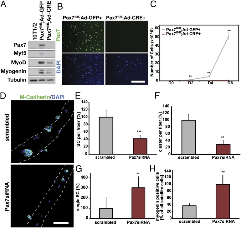
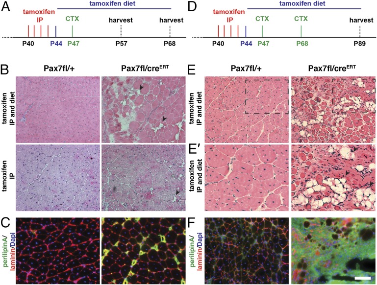
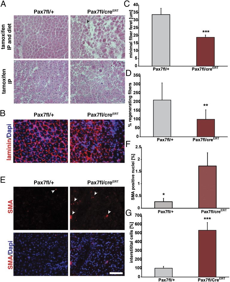
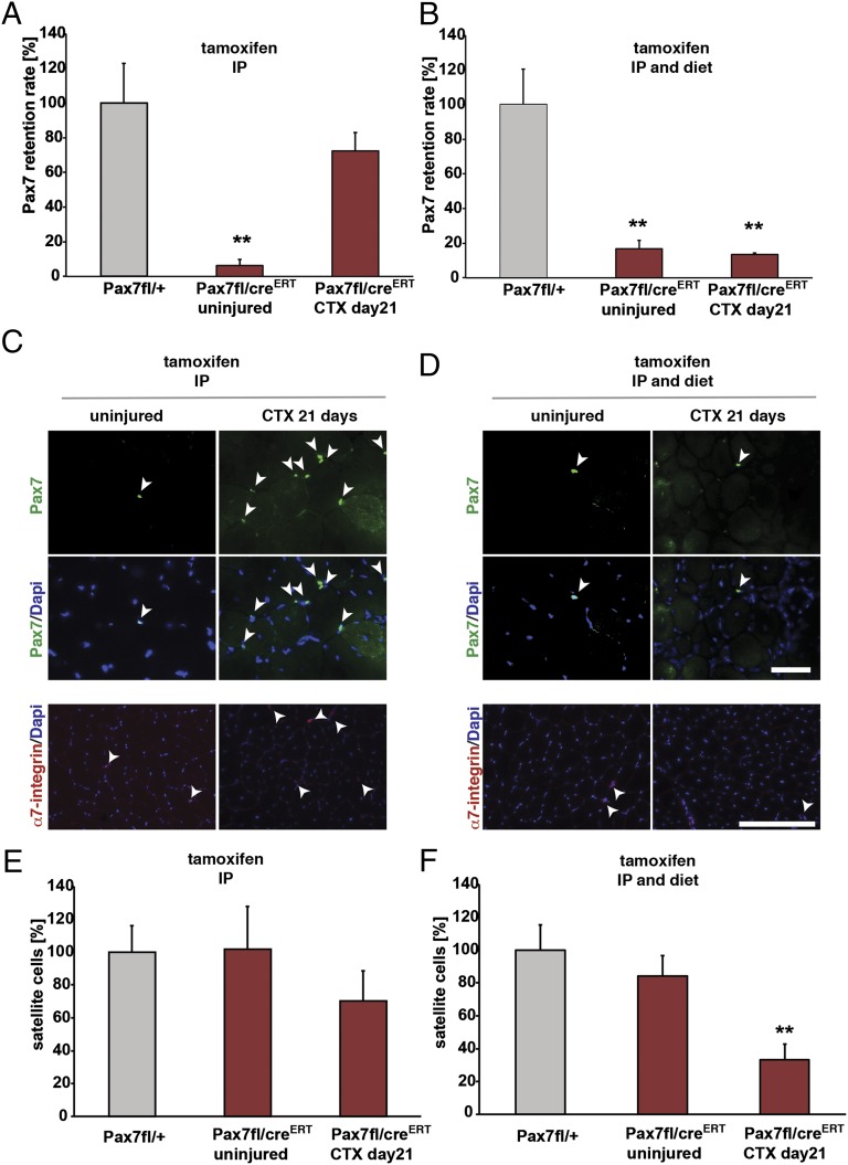
Extensive analyses of mice carrying null mutations in paired box 7 (Pax7) have confirmed the progressive loss of the satellite cell lineage in skeletal muscle, resulting in severe muscle atrophy and death. A recent study using floxed alleles and tamoxifen-induced inactivation concluded that after 3 wk of age, Pax7 was entirely dispensable for satellite cell function. Here, we demonstrate that Pax7 is an absolute requirement for satellite cell function in adult skeletal muscle. Following Pax7 deletion, satellite cells and myoblasts exhibit cell-cycle arrest and dysregulation of myogenic regulatory factors. Maintenance of Pax7 deletion through continuous tamoxifen administration prevented regrowth of Pax7-expressing satellite cells and a profound muscle regeneration deficit that resembles the phenotype of skeletal muscle following genetically engineered ablation of satellite cells. Therefore, we conclude that Pax7 is essential for regulating the expansion and differentiation of satellite cells during both neonatal and adult myogenesis.

Keywords: CreERT2; stem cell.


Conflict of interest statement

The authors declare no conflict of interest.

Figures

Fig. 1.
Fig. 1.
Pax7 deletion results in cell-cycle arrest and precocious differentiation. (A) Infection of Pax7fl/fl myoblasts with an adenovirus encoding the Cre gene leads to depletion of Pax7 expression and loss of Myf5 protein expression. (B) Infection of Pax7fl/fl myoblasts with an adenovirus encoding the Cre gene leads to depletion of Pax7 expression. Pax7 immunostaining is depicted in green. Nuclei are counterstained with DAPI. (Scale bar: 100 µm.) (C) Ad-Cre-mediated Pax7 deletion results in growth arrest in primary myoblasts, relative to control (Ad-GFP) myoblasts; n = 3, **P < 0.01. (D) The siRNA-mediated depletion of Pax7 expression in satellite cells leads to reduced numbers of satellite cells (marked by M-Cadherin staining in green) after 72 h of culture. Nuclei are counterstained with DAPI. (Scale bar: 100 µm.) (E) Following depletion of Pax7 in satellite cells on single myofibers, the number of satellite cells per fiber is significantly reduced; n = 4, ***P < 0.001. (F) Numbers of clusters (three or more satellite cells on a fiber attached to each other) are reduced following treatment with Pax7 siRNA compared with scrambled control; n = 4, ***P < 0.01. (G) Increase in the number of single satellite cells on myofibers following 72 h in culture and treatment with Pax7siRNA; n = 4, ***P < 0.01. (H) Numbers of myogenin-positive satellite cells increase after 72 h in culture following depletion of Pax7 expression with Pax7 siRNA; n = 4, ***P < 0.01.
Fig. 2.
Fig. 2.
Inactivation of Pax7 in adult satellite cells markedly impairs muscle regeneration. (A) Schematic showing the experimental regime for single injury used in this study. (B) H&E staining of tibialis anterior muscle 21 d after acute injury induced by CTX injection. Arrowheads point to fat depositions. (C) Immunostaining for perilipin (in green) demonstrates increased adipogenesis in Pax7fl/CreERT2 mice, laminin staining is shown in red, and nuclei are counterstained with DAPI (in blue). (D) Schematic showing the experimental regime for double CTX injury used in this study. (E) H&E staining of tibialis anterior muscle 21 d after the second injury induced by CTX injection. Arrowheads point to fat depositions. (E’) A magnification of E. (F) Immunostaining for perilipin (in green) demonstrates increased adipogenesis in Pax7fl/CreERT2 mice, laminin staining is shown in red, and nuclei are counterstained with DAPI (in blue). (Scale bar: 100 µm.)
Fig. 3.
Fig. 3.
Significantly impaired muscle regeneration with continuous tamoxifen treatment. (A) H&E staining 10 d after acute injury reveals markedly impaired regeneration following continuous tamoxifen treatment. (Scale bar: 100 μm.) (B) Immunostaining for laminin (in red) showing decreased numbers of fibers and also reduced fiber feret. Nuclei are counterstained with DAPI (in blue). (Scale bar: 100 μm.) (C) Quantification of the minimal fiber feret of muscles at 10 d after acute injury; n= 6, ***P < 0.001. (D) Quantification of the amount of regenerating fibers at 10 d after CTX injury; n = 6, *P < 0.01. (E) Immunostaining for smooth muscle actin (SMA, in red) demonstrating increased numbers of SMA-positive nuclei in Pax7fl/CreERT2 animals compared with Pax7fl/+ animals at 10 d after acute injury. Nuclei are counterstained with DAPI (in blue). (Scale bar: 100 μm.) (F) Quantification of SMA-positive nuclei relative to the total number of nuclei; n = 6, *P < 0.05. (G) Quantification of the numbers of interstitial cells, n = 4, ***P < 0.001.
Fig. 4.
Fig. 4.
Continuous tamoxifen treatment prevents grow-back of residual Pax7-expressing satellite cells. (A) Quantification of Pax7-expressing nuclei in Pax7fl/CreERT2 mice induced with tamoxifen before and after acute injury; n = 3, **P < 0.01. (B) Quantification of Pax7-expressing nuclei in Pax7fl/CreERT2 mice with continuous tamoxifen application before and after acute injury; n = 3, **P < 0.01. (C) Pax7 expression is lost after tamoxifen-induced excision in Pax7fl/CreERT2 mice (IP injection) but reoccurs after acute injury, Pax7 immunostaining is shown in green, and nuclei are counterstained with DAPI (in blue). Staining for α7-integrin is shown in red, and nuclei are counterstained with DAPI (in blue). (D) Pax7 deletion is only maintained when tamoxifen is continuously administered during regeneration of CTX-injured muscles, Pax7 immunostaining is shown in green, and nuclei are counterstained with DAPI (in blue). Staining for α7-integrin is shown in red, and nuclei are counterstained with DAPI (in blue). (E) Quantification of numbers of satellite cells (marked by expression of α7-integrin) in Pax7fl/CreERT2 mice induced with tamoxifen before and after acute injury; n = 3. (F) Quantification of numbers of satellite cells (marked by expression of α7-integrin) in Pax7fl/CreERT2 mice with continuous tamoxifen application before and after acute injury; n = 3, **P < 0.01. (Scale bar: 50 µm.)

References

    1. Relaix F, et al. Pax3 and Pax7 have distinct and overlapping functions in adult muscle progenitor cells. J Cell Biol. 2006;172(1):91–102. - PMC - PubMed
    1. Lepper C, Partridge TA, Fan CM. An absolute requirement for Pax7-positive satellite cells in acute injury-induced skeletal muscle regeneration. Development. 2011;138(17):3639–3646. - PMC - PubMed
    1. Sambasivan R, et al. Pax7-expressing satellite cells are indispensable for adult skeletal muscle regeneration. Development. 2011;138(17):3647–3656. - PubMed
    1. Seale P, Ishibashi J, Scimè A, Rudnicki MA. Pax7 is necessary and sufficient for the myogenic specification of CD45+:Sca1+ stem cells from injured muscle. PLoS Biol. 2004;2(5):E130. - PMC - PubMed
    1. Oustanina S, Hause G, Braun T. Pax7 directs postnatal renewal and propagation of myogenic satellite cells but not their specification. EMBO J. 2004;23(16):3430–3439. - PMC - PubMed

Publication types