. 2013 Sep 16;8(9):e73585.
doi: 10.1371/journal.pone.0073585. eCollection 2013.

Long-term evolution of brainstem electrical evoked responses to sound after restricted ablation of the auditory cortex

Affiliations

Long-term evolution of brainstem electrical evoked responses to sound after restricted ablation of the auditory cortex

Verónica Lamas et al. PLoS One. .

Erratum in

  • PLoS One. 2013;8(11). doi:10.1371/annotation/07ec3908-23dd-4c31-926d-9d73a2815032

Abstract

Introduction: This study aimed to assess the top-down control of sound processing in the auditory brainstem of rats. Short latency evoked responses were analyzed after unilateral or bilateral ablation of auditory cortex. This experimental paradigm was also used towards analyzing the long-term evolution of post-lesion plasticity in the auditory system and its ability to self-repair.

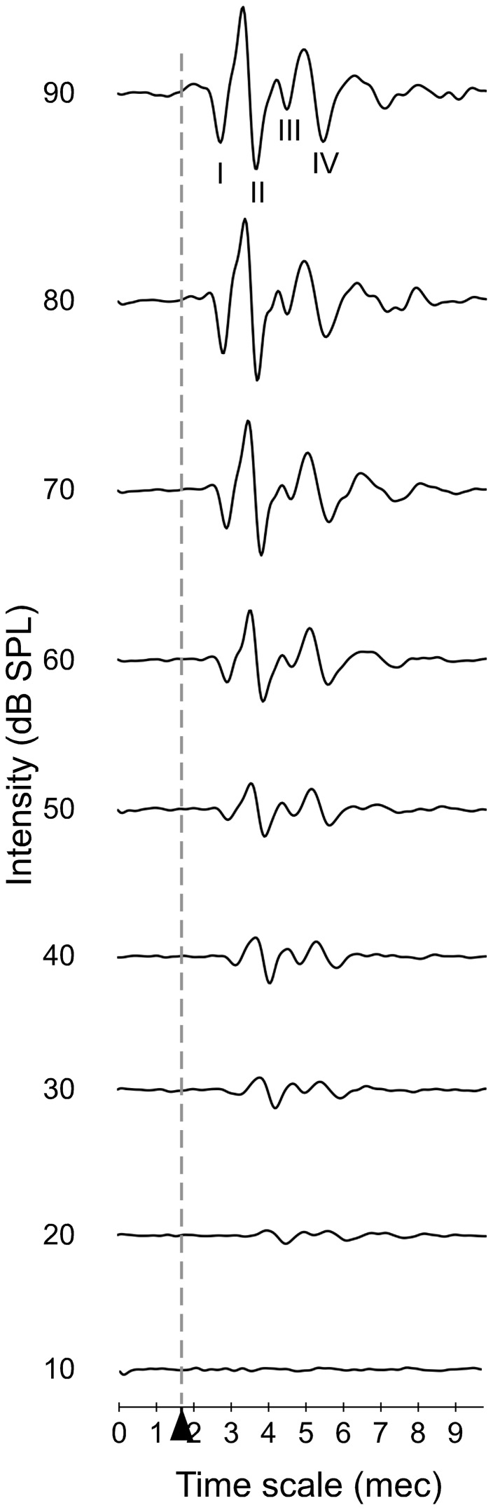
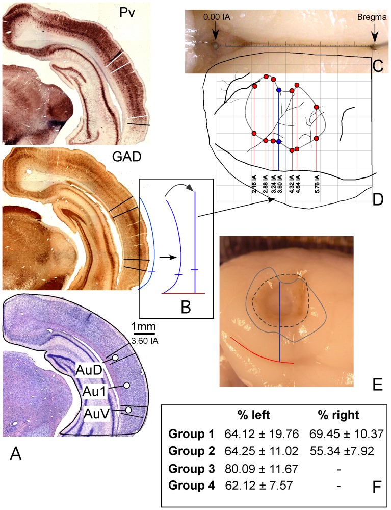
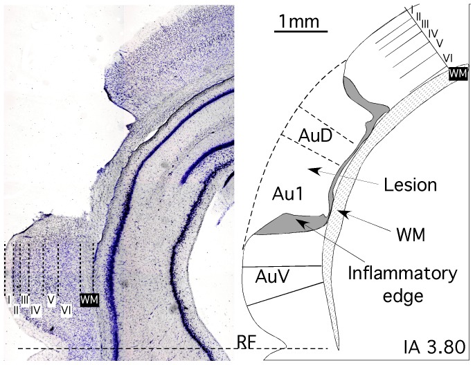
Method: Auditory cortex lesions were performed in rats by stereotactically guided fine-needle aspiration of the cerebrocortical surface. Auditory Brainstem Responses (ABR) were recorded at post-surgery day (PSD) 1, 7, 15 and 30. Recordings were performed under closed-field conditions, using click trains at different sound intensity levels, followed by statistical analysis of threshold values and ABR amplitude and latency variables. Subsequently, brains were sectioned and immunostained for GAD and parvalbumin to assess the location and extent of lesions accurately.

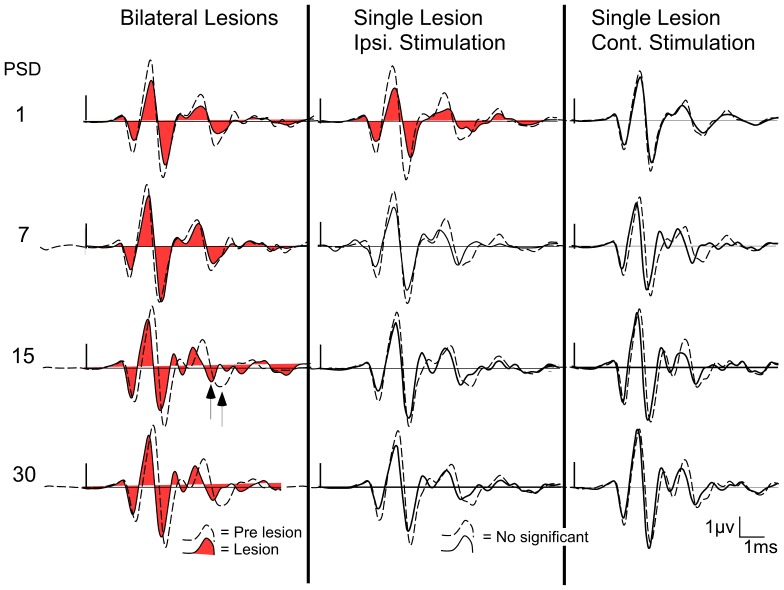
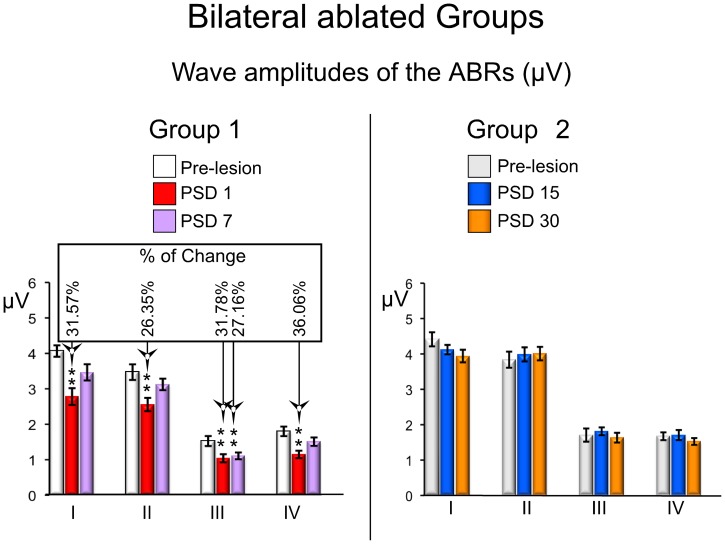
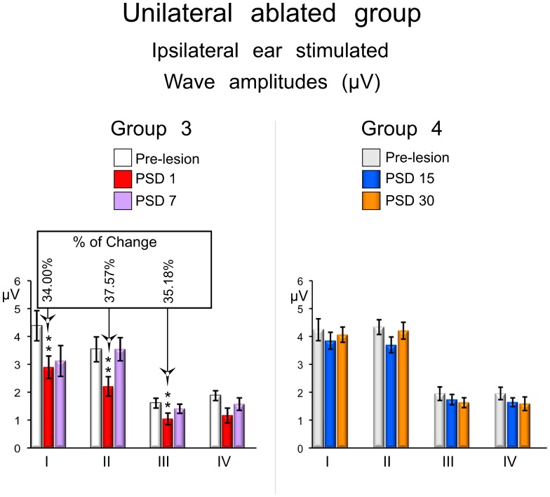
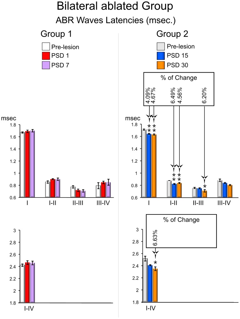
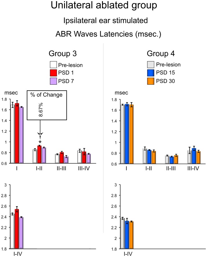
Results: Alterations in ABR variables depended on the type of lesion and post-surgery time of ABR recordings. Accordingly, bilateral ablations caused a statistically significant increase in thresholds at PSD1 and 7 and a decrease in waves amplitudes at PSD1 that recover at PSD7. No effects on latency were noted at PSD1 and 7, whilst recordings at PSD15 and 30 showed statistically significant decreases in latency. Conversely, unilateral ablations had no effect on auditory thresholds or latencies, while wave amplitudes only decreased at PSD1 strictly in the ipsilateral ear.

Conclusion: Post-lesion plasticity in the auditory system acts in two time periods: short-term period of decreased sound sensitivity (until PSD7), most likely resulting from axonal degeneration; and a long-term period (up to PSD7), with changes in latency responses and recovery of thresholds and amplitudes values. The cerebral cortex may have a net positive gain on the auditory pathway response to sound.


Conflict of interest statement

Competing Interests: The authors have declared that no competing interests exist.

Figures

Figure 1
Figure 1. An example of the ABR waveforms obtained from a control animal during recordings from 10 to 90(msec).
The stimulus onset starts at 1.7(Alvarado et al. 2012), wave II is the largest wave in rat ABR recordings whereas wave III is the smallest.
Figure 2
Figure 2. Procedure for the location and extent of lesions in the rat brain AC.
A. Coronal caudal sections at a similar rostro-caudal level stained with Parvalbumin (Pv), Glutamic Acid Decarboxilase (GAD) and Nissl (interaural 3.60 – similar to Patxinos and Watson). In the immunostained sections, positive pyramidal cells and neuropil of layers III and V enable to define the AC boundaries (highlighted on the photographs with lines perpendicular to the surface of the section). B. Outline of the section contour after labeling the AC boundary. The straight line (in bold) on the right side of the curved arrow was drawn with the same length as the auditory cortex perimeter, using the Canvas software. After scaling the line to compensate the shrinkage of the section, the coordinates were transferred to the macroscopic image of the brain (thick arrow). C. Photograph of the brain midline with sterotactically (Patxinos and Watson coordinates) implanted needles in 0.00 IA and Bregma. D. AC map after transferring the coordinates of AC boundaries from seven rostrocaudal sectioning levels. E. Overlapping map of the cortex on a lesioned brain, positioned into a matrix to calculate the extension and location of the ablated surface relative to the AC boundaries. F. Results of the percentage of AC affected by lesions in the experimental groups.
Figure 3
Figure 3. Example of section from animals received lesions.
Left, a Nissl stained section showing the different cortical layers and the ablated area. Right, the corresponding drowing line, showing the AC subdivisions affected by the ablation. Note that lesion affect all AC layers but not the underlying white matter (WM).
Figure 4
Figure 4. Average ABRs waveform before and after cortical ablation from all experimental groups (n = 7), at different post-surgery days (PSD): at left, ABR waveforms from animals with bilateral AC lesions; in the middle, ABR waveforms of the ear ipsilateral to the AC ablation from the unilaterally ablated animals; at right, ABR waveforms of the ear contralateral to the AC ablation from the unilaterally ablated animals.
Figure 5
Figure 5. ABRs changes in waves amplitudes from the bilateral ablated experimental groups.
The mean±SEM amplitude of waves in microvolts (µV) is shown in the graphs. * p<0.05. ** p<0.01. *** p<0.001. The percentage of change (Alvarado et al 2007) regarding the pre-lesion condition is shown in the top box. Note that all mean values of waves decrease in relation to the pre-lesion condition in the short-term (PSD1 and 7) periods, and no significant changes were observed at long-term (PSD 15 and 30).
Figure 6
Figure 6. ABRs changes in waves amplitudes from the unilateral ablated experimental groups.
The mean±SEM amplitude of waves in microvolts (µV) is shown in the graphs. * p<0.05. ** p<0.01. *** p<0.001. The percentage of change (Alvarado et al 2007) regarding the pre-lesion condition is shown in the top box. Note that mean values of waves I, II and III decrease in relation to the pre-lesion condition in the short-term (PSD1 and 7) periods, and no significant changes was observed at long-term (PSD 15 and 30).
Figure 7
Figure 7. ABRs changes in waves latencies from the bilateral ablated experimental groups.
The mean±SEM of significant latencies of all intervals of waves in milliseconds are show on the graphs. * p<0.05. ** p<0.01. *** p<0.001. The percentage of change (Alvarado et al 2007) regarding the pre-lesion condition is shown in the top box. Latencies are significantly shortened in recordings conducted in the long-term (PSD 15 and 30).
Figure 8
Figure 8. ABRs changes in waves latencies from the unilateral ablated experimental groups.
The mean±SEM of significant latencies of all intervals of waves in milliseconds are show on the graphs. * p<0.05. ** p<0.01. *** p<0.001. The percentage of change (Alvarado et al 2007) regarding the pre-lesion condition is shown in the top box. Latencies of wave I–II are significantly increased in recordings conducted in the sort-term (PSD 1).

References

    1. Rasmussen GL (1953) Further observations of the efferent cochlear bundle. J Comp Neurol 99: 61–74. - PubMed
    1. Galambos R (1956) Suppression of auditory nerve activity by stimulation of efferent fibers to cochlea. J Neurophysiol 19: 424–437. - PubMed
    1. Coomes DL, Schofield BR (2004) Projections from the auditory cortex to the superior olivary complex in guinea pigs. Eur J Neurosci 19: 2188–2200. - PubMed
    1. Mulders WH, Robertson D (2000) Evidence for direct cortical innervation of medial olivocochlear neurones in rats. Hear Res 144: 65–72. - PubMed
    1. Feliciano M, Saldana E, Mugnaini E (1995) Direct projections from the rat primary auditory neocortex to nucleus sagulum, paralemniscal regions, superior olivary complex and cochlear nuclei. Auditory Neuroscience 1: 287–308.

Publication types

LinkOut - more resources