. 2013 Sep 16;8(9):e75076.
doi: 10.1371/journal.pone.0075076. eCollection 2013.

Insight into the intermolecular recognition mechanism between Keap1 and IKKβ combining homology modelling, protein-protein docking, molecular dynamics simulations and virtual alanine mutation

Affiliations

Insight into the intermolecular recognition mechanism between Keap1 and IKKβ combining homology modelling, protein-protein docking, molecular dynamics simulations and virtual alanine mutation

Zheng-Yu Jiang et al. PLoS One. .

Abstract

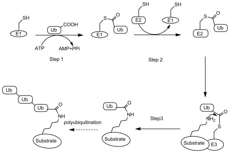
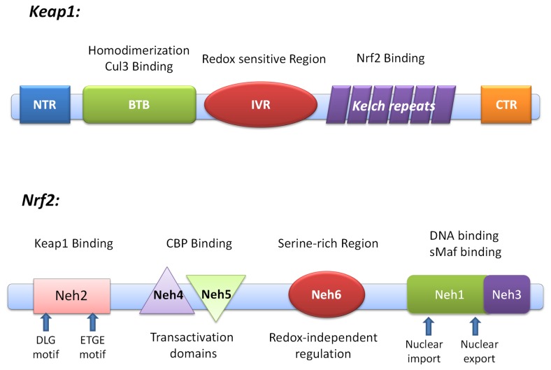
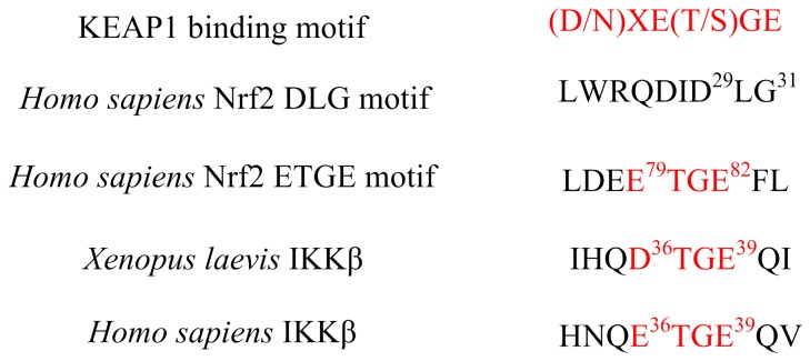
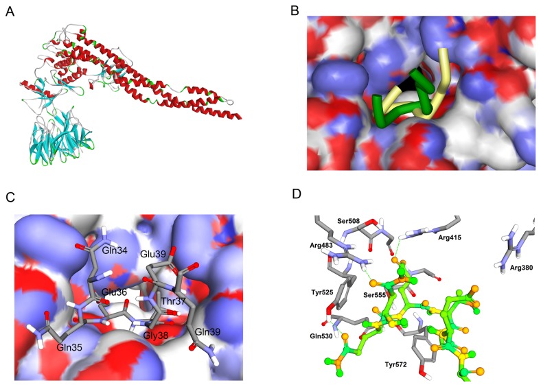
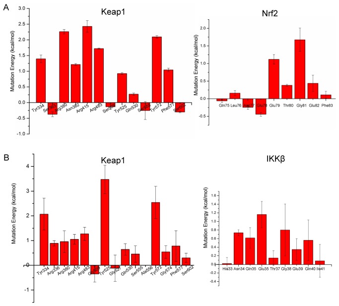
Degradation of certain proteins through the ubiquitin-proteasome pathway is a common strategy taken by the key modulators responsible for stress responses. Kelch-like ECH-associated protein-1(Keap1), a substrate adaptor component of the Cullin3 (Cul3)-based ubiquitin E3 ligase complex, mediates the ubiquitination of two key modulators, NF-E2-related factor 2 (Nrf2) and IκB kinase β (IKKβ), which are involved in the redox control of gene transcription. However, compared to the Keap1-Nrf2 protein-protein interaction (PPI), the intermolecular recognition mechanism of Keap1 and IKKβ has been poorly investigated. In order to explore the binding pattern between Keap1 and IKKβ, the PPI model of Keap1 and IKKβ was investigated. The structure of human IKKβ was constructed by means of the homology modeling method and using reported crystal structure of Xenopus laevis IKKβ as the template. A protein-protein docking method was applied to develop the Keap1-IKKβ complex model. After the refinement and visual analysis of docked proteins, the chosen pose was further optimized through molecular dynamics simulations. The resulting structure was utilized to conduct the virtual alanine mutation for the exploration of hot-spots significant for the intermolecular interaction. Overall, our results provided structural insights into the PPI model of Keap1-IKKβ and suggest that the substrate specificity of Keap1 depend on the interaction with the key tyrosines, namely Tyr525, Tyr574 and Tyr334. The study presented in the current project may be useful to design molecules that selectively modulate Keap1. The selective recognition mechanism of Keap1 with IKKβ or Nrf2 will be helpful to further know the crosstalk between NF-κB and Nrf2 signaling.


Conflict of interest statement

Competing Interests: The authors have declared that no competing interests exist.

Figures

Figure 1
Figure 1. Ubiquitination of a protein substrate through E1→E2→E3.
Step1: E1 activates Ub C terminus to form a thioester intermediate by coupling ATP hydrolysis; Step2: the activated Ub is transferred to E2 also in the thioester form; Steps3: the E3 ligase catalyzes the transfer of one Ub molecule at a time or a Ub chain to a protein target.
Figure 2
Figure 2. Domain structure of Nrf2 and Keap1.
Keap1 possesses four characteristic domains: the Broad complex, Tramtrack, and Bric-a-Brac (BTB); the intervening region (IVR); the double glycine repeat or Kelch repeat (DGR); and the C-terminal region (CTR). The DGR and CTR domains, also known as DC domain, mediate interaction with Neh2 domain of Nrf2. Nrf2 can be divided into six highly homologous regions (Neh1 to Neh6 domains) through comparing the human and chicken Nrf2 amino acid sequences. Neh2 is responsible for interacting with Keap1.
Figure 3
Figure 3. Sequence comparison of Keap1 binding motif.
The Asn34 and Gln35 are located before ETGE in human IKKβ, while these two residues change to the Asp77 and Glu78 in Nrf2.
Figure 4
Figure 4. Homology Modeling of Homo sapiens IKKβ.
(A) Domain structure of IKKβ. IKKβ can be divided into three function domains: a kinase domain (KD), an ubiquitin-like domain (ULD) and an elongated, α-helical scaffold/dimerization domain (SDD) colored as green, red and yellow. (B) Ribbon diagram of Homo sapiens IKKβ from Homology Modeling. KD (green), ULD (red) and SDD (yellow) are labeled. (C) Superimpose of Homo sapiens IKKβ and Xenopus laevis IKKβ. The Keap1 binding motif is colored as azure. In general, the structure of Homo sapiens IKKβ (yellow) and Xenopus laevis IKKβ (red) are quite similar. Both of the Keap1 binding motifs (blue) form the β-turn structure. (D) & (E) Structure difference of Keap1 binding motif between Homo sapiens IKKβ (red) and Xenopus laevis IKKβ (blue). The most different residues were labeled in the picture.
Figure 5
Figure 5. Protein-Protein Docking of IKKβ and Keap1.
(A) Complex of IKKβ and Keap1 shown as ribbon diagram. The Keap1 binding motif of IKKβ fits into an identical pocket of Keap1. (B) Superimpose of IKKβ (Dark green) and Nrf2 (yellow, PDB ID: 2FLU) ETGE motif in the Keap1 cavity. IKKβ and Nrf2 occupy almost the same part of Keap1 cavity. However, the structures of ETGE motif show some differences. In the case of IKKβ, the two antiparallel β-strands, forming the turn region, are closer than the Nrf2 ETGE motif. (C) The top view of Keap1-IKKβ complex. The IKKβ ETGE motif is represented in sticks and the surface of Keap1 is colored by partial charge. Both of the two side chain carboxyl groups point to the positively charged surface. (D) Interacting amino acid residues on the IKKβ and Keap1. The interacting region of Keap1 is represented in sticks and the regions of IKKβ are shown in ball and sticks.
Figure 6
Figure 6. MD simulation of Keap1 DC domain.
(A) The six kelch repeats that comprise the DGR domain of Keap1 form a highly symmetric, 6-bladed-propeller structure (Blade I: residue 598-609 and 327-358, blue; Blade II: residue 359-409, red; Blade III: residue 410-456, purple; Blade IV: residue 457-503, yellow; Blade V: 504-550, green; Blade VI: residue 551-597, orange PDB code: 2FLU). (B) Analysis of Root Mean Square Deviation of backbone atoms during molecular dynamics simulation (red for the first run, blue for the second run and black for the third run). (C) & (D) Representative structures of triplicate MD simulations (coloured as azure, yellow and green) superimposed to the starting structure (red). Though the whole structure of Keap1 DC domain is stable, all six blades of the β-propeller tend to be more open and loose.
Figure 7
Figure 7. MD simulation of IKKβ-Keap1 complex.
(A) RMSD value of backbone atoms respect to the starting structure (yellow for the first run, gray for the second run and blue for the third run). (B) Superimposition of three representative structures of triplicate MD simulations (coloured as red, yellow and blue). Only the ETGE motif was shown in the figure. The Keap1 DC domain is stable during the simulation. (C) Interaction model of Keap1-Nrf2. (D) (E) & (F) Binding model of three representative structures of triplicate MD simulations. The residues of Keap1 are represented as sticks and the residues of ETGE motif are represented as ball and sticks. Hydrogen bonds are shown as green dashed line.
Figure 8
Figure 8. Virtual Alanine Mutation of binding residues.
The Y axis is the weighted mutation energy (unit: kcal/mol) and the X axis is the name of mutated amino acid. (A) Mean values of Virtual Alanine Mutation of Nrf2-Keap1 complex with standard deviation. (B) Mean values of Virtual Alanine Mutation of Keap1-IKKβ complex obtained from triplicate MD simulations with standard deviation.

References

    1. Spasser L, Brik A (2012) Chemistry and biology of the ubiquitin signal. Angew Chem Int Ed Engl 51: 6840-6862. doi:10.1002/anie.201200020. PubMed: 22696461. - DOI - PubMed
    1. Pickart CM (2001) Mechanisms underlying ubiquitination. Annu Rev Biochem 70: 503-533. doi:10.1146/annurev.biochem.70.1.503. PubMed: 11395416. - DOI - PubMed
    1. Weissman AM (2001) Themes and variations on ubiquitylation. Nat Rev Mol Cell Biol 2: 169-178. doi:10.1038/35056563. PubMed: 11265246. - DOI - PubMed
    1. Haas AL, Rose IA (1982) The mechanism of ubiquitin activating enzyme. A kinetic and equilibrium analysis. J Biol Chem 257: 10329-10337. PubMed: 6286650. - PubMed
    1. Hochstrasser M (2006) Lingering mysteries of ubiquitin-chain assembly. Cell 124: 27-34. doi:10.1016/j.cell.2005.12.025. PubMed: 16413479. - DOI - PubMed

Publication types

MeSH terms

Substances

Associated data