

A popliteal giant synovial osteochondroma mimicking a parosteal osteosarcoma
- PMID: 24066980
- PMCID: PMC3850780
- DOI: 10.1186/1477-7819-11-241
A popliteal giant synovial osteochondroma mimicking a parosteal osteosarcoma
Abstract
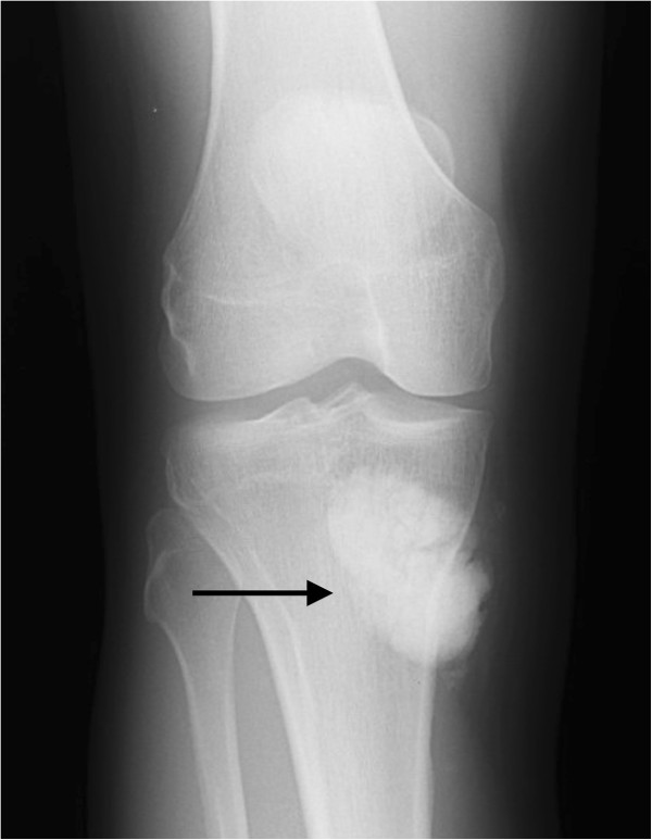
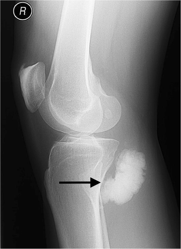
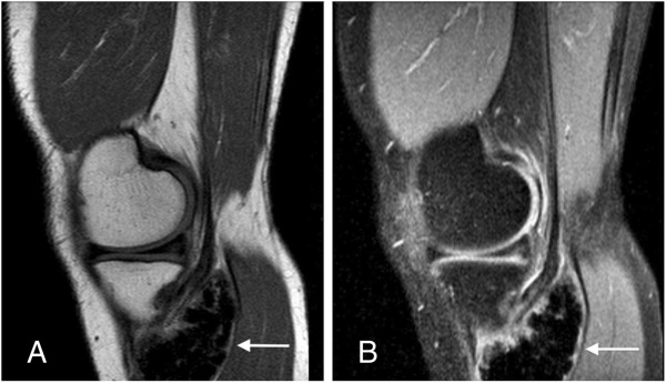
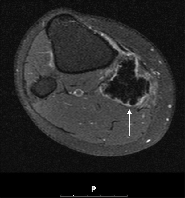
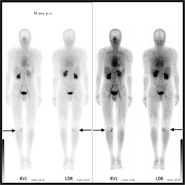
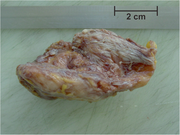
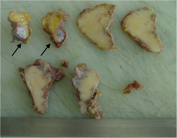
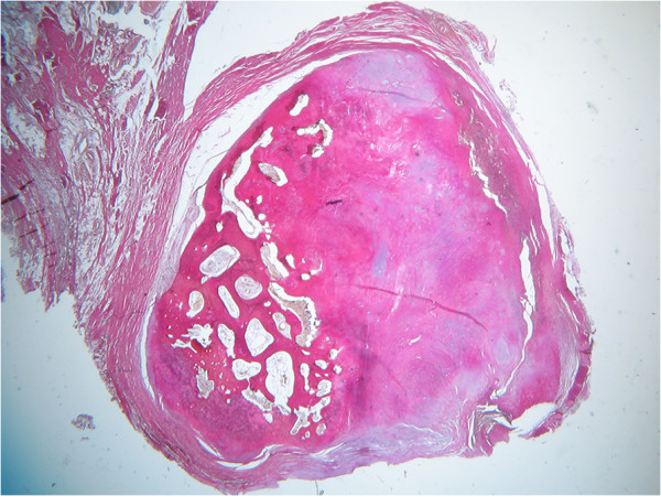
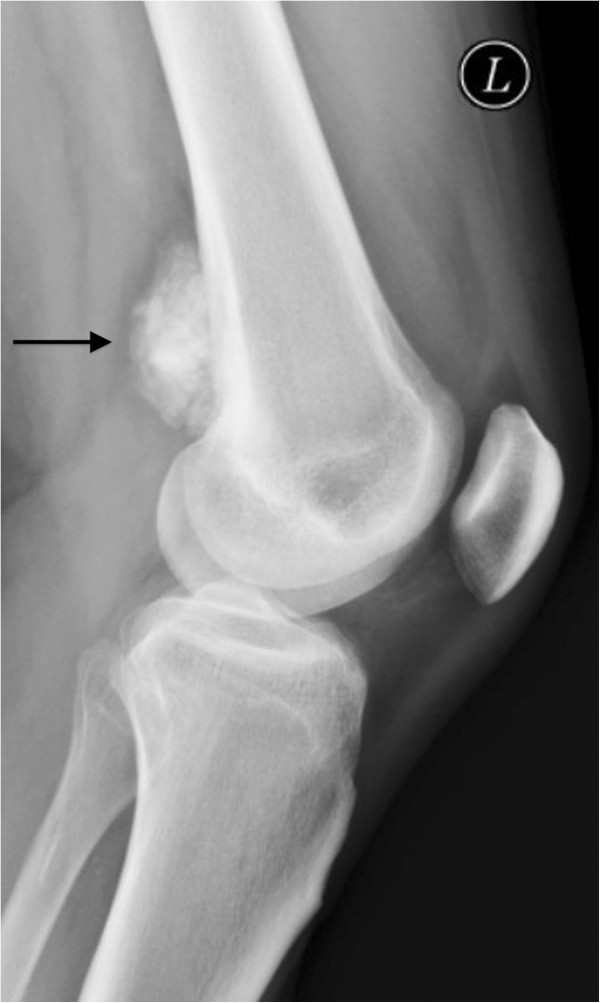
Both giant synovial osteochondroma and parosteal osteosarcoma are rare musculo-skeletal tumors, often localized in the vicinity of the knee. Misdiagnosis of a malignant bone tumor can entail fatal consequences. Etiology of giant synovial osteochondroma is widely unsolved but is believed to originate from synovial chondromatosis, a mostly benign metaplasia of the synovial membrane. Parosteal osteosarcoma is a low-grade surface osteosarcoma with a propensity of local recurrence and the potential of distant metastasis and therefore requiring a different therapeutical approach. We report the case of a popliteal giant osteochondroma mimicking a parosteal osteosarcoma. Relevant facts of this rare entity regarding pathogenesis, treatment, and differential diagnoses will be discussed.
Figures

References
-
- Fletcher C, Unni K, Mertens F. World health organization classification of tumours: pathology and genetics of tumours of soft tissue and bone. Malignant fibrous histiocytoma of bone. Lyon: IARC Press; 2002.
-
- Grimer R. WHO Classification of Tumours of Soft Tissue and Bone, Volume 5. 4. Geneva: WHO Press; 2013.
-
- Freyschmidt JOH, Jundt G. Knochenutmoren mit kiefertumoren: klinik-radiologie-pathologie. Berlin: Springer; 2010.
-
- Mosher JF Jr, Kettelkamp DB, Campbell CJ. Intracapsular or para-articular chondroma. A report of three cases. J Bone Joint Surg Am. 1966;48:1561–1569. - PubMed
Publication types
MeSH terms
LinkOut - more resources
Full Text Sources
Other Literature Sources
Medical
