Review
. 2013 Sep 25:11:242.
doi: 10.1186/1477-7819-11-242.

Primary angiosarcoma of the small intestine with metastasis to the liver: a case report and review of the literature

Affiliations
Review

Primary angiosarcoma of the small intestine with metastasis to the liver: a case report and review of the literature

Qingqiang Ni et al. World J Surg Oncol. .

Abstract

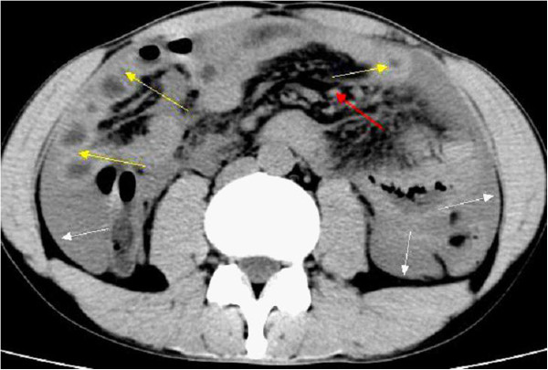
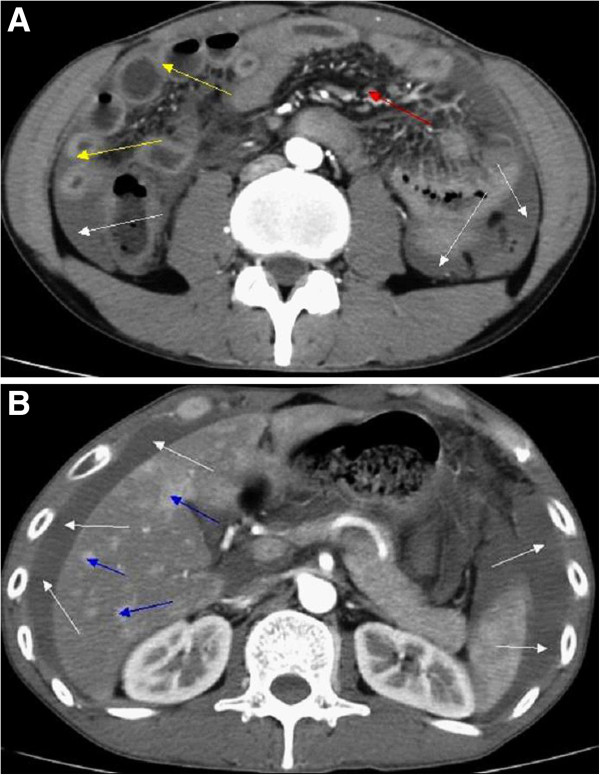
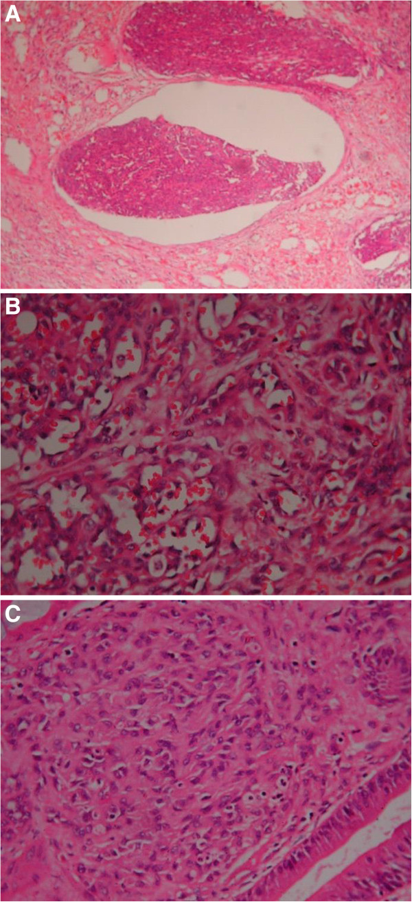
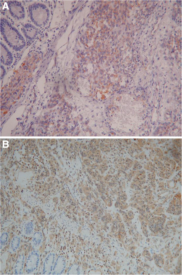
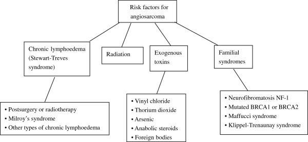
Angiosarcoma is a rare disease with a poor prognosis; significantly, patients with intestinal angiosarcomas who survive over 1 year after diagnosis are extraordinarily rare. This article describes the case of a 33-year-old gentleman who presented with abdominal pain of 4 months duration, which had increased in severity 2 weeks prior to presentation. After a complicated diagnostic and therapeutic process, the diagnosis of primary angiosarcoma of the small intestine with metastasis to the liver was made by pathological and immunohistochemical examinations. We reviewed previous cases of angiosarcoma described in the English literature to determine their risk factors, diagnosis and treatment, and we found that angiosarcoma is extremely rare, especially in the small intestine. To the best of our knowledge, this may be the youngest case of primary angiosarcoma of the small intestine with metastasis to the liver reported in the English literature.


Figures

Figure 1
Figure 1
Computed tomography scans of the whole abdomen showed a significantly thickened intestinal wall (yellow arrows) located at the end of the jejunum and the proximal ileum, excessive ascites (white arrows), a few enlarged lymph nodes in the abdomen (red arrow) and the location of the primary angiosarcoma of the small intestine (black arrow).
Figure 2
Figure 2
Enhanced computed tomography scan. (A) A significantly thickened intestinal wall (yellow arrows) located in the end of the jejunum and the proximal ileum, excessive ascites (white arrows) and a few enlarged lymph nodes in the abdomen (red arrow). (B) Different degrees of enhancement were noted in the hepatic arterial phase (blue arrows).
Figure 3
Figure 3
Microscopic examination. (A) Tumour tissue can be observed invading the vascular cavity (hematoxylin and eosin stain; ×100). (B) Tumor blood vessels were abundant, with the tumor cells surrounding them (hematoxylin and eosin stain; ×400). (C) The tumor cells were arranged in a slit-shaped pattern (hematoxylin and eosin stain; ×400).
Figure 4
Figure 4
Immunohistochemistry results. The tumor cells were positive for (A) CD31 (hematoxylin and eosin stain; ×400) and (B) vimentin (hematoxylin and eosin stain; ×200).
Figure 5
Figure 5
Risk factors for angiosarcoma.

References

    1. Al Ali J, Ko HH, Owen D, Steinbrecher UP. Epithelioid angiosarcoma of the small bowel. Gastrointest Endosc. 2006;64:1018–1021. doi: 10.1016/j.gie.2006.04.020. - DOI - PubMed
    1. Young RJ, Brown NJ, Reed MW, Hughes D, Woll PJ. Angiosarcoma. Lancet Oncol. 2010;11:983–991. doi: 10.1016/S1470-2045(10)70023-1. - DOI - PubMed
    1. Naka N, Ohsawa M, Tomita Y, Kanno H, Uchida A, Aozasa K. Angiosarcoma in Japan. A review of 99 cases. Cancer. 1995;75:989–996. doi: 10.1002/1097-0142(19950215)75:4<989::AID-CNCR2820750414>3.0.CO;2-K. - DOI - PubMed
    1. Yoshida K, Ito F, Nakazawa H, Maeda Y, Tomoe H, Aiba M. A case of primary renal angiosarcoma. Rare Tumors. 2009;1:e28. - PMC - PubMed
    1. Mohammed A, Aliyu HO, Liman AA, Abdullahi K, Abubakar N. Angiosarcoma of the small intestine. Ann Afr Med. 2011;10:246–248. doi: 10.4103/1596-3519.84702. - DOI - PubMed