. 2013 Oct 29;52(43):7689-701.
doi: 10.1021/bi401132w. Epub 2013 Oct 14.

Physical characterization of the manganese-sensing pneumococcal surface antigen repressor from Streptococcus pneumoniae

Affiliations

Physical characterization of the manganese-sensing pneumococcal surface antigen repressor from Streptococcus pneumoniae

John P Lisher et al. Biochemistry. .

Abstract

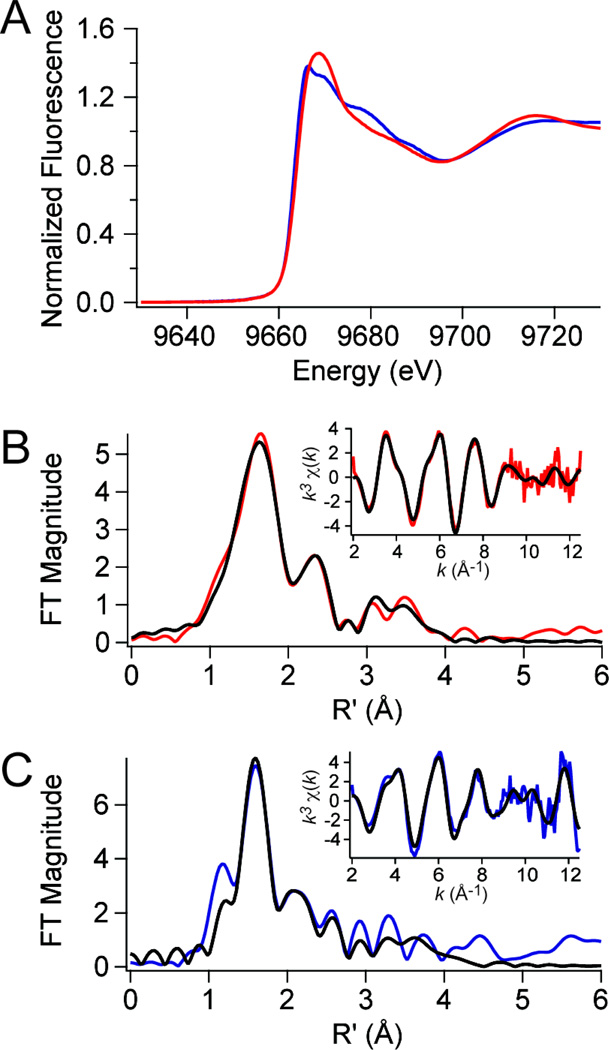
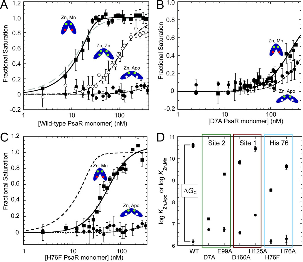
Transition metals, including manganese, are required for the proper virulence and persistence of many pathogenic bacteria. In Streptococcus pneumoniae (Spn), manganese homeostasis is controlled by a high-affinity Mn(II) uptake complex, PsaBCA, and a constitutively expressed efflux transporter, MntE. psaBCA expression is transcriptionally regulated by the DtxR/MntR family metalloregulatory protein pneumococcal surface antigen repressor (PsaR) in Spn. Here, we present a comprehensive analysis of the metal and DNA binding properties of PsaR. PsaR is a homodimer in the absence and presence of metals and binds two manganese or zinc atoms per protomer (four per dimer) in two pairs of structurally distinct sites, termed site 1 and site 2. Site 1 is likely filled with Zn(II) in vivo (K(Zn1) ≥ 10¹³ M⁻¹; K(Mn1) ≈ 10⁸ M⁻¹). The Zn(II)-site 1 complex adopts a pentacoordinate geometry as determined by X-ray absorption spectroscopy containing a single cysteine and appears to be analogous to the Cd(II) site observed in Streptococcus gordonii ScaR. Site 1 is necessary but not sufficient for full positive allosteric activation of DNA operator binding by metals as measured by ΔGc, the allosteric coupling free energy, because site 1 mutants show an intermediate ΔGc. Site 2 is the primary regulatory site and governs specificity for Mn(II) over Zn(II) in PsaR, where ΔGc(Zn,Mn) >> ΔGc(Zn,Zn) despite the fact that Zn(II) binds site 2 with an affinity 40-fold higher than that of Mn(II); i.e., K(Zn2) > K(Mn2). Mutational studies reveal that Asp7 in site 2 is a critical ligand for Mn(II)-dependent allosteric activation of DNA binding. These findings are discussed in the context of other well-studied DtxR/MntR Mn(II)/Fe(II) metallorepressors.


Figures

Figure 1
Figure 1
Representative multi-angle light scattering (MALS) traces of wild-type PsaRapo,apo (left) and PsaRZn,apo (right). Red trace, refractive index; blue trace, Raleigh ratio. The red peak at ≈40 min in each panel marks the included volume of the column and is not protein. Green trace, determined molar mass (kDa) across the main eluting species. Parameters for all metallated derivatives are complied in Table 1.
Figure 2
Figure 2
Representative isotherms obtained when a mixture of wild-type apo-PsaR and magfura-2 (mf2) (A,B; open symbols A325; filled symbols, A360) or quin-2 (C; filled symbols, A265) is titrated with the indicated metal salt. (A) Zn(II) titration into mag-fura-2 (12 µM) and PsaRapo,apo (23 µM monomer). (B) Mn(II) titration into a solution of mag-fura-2 (10 µM) and PsaR (17 µM monomer). (C) Zinc titration into a solution of quin-2 (12 µM) and PsaR (10 µM monomer). The continuous lines represent a nonlinear global least squares fit employing either a two-site (mag-fura-2) or single site (quin-2) competition model with the optimized binding parameters from multiple experiments compiled in Table 2. The broken lines in panels B and C represent simulated curves with each KMn (panel B) or KZn (panel C) 10-fold higher or 10-fold lower than the optimized value, for visual comparison with the fitted curves (Table 2).
Figure 3
Figure 3
Zn K-edge X-ray absorption spectroscopy of wild-type PsaRZn,apo (red traces) and PsaRZn,Mn (blue traces). (A) Zinc XANES spectra for each species. (B) Fourier-transformed EXAFS data (main panel) obtained for PsaRZn,apo (red trace) superimposed on best-fit coordination complex fits (black lines) as described by the parameters in Table 3. Inset, unfiltered k3-weighted EXAFS spectrum and fits for PsaRZn,apo. (C) Fourier-transformed EXAFS data (main panel) obtained for PsaRZn,Mn (blue trace) superimposed on best-fit coordination complex fits (black lines) (see Table 3). Inset, unfiltered k3-weighted EXAFS spectrum and fits for PsaRZn,Mn.
Figure 4
Figure 4
Close-up view of the Cd(II) site 1 in each of the two subunits (A,B) in Sgo ScaR (PDB code 3HRT) (ligating residues shaded orange; E80, C123, H125 and D160), emphasizing the physical location of H76 and immediately adjacent candidate regulatory site 2 ligands D7, E99, E102 and H103 (shaded yellow). Side-chains are shown in stick in elemental shading; Cd(II) ion, green sphere. Note that the homodimer is strongly asymmetric and this asymmetry extends to the metal site region which adopts distinct structures in each protomer. D7 makes much closer approach to the other candidate site 2 ligands in the α5 helix in the right (panel B) protomer relative to the left (panel A). Note also metal coordination bonds are unreasonably long in all cases, likely attributed to the modest resolution (2.7 Å) of the structure.
Figure 5
Figure 5
DNA binding isotherms for various metallated states of wild-type PsaR (A), D7A PsaR (B) and H76F PsaR (C). In each case, data for PsaRZn,apo and PsaRZn,Zn and PsaRZn,Mn species are shown in filled circles, open circles and filled squares respectively. The smooth curves through each data set correspond to that derived from a dissociable dimer binding model (see Materials and Methods for details) with KDNAi vales complied in Table 4. The grey dashed line in panel (A) shows the fit for a nondissociable dimer model for PsaR binding (see Table 4). The dashed line in panel (C) shows the fit for wild-type PsaRZn,Mn for comparison only. (D) Summary of the DNA binding affinities of PsaRZn,apo (filled circles) and PsaRZn,Mn (fillled squares) for wild-type PsaR and indicated mutants with the vertical separation between these Ki values proportional to ΔGc from ΔGcMn = −RTln(KDNAZn,Mn/KDNAZn,apo) or for Zn(II), ΔGcZn = −RTln(KDNAZn,Zn/KDNAZn,apo). ΔGc is equivalent to a ΔΔG, or the difference in PsaO DNA binding free energy between the appropriate metallated form (PsaRZn,Mn or PsaRMn,Mn) and PsaRZn,apo.
Figure 6
Figure 6
Comparison of the Mn(II)-bound chelate regions of wild-type MntR (2F5D) (A) and E11K MntR (4HX4) (B). The A and C Mn(II) ions are indicated in each panel and all residues are shown in stick representation. Coordination bonds are represented by black dashed lines, with potential electrostatic interactions deriving the εNH2 group of Lys11in E11K PsaR shown as green dashed lines in panel B. All residues are conserved in PsaR with identical residue numbers except that His77 is His76 in PsaR (see text for details). E11K MntR (B) is an excellent model for the Mn(II)-bound in site 2 in PsaR (this work).

Similar articles

Cited by

References

    1. Andreini C, Bertini I, Cavallaro G, Holliday GL, Thornton JM. Metal ions in biological catalysis: from enzyme databases to general principles. J Biol Inorg Chem. 2008;13:1205–1218. - PubMed
    1. Waldron KJ, Rutherford JC, Ford D, Robinson NJ. Metalloproteins and metal sensing. Nature. 2009;460:823–830. - PubMed
    1. Klein JS, Lewinson O. Bacterial ATP-driven transporters of transition metals: physiological roles, mechanisms of action, and roles in bacterial virulence. Metallomics. 2011;3:1098–1108. - PubMed
    1. Ma Z, Jacobsen FE, Giedroc DP. Coordination Chemistry of Bacterial Metal Transport and Sensing. Chem Rev. 2009;109:4644–4681. - PMC - PubMed
    1. Imlay JA. The molecular mechanisms and physiological consequences of oxidative stress: lessons from a model bacterium. Nat Rev Microbiol. 2013;11:443–454. - PMC - PubMed

Publication types

MeSH terms

LinkOut - more resources