. 2013 Sep 26:10:119.
doi: 10.1186/1742-2094-10-119.

Dysregulation of the complement cascade in the hSOD1G93A transgenic mouse model of amyotrophic lateral sclerosis

Affiliations

Dysregulation of the complement cascade in the hSOD1G93A transgenic mouse model of amyotrophic lateral sclerosis

John D Lee et al. J Neuroinflammation. .

Abstract

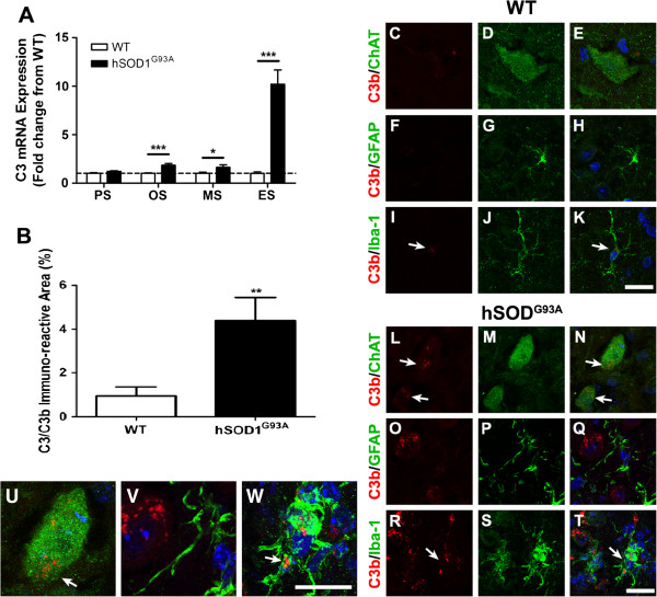
Background: Components of the innate immune complement system have been implicated in the pathogenesis of amyotrophic lateral sclerosis (ALS); however, a comprehensive examination of complement expression in this disease has not been performed. This study therefore aimed to determine the expression of complement components (C1qB, C4, factor B, C3/C3b, C5 and CD88) and regulators (CD55 and CD59a) in the lumbar spinal cord of hSOD1(G93A) mice during defined disease stages.

Methods: hSOD1(G93A) and wild-type mice were examined at four different ages of disease progression. mRNA and protein expression of complement components and regulators were examined using quantitative PCR, western blotting and ELISA. Localisation of complement components within lumbar spinal cord was investigated using immunohistochemistry. Statistical differences between hSOD1(G93A) and wild-type mice were analysed using a two-tailed t-test at each stage of disease progression.

Results: We found several early complement factors increased as disease progressed, whilst complement regulators decreased; suggesting overall increased complement activation through the classical or alternative pathways in hSOD1(G93A) mice. CD88 was also increased during disease progression, with immunolocalisation demonstrating expression on motor neurons and increasing expression on microglia surrounding the regions of motor neuron death.

Conclusions: These results indicate that local complement activation and increased expression of CD88 may contribute to motor neuron death and ALS pathology in the hSOD1(G93A) mouse. Hence, reducing complement-induced inflammation could be an important therapeutic strategy to treat ALS.


Figures

Figure 1
Figure 1
Expression and localisation of C1q in hSOD1G93A and wild-type mice during disease progression. (A) mRNA expression profile of C1qB in lumbar spinal cord of hSOD1G93A mice relative to wild-type (WT) mice. Dashed line, baseline expression in WT controls at each time point. (B) Degree of immunolabelling for C1q significantly increased in the lumbar spinal cord of hSOD1G93A mice at end stage when compared with WT mice. (A, B) Data expressed as mean ± standard error of the mean (n = 6 mice/group; *P <0.05, **P <0.01, ***P <0.001, Student t test). (C) to (T) Double immunolabelling of C1q (red) with cellular markers (green) for motor neurons (ChAT; (C) to (E) WT mice, (L) to (N) hSOD1G93A mice), astrocyte (glial fibrillary acidic protein (GFAP); (F) to (H) WT mice, (O) to (Q) hSOD1G93A mice), and microglia (Iba-1; (I) to (K) WT mice, (R) to (T) hSOD1G93A mice) in the ventral lumbar spinal cord of WT and hSOD1G93A mice (end stage). There was minimal expression of C1q in WT (C, F and I) with marked increase in hSOD1G93A mice (L, O and R). In hSOD1G93A mice, C1q was co-localised with ChAT-positive motor neurons (white arrows in (L) and (N) (detailed in U)). There was little to no co-localisation of C1q with GFAP-positive astrocytes (Q (detailed in V)), and minimal co-localisation with Iba-1-labelled microglia (white arrows in R and T (detailed in W)). PS, pre-symptomatic; OS, onset; MS, mid-symptomatic; ES, end-stage. Scale bar for all panels = 20 μm.
Figure 2
Figure 2
Altered expression of complement components in hSOD1G93A and wild-type mice at different ages of disease progression. (A) mRNA expression of CD55 in the lumbar spinal cord of hSOD1G93A transgenic mice relative to age-matched wild-type (WT) mice at four different ages. (B) Representative western blot of CD55 with glyceraldehyde-3-phosphate dehydrogenase (GAPDH) in the lumbar spinal cord of hSOD1G93A mice (SOD1) relative to age-matched WT mice, at different ages of disease progression. (C) Protein expression of CD55 determined by semi-quantitative densitometry in the lumbar spinal cord of hSOD1G93A mice relative to age-matched WT mice at four different ages. (D) to (F) mRNA expressions of C4 (classical pathway, D), factor B (alternative pathway, E) and CD59a (regulator, F) in lumbar spinal cord of hSOD1G93A mice relative to age-matched WT mice at four different ages. (A, C, D, E, F) Dashed lines, baseline expressions in WT controls at each time point. Data expressed as mean ± standard error of the mean (n = 6 mice/group; *P <0.05, **P <0.01, ***P <0.001, Student t test).
Figure 3
Figure 3
Localisation and expression of C3/C3b in hSOD1G93A and wild-type mice during disease progression. (A) mRNA expression profile of C3 in lumbar spinal cord of hSOD1G93A mice relative to wild-type (WT) mice. Dashed line, baseline expression in WT controls at each time point. (B) Degree of immunolabelling for C3b significantly increased in the lumbar spinal cord of hSOD1G93A mice at end stage when compared with WT mice. (A, B) Data expressed as mean ± standard error of the mean (n = 6 mice/group; *P <0.05, **P <0.01, ***P <0.001, Student t test). (C) to (T) Double immunolabelling of C3b (red) with cellular markers (green) for motor neurons (ChAT; (C) to (E) WT mice, (L) to (N) hSOD1G93A mice), astrocyte (glial fibrillary acidic protein (GFAP); (F) to (H) WT mice, (O) to (Q) for hSOD1G93A mice), and microglia (Iba-1; (I) to (K) WT mice, (R) to (T) hSOD1G93A mice) in the ventral lumbar spinal cord of WT and hSOD1G93A mice (end stage). C3b immunolabelling was absent on motor neurons in WT mice (C to E), but was present on motor neurons in hSOD1G93A mice (white arrows in L and N (detailed in U)). There was minimal co-localisation of C3b with Iba-1-labelled microglia in WT (white arrows in I and K). In hSOD1G93A mice immunolabelling of C3b was evident in Iba-1-labelled microglia (white arrows, R and T (detailed in W)). There was no co-localisation with C3b and GFAP-positive astrocytes in WT and hSOD1G93A mice (F to H for WT, O to Q (detailed on V) for hSOD1G93A mice). PS, pre-symptomatic; OS, onset; MS, mid-symptomatic and ES, end-stage. Scale bars for all panels = 20 μM.
Figure 4
Figure 4
Expression and localisation of C5 and C5a in hSOD1G93A and wild-type mice during disease progression. (A) mRNA expression profile of C5 in lumbar spinal cord of hSOD1G93A mice relative to wild-type (WT) mice. Dashed line, baseline expression in WT controls at each time point. (B) Protein expression of C5a in the lumbar spinal cord of hSOD1G93A mice has decreased by end stage (ES) when compared with WT mice. (A, B) Data expressed as mean ± standard error of the mean (n = 6 mice/group; *P <0.05, Student t test). (C) to (H) Double immunolabelling for C5 (red) with cellular makers (green) for motor neurons (ChAT; C and D, arrows), astrocytes (glial fibrillary acidic protein (GFAP), E and F), and microglia (CD11b; G and H), in the ventral lumbar spinal cord region of hSOD1G93A mice (D, F and H) and WT mice (C, E and G) at end stage. Co-localisation of C5 with these cellular markers is seen as a merge of green and red (for example, white arrows in C, D, and H). PS, pre-symptomatic; OS, onset; MS, mid-symptomatic; ES, end-stage. Scale bar for C to H = 20 μm.
Figure 5
Figure 5
Expression and localisation of CD88 in hSOD1G93A and wild-type mice at four different ages. (A) mRNA expression of CD88 in the lumbar spinal cord of hSOD1G93A mice relative to age-matched wild-type (WT) mice at four different ages. (B) Representative western blot of CD88 with glyceraldehyde-3-phosphate dehydrogenase (GAPDH) in the lumbar spinal cord of hSOD1G93A (SOD1) mice relative to age-matched WT mice at different ages. (C) Protein expression of CD88 determined by semi-quantitative densitometry in the lumbar spinal cord of hSOD1G93A mice relative to age-matched WT mice at four different ages. (A), (C) Dashed lines, baseline expression in WT controls at each time point; data expressed as mean ± standard error of the mean (n = 6 mice/group; *P <0.05, **P <0.01, ***P <0.001, Student t test). (D) to (U) Double immunolabelling of CD88 (red) with cellular markers (green) for motor neurons (ChAT; (D) to (F) WT mice, (M) to (O) hSOD1G93A mice), microglia (Iba-1; (G) to (I) WT mice, (P) to (R) hSOD1G93A mice), and astrocytes (glial fibrillary acidic protein (GFAP); (J) to (L) WT mice, (S) to (U) for hSOD1G93A mice) in the ventral lumbar spinal cord of WT and hSOD1G93A mice at end stage. CD88 was co-localised with ChAT-positive motor neurons (F, O, white arrows). (D′) CD88 mRNA transcript within lumbar motor neurons (determine by large cell size and location within the ventral horn). In hSOD1G93A mice, immunolabelling of CD88 also evident on other cell types, indicated by lack of co-localisation with anti-ChAT (yellow arrows in M and O). (G), (I) White arrows, small amount of CD88 staining within nonactivated microglia in WT mice, with increased CD88 expression on activated microglia in hSOD1G93A mice (P and R, white arrows). PS, pre-symptomatic; OS, onset; MS, mid-symptomatic; ES, end stage. Scale bars for all panels = 20 μM.

Similar articles

Cited by

References

    1. Bruijn LI, Miller TM, Cleveland DW. Unraveling the mechanisms involved in motor neuron degeneration in ALS. Annu Rev Neurosci. 2004;27:723–749. doi: 10.1146/annurev.neuro.27.070203.144244. - DOI - PubMed
    1. Cozzolino M, Ferri A, Carri MT. Amyotrophic lateral sclerosis: from current developments in the laboratory to clinical implications. Antioxid Redox Signal. 2008;10:405–443. doi: 10.1089/ars.2007.1760. - DOI - PubMed
    1. Woodruff TM, Costantini KJ, Taylor SM, Noakes PG. Role of complement in motor neuron disease: animal models and therapeutic potential of complement inhibitors. Adv Exp Med Biol. 2008;632:143–158. - PubMed
    1. Lee JD, Lee JY, Taylor SM, Noakes PG, Woodruff TM. In: Amyotrophic Lateral Sclerosis. Maurer MH, editor. Croatia: InTech; 2012. Innate Immunity in ALS; pp. 393–412.
    1. Almer G, Guegan C, Teismann P, Naini A, Rosoklija G, Hays AP, Chen C, Przedborski S. Increased expression of the pro-inflammatory enzyme cyclooxygenase-2 in amyotrophic lateral sclerosis. Ann Neurol. 2001;49:176–185. doi: 10.1002/1531-8249(20010201)49:2<176::AID-ANA37>3.0.CO;2-X. - DOI - PubMed

Publication types

MeSH terms