

Combined rod and cone transduction by adeno-associated virus 2/8
- PMID: 24067103
- PMCID: PMC3868409
- DOI: 10.1089/hum.2013.154
Combined rod and cone transduction by adeno-associated virus 2/8
Abstract
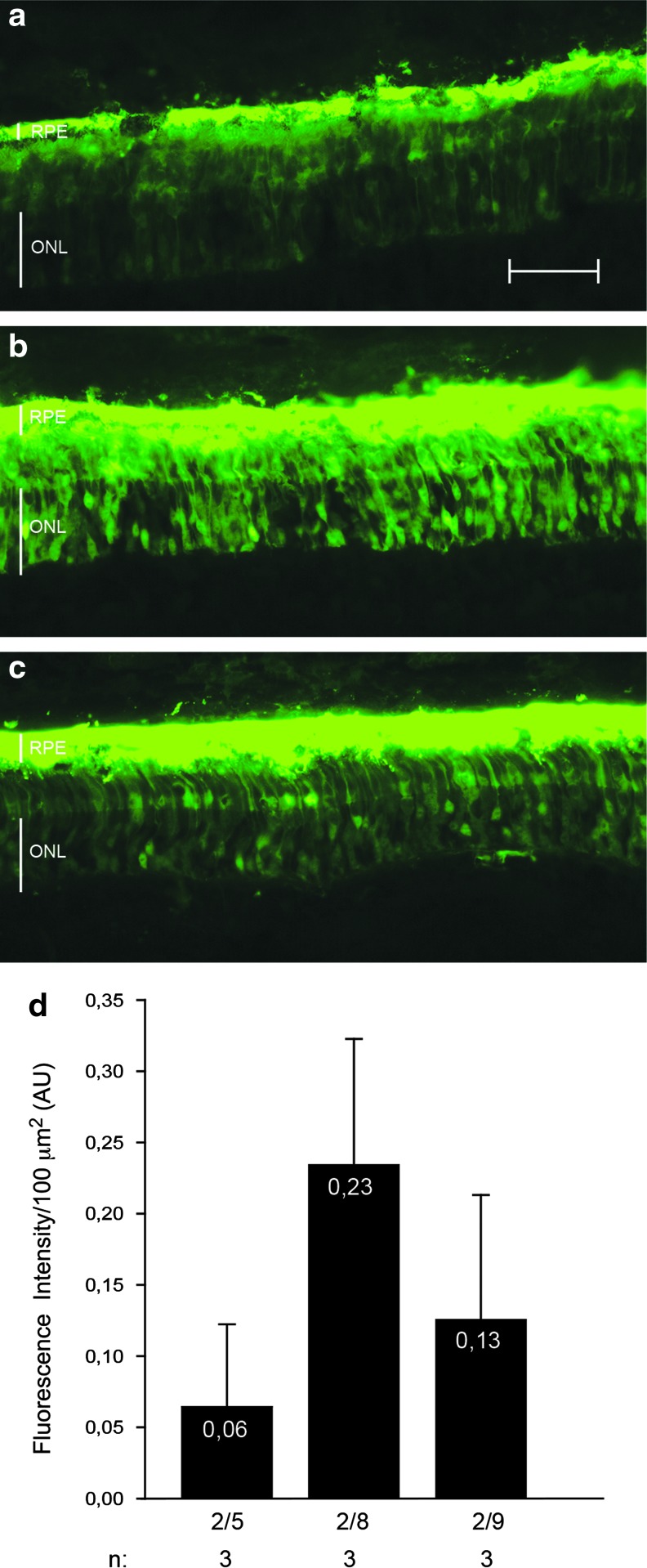
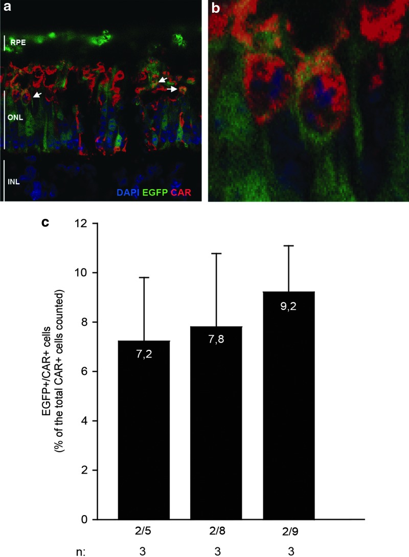
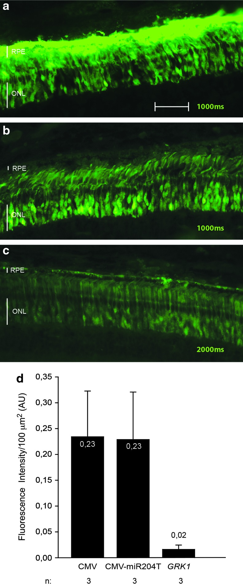
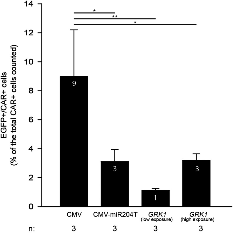
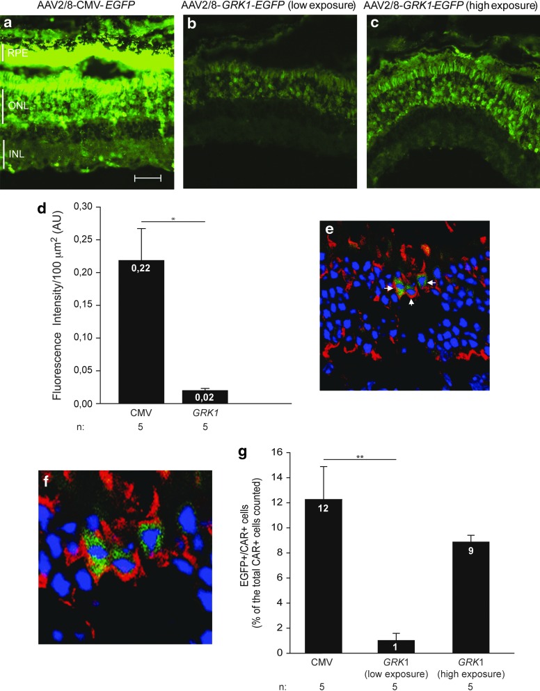
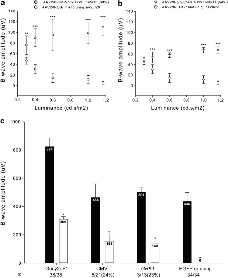
Gene transfer to both cone and rod photoreceptors (PRs) is essential for gene therapy of inherited retinal degenerations that are caused by mutations in genes expressed in both PR types. Vectors based on the adeno-associated virus (AAV) efficiently transduce PRs of different species. However, these are predominantly rods and little is known about the ability of the AAV to transduce cones in combination with rods. Here we show that AAV2/8 transduces pig cones to levels that are similar to AAV2/9, and the outer nuclear layer (mainly rods) to levels that are on average higher, although not statistically significant, than both AAV2/5 and AAV2/9. We additionally found that the ubiquitous cytomegalovirus (CMV), but not the PR-specific GRK1 promoter, transduced pig cones efficiently, presumably because GRK1 is not expressed in pig cones as observed in mice and humans. Indeed, the GRK1 and CMV promoters transduce a similar percentage of murine cones with the CMV reaching the highest expression levels. Consistent with this, the AAV2/8 vectors with either the CMV or the GRK1 promoter restore cone function in a mouse model of Leber congenital amaurosis type 1 (LCA1), supporting the use of AAV2/8 for gene therapy of LCA1 as well as of other retinal diseases requiring gene transfer to both PR types.
Figures
Similar articles
-
Rod Outer Segment Development Influences AAV-Mediated Photoreceptor Transduction After Subretinal Injection.Hum Gene Ther. 2017 Jun;28(6):464-481. doi: 10.1089/hum.2017.020. Hum Gene Ther. 2017. PMID: 28510482 Free PMC article.
-
AAV9 targets cone photoreceptors in the nonhuman primate retina.PLoS One. 2013;8(1):e53463. doi: 10.1371/journal.pone.0053463. Epub 2013 Jan 30. PLoS One. 2013. PMID: 23382846 Free PMC article.
-
AAV-mediated gene therapy in the guanylate cyclase (RetGC1/RetGC2) double knockout mouse model of Leber congenital amaurosis.Hum Gene Ther. 2013 Feb;24(2):189-202. doi: 10.1089/hum.2012.193. Hum Gene Ther. 2013. PMID: 23210611 Free PMC article.
-
An Update on Gene Therapy for Inherited Retinal Dystrophy: Experience in Leber Congenital Amaurosis Clinical Trials.Int J Mol Sci. 2021 Apr 26;22(9):4534. doi: 10.3390/ijms22094534. Int J Mol Sci. 2021. PMID: 33926102 Free PMC article. Review.
-
Therapeutic strategy for handling inherited retinal degenerations in a gene-independent manner using rod-derived cone viability factors.C R Biol. 2014 Mar;337(3):207-13. doi: 10.1016/j.crvi.2013.12.002. Epub 2014 Feb 20. C R Biol. 2014. PMID: 24702847 Review.
Cited by
-
Advances in Gene Therapy for Diseases of the Eye.Hum Gene Ther. 2016 Aug;27(8):563-79. doi: 10.1089/hum.2016.040. Epub 2016 Jun 13. Hum Gene Ther. 2016. PMID: 27178388 Free PMC article. Review.
-
Assessment of Adeno-Associated Virus Serotype Tropism in Human Retinal Explants.Hum Gene Ther. 2018 Apr;29(4):424-436. doi: 10.1089/hum.2017.179. Epub 2018 Feb 23. Hum Gene Ther. 2018. PMID: 29160116 Free PMC article.
-
Current trends in gene therapy for retinal diseases (Review).Exp Ther Med. 2022 Jan;23(1):26. doi: 10.3892/etm.2021.10948. Epub 2021 Nov 5. Exp Ther Med. 2022. PMID: 34815778 Free PMC article. Review.
-
Leber Congenital Amaurosis (LCA): Potential for Improvement of Vision.Invest Ophthalmol Vis Sci. 2019 Apr 1;60(5):1680-1695. doi: 10.1167/iovs.19-26672. Invest Ophthalmol Vis Sci. 2019. PMID: 31009524 Free PMC article. Review. No abstract available.
-
Vector platforms for gene therapy of inherited retinopathies.Prog Retin Eye Res. 2014 Nov;43:108-28. doi: 10.1016/j.preteyeres.2014.08.001. Epub 2014 Aug 12. Prog Retin Eye Res. 2014. PMID: 25124745 Free PMC article. Review.
References
Publication types
MeSH terms
Supplementary concepts
Grants and funding
LinkOut - more resources
Full Text Sources
Other Literature Sources
Medical
Research Materials
