. 2013 Nov 8;288(45):32184-32193.
doi: 10.1074/jbc.M113.514356. Epub 2013 Sep 25.

Interferon-induced transmembrane protein 3 is a type II transmembrane protein

Affiliations

Interferon-induced transmembrane protein 3 is a type II transmembrane protein

Charles C Bailey et al. J Biol Chem. .

Abstract

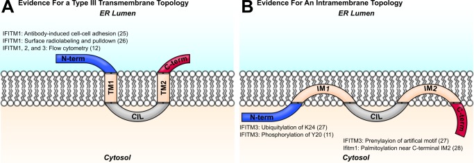
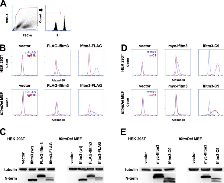
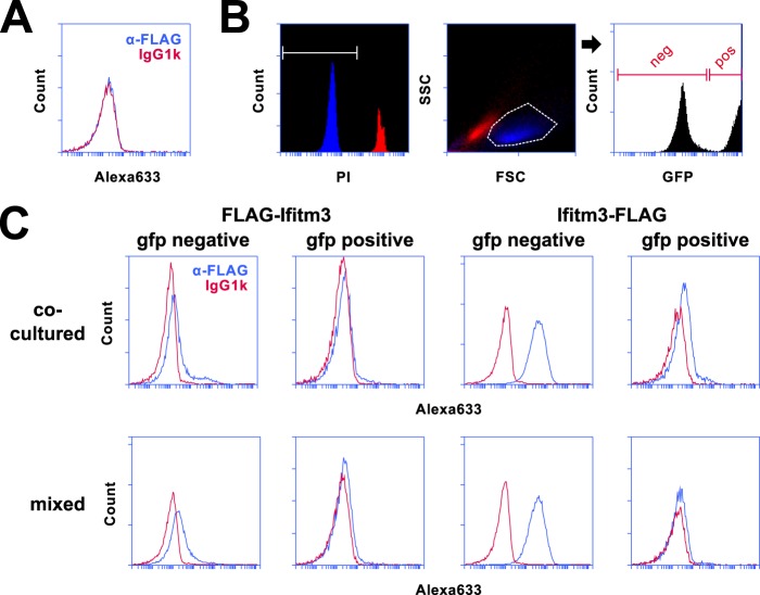
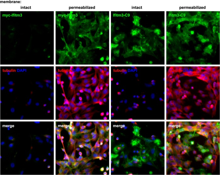
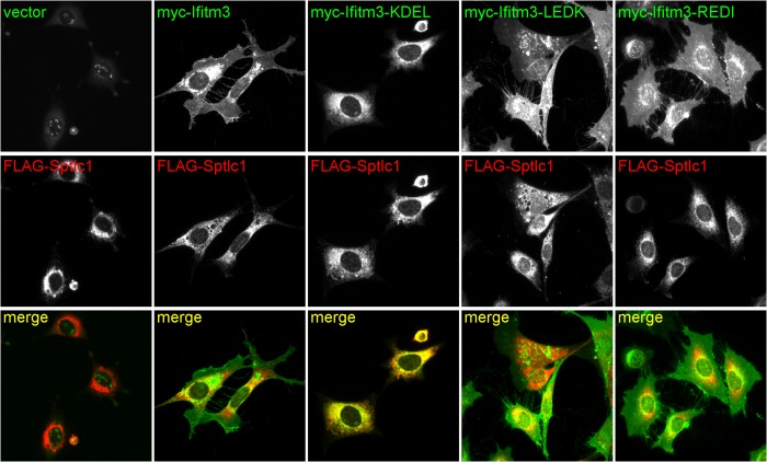
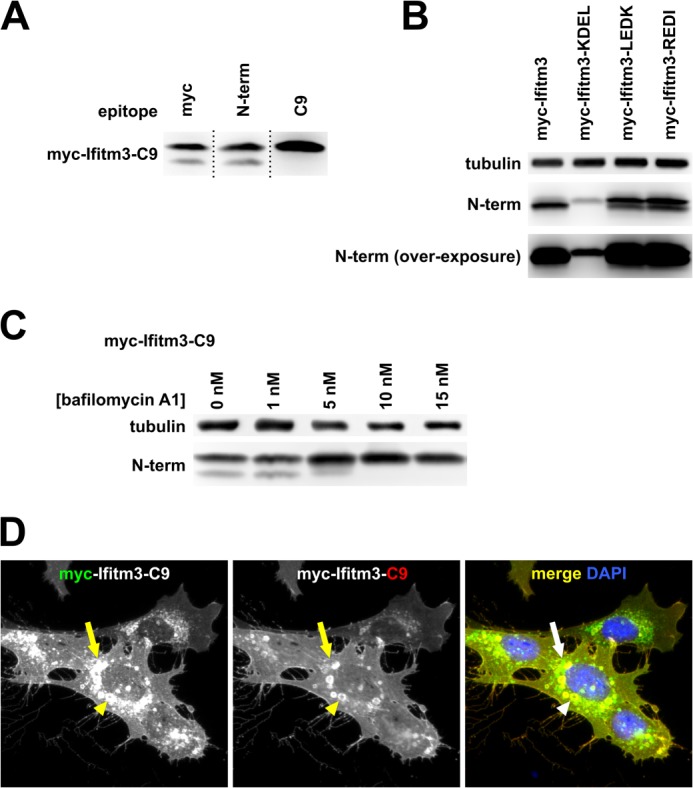
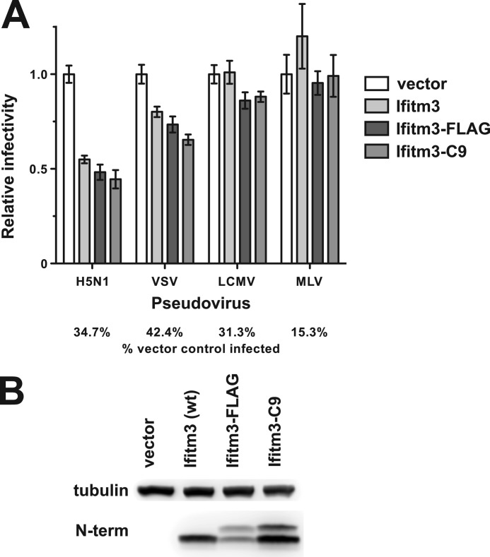
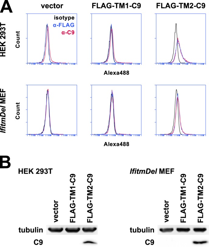
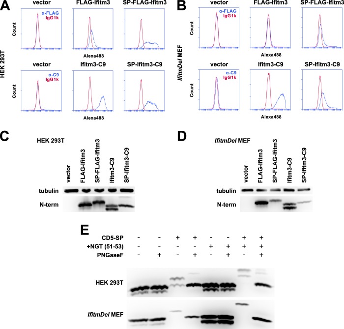
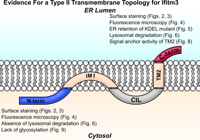
The interferon-induced transmembrane (IFITM) proteins are a family of small membrane proteins that inhibit the cellular entry of several genera of viruses. These proteins had been predicted to adopt a two-pass, type III transmembrane topology with an intracellular loop, two transmembrane helices (TM1 and TM2), and extracellular N and C termini. Recent work, however, supports an intramembrane topology for the helices with cytosolic orientation of both termini. Here we determined the topology of murine Ifitm3. We found that the N terminus of Ifitm3 could be stained by antibodies at the cell surface but that this conformation was cell type-dependent and represented a minority of the total plasma membrane pool. In contrast, the C terminus was readily accessible to antibodies at the cell surface and extracellular C termini comprised most or all of those present at the plasma membrane. The addition of a C-terminal KDEL endoplasmic reticulum retention motif to Ifitm3 resulted in sequestration of Ifitm3 in the ER, demonstrating an ER-luminal orientation of the C terminus. C-terminal, but not N-terminal, epitope tags were also degraded within lysosomes, consistent with their luminal orientation. Furthermore, epitope-tagged Ifitm3 TM2 functioned as a signal anchor sequence when expressed in isolation. Collectively, our results demonstrate a type II transmembrane topology for Ifitm3 and will provide insight into its interaction with potential targets and cofactors.

Keywords: Flow Cytometry; Ifitm3; Interferon; Membrane Proteins; Microscopic Imaging; Virus Entry.


Figures

FIGURE 1.
FIGURE 1.
Two models of IFITM protein topology are shown. Evidence for the orientation of each terminus with respect to the membrane is shown with the relevant citation. A, IFITM proteins were predicted to assume a two-pass type III transmembrane topology, with two transmembrane domains (TM1 and TM2) flanking the conserved intracellular loop (CIL). Early studies identified IFITM1 N termini in the extracellular space, and other investigators have detected IFITM N termini at the plasma membrane by flow cytometry. (See figure for references.) B, more recent studies support a topology with two intramembrane domains (IM1 and IM2) and cytosolic N and C termini. Post-translational modifications of the IFITM3 N terminus imply access of the N terminus to cytosolic enzymes. Evidence for an intracellular C terminus comes from studies of lipidation motif reporters. (See figure for references.)
FIGURE 2.
FIGURE 2.
FLAG-tagged Ifitm3 N and C termini can be detected at the surface of intact cells. A, the gating strategy used to produce subsequent histograms is shown here. To confirm staining of only extracellular FLAG antigen, PI-positive cells were excluded from analysis. SSC-A, side scatter; FSC-A, forward scatter. B, staining for the FLAG epitopes on 293T cells and MEFs is shown in blue. Fluorescence staining from the isotype control is shown in red. Alexa488, Alexa Fluor 488. C, expression levels of wild type and N- and C-terminally FLAG-tagged Ifitm3 were determined by Western blot with an antibody against the native Ifitm3 N terminus. D, the experiment shown in A was repeated with N-terminal Myc and C-terminal C9 tags in place of the FLAG epitopes. Myc staining is shown in blue, and C9 staining is shown in red. E, total expression of the constructs was determined by Western blot. The double band pattern for C-terminally tagged Ifitm3 constructs is explored in detail in Fig. 6.
FIGURE 3.
FIGURE 3.
Surface staining of N and C termini is not an artifact of membrane adherent cellular debris. A, staining of Ifitm3/eGFP+ with anti-FLAG (blue) and isotype control (red) antibodies is shown. Alexa633, Alexa Fluor 633. B, PI (intact) and PI+ (permeabilized) 293T cell populations were mapped onto a forward/side scatter plot (FSC/SSC). Intact 293T cells (blue) could be clearly distinguished from cells with compromised membrane integrity (red) by scatter gating alone. Scatter gating was used in lieu of PI staining to ensure analysis of only intact cells for during subsequent experiments. Cells were further sorted based on eGFP expression as shown in the rightmost histogram. C, cells expressing either FLAG-Ifitm3 or Ifitm3-FLAG were co-cultured with Ifitm3/eGFP+ cells for 18 h (top panels) or mixed with Ifitm3/eGFP+ cells immediately prior to surface staining (bottom panels). FLAG staining is shown in blue, and isotype control staining is shown in red.
FIGURE 4.
FIGURE 4.
In contrast to Ifitm3 N termini, the C termini are mostly or entirely extracellular. MEFs expressing either Myc-Ifitm3 or Ifitm3-C9 were first fixed and then either permeabilized with detergent or left with membranes intact prior to indirect immunofluorescence. Ifitm3 epitope tag staining is shown in green (top panels). Membrane integrity was assessed by co-staining for α-tubulin (middle panels, red). Nuclei were counterstained with DAPI (blue). Exposure times for each channel were identical, and contrast adjustment was applied uniformly to all panels for each color channel.
FIGURE 5.
FIGURE 5.
The addition of a C-terminal KDEL motif to Myc-Ifitm3 results in its retention in the ER. Top panels show Myc (N-terminal) staining of cells expressing Ifitm3 with or without the indicated C-terminal additions. Middle panels show staining for FLAG-tagged Sptlc1 (an ER marker). Myc-Ifitm3-KDEL was poorly expressed in comparison with the other constructs, so the brightness of this panel was adjusted independently to better visualize its co-localization with Sptlc1. The nucleolar Myc staining is an artifact of the 9E11 antibody and has been previously described (17).
FIGURE 6.
FIGURE 6.
Ifitm3 C termini are degraded in lysosomes. A, lysates from MEFs expressing an N- and C-terminally tagged Ifitm3 (myc-Ifitm3-C9) were examined by Western blot with staining for the indicated epitopes. Dotted lines indicate the sites of reassembly of the membrane after staining. B, Western blot for Myc-Ifitm3 and Myc-Ifitm3-LEDK and -REDI is shown at two exposures to highlight the double band pattern for Ifitm3-LEDK and REDI (upper) and confirm the lack of a second band for Ifitm3-KDEL (overexposed, lower). C, bafilomycin A1 treatment inhibits degradation of the C-terminal tag as assessed by Western blot. D, MEFs expressing Myc-Ifitm3-C9 were co-stained for Myc (green) and C9 (red) epitopes. The plasma membrane is yellow/orange, indicating co-localization of the two epitopes (full-length Myc-Ifitm3-C9). Green cytoplasmic puncta (arrow indicates an example) show the sites of removal of the C9 tag. The C9 tag remained intact within the enlarged vacuoles that accompany IFITM protein overexpression (arrowhead).
FIGURE 7.
FIGURE 7.
C-terminal tags do not impair the restriction activity or expression of Ifitm3. A, MEFs were transduced with a vector control or the indicated Ifitm3 constructs. Transduced MEFs were then infected with eGFP-encoding retroviruses pseudotyped with the envelope glycoproteins of four different viruses. Bars show the proportion of the percentage of infected (eGFP+) cells expressing each Ifitm3 construct relative to percentage of infected vector-transduced cells infected. Error bars show the median and range of triplicate wells. VSV, vesicular stomatitis virus; LCMV, lymphocytic choriomeningitis virus. B, expression levels of the constructs were determined by Western blot against the native Ifitm3 N terminus.
FIGURE 8.
FIGURE 8.
The second hydrophobic region of Ifitm3 acts as a signal anchor sequence. A, the first and second predicted intra/transmembrane domains were subcloned from Ifitm3 and expressed with the addition of N-terminal FLAG and C-terminal C9 tags (FLAG-TM1-C9 and FLAG-TM2-C9). 293T cells or MEFs expressing these constructs were stained by indirect immunofluorescence for both tags in addition to an isotype control antibody. FLAG-TM2-C9 but not FLAG-TM1-C9 could be detected at the surface of both cell types by flow cytometry. Permeabilized cells were excluded from analysis by PI gating as shown in Fig. 2. Alexa488, Alexa Fluor 488. B, FLAG-TM2-C9 but not FLAG-IM1-C9 could be detected by Western blot.
FIGURE 9.
FIGURE 9.
The addition of a signal peptide to the Ifitm3 N terminus mimics a type III topology. A, 293T cells expressing FLAG-Ifitm3, Ifitm3-C9, or the same constructs with the addition of an N-terminal CD5 signal peptide were stained for the appropriate epitopes and analyzed by flow cytometry. Alexa488, Alexa Fluor 488. B, MEFs were analyzed by flow cytometry as above. C, a Western blot for the native Ifitm3 N terminus shows the relative expression of each construct in 293T cells. Lysates were treated with peptide-N-glycosidase F to remove N-linked glycosylation of the N terminus. D, the relative expression of each construct in MEFs is shown by Western blot. E, the N terminus of Ifitm3 can be glycosylated but is not normally accessible to cellular glycosylation machinery. Cells were transfected (293T, top) or transduced (MEF, bottom) with constructs expressing C9-tagged Ifitm3 with or without a CD5 signal peptide (CD5-SP) and with or without introduction of an additional NGT motif (+NGT (51–53)). Lysates were treated with peptide-N-glycosidase F (PNGaseF) or mock-treated as indicated and analyzed by Western blot with antibodies against the native N terminus.
FIGURE 10.
FIGURE 10.
Ifitm3 is a type II transmembrane protein. Ifitm3 contains an N-terminal intramembrane domain (IM1) and C-terminal transmembrane domain (TM2) flanking the conserved intracellular loop (CIL) with an extracellular C terminus and intracellular N terminus. Lines of evidence from this study supporting a type II topology are listed next to the N and C termini of the molecule.

References

    1. Zhang Z., Liu J., Li M., Yang H., Zhang C. (2012) Evolutionary dynamics of the interferon-induced transmembrane gene family in vertebrates. PLoS One 7, e49265. - PMC - PubMed
    1. Diamond M. S., Farzan M. (2013) The broad-spectrum antiviral functions of IFIT and IFITM proteins. Nat. Rev. Immunol. 13, 46–57 - PMC - PubMed
    1. Schoggins J. W., Wilson S. J., Panis M., Murphy M. Y., Jones C. T., Bieniasz P., Rice C. M. (2011) A diverse range of gene products are effectors of the type I interferon antiviral response. Nature 472, 481–485 - PMC - PubMed
    1. Huang I.-C., Bailey C. C., Weyer J. L., Radoshitzky S. R., Becker M. M., Chiang J. J., Brass A. L., Ahmed A. A., Chi X., Dong L., Longobardi L. E., Boltz D., Kuhn J. H., Elledge S. J., Bavari S., Denison M. R., Choe H., Farzan M. (2011) Distinct patterns of IFITM-mediated restriction of filoviruses, SARS coronavirus, and influenza A virus. PLoS Pathog. 7, e1001258. - PMC - PubMed
    1. Jiang D., Weidner J. M., Qing M., Pan X.-B., Guo H., Xu C., Zhang X., Birk A., Chang J., Shi P.-Y., Block T. M., Guo J.-T. (2010) Identification of five interferon-induced cellular proteins that inhibit West Nile virus and dengue virus infections. J. Virol. 84, 8332–8341 - PMC - PubMed

Publication types