. 2013 Nov;140(21):4362-74.
doi: 10.1242/dev.098061. Epub 2013 Sep 25.

Semicircular canal morphogenesis in the zebrafish inner ear requires the function of gpr126 (lauscher), an adhesion class G protein-coupled receptor gene

Affiliations

Semicircular canal morphogenesis in the zebrafish inner ear requires the function of gpr126 (lauscher), an adhesion class G protein-coupled receptor gene

Fan-Suo Geng et al. Development. 2013 Nov.

Abstract

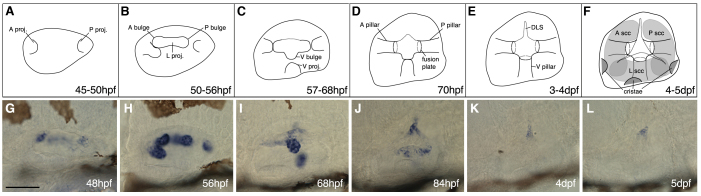
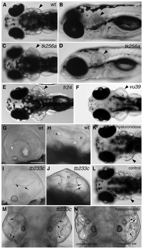
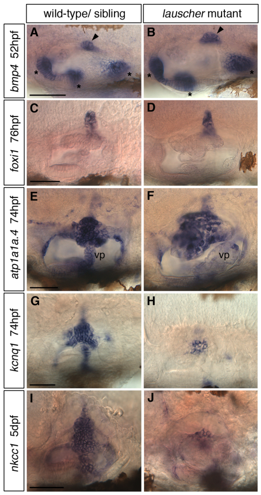
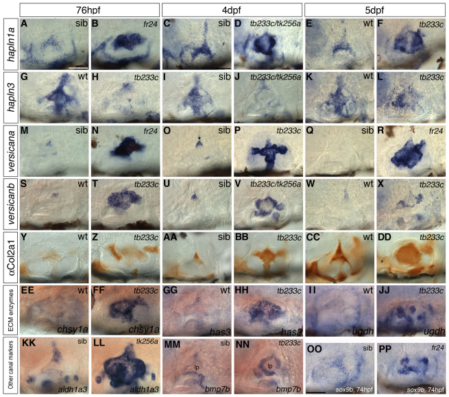
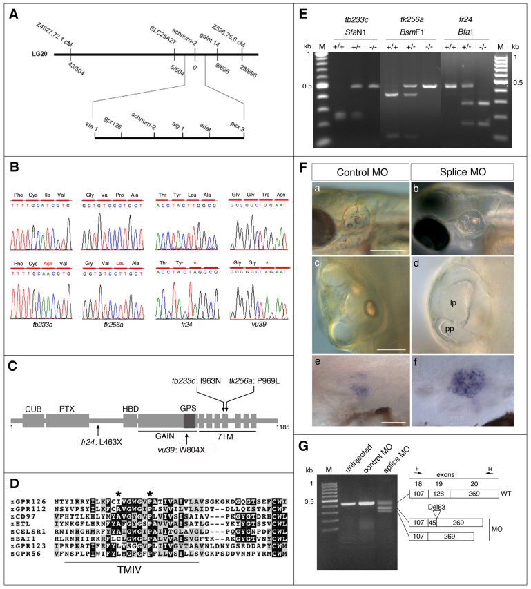
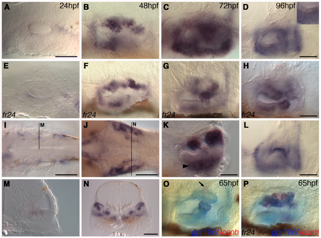
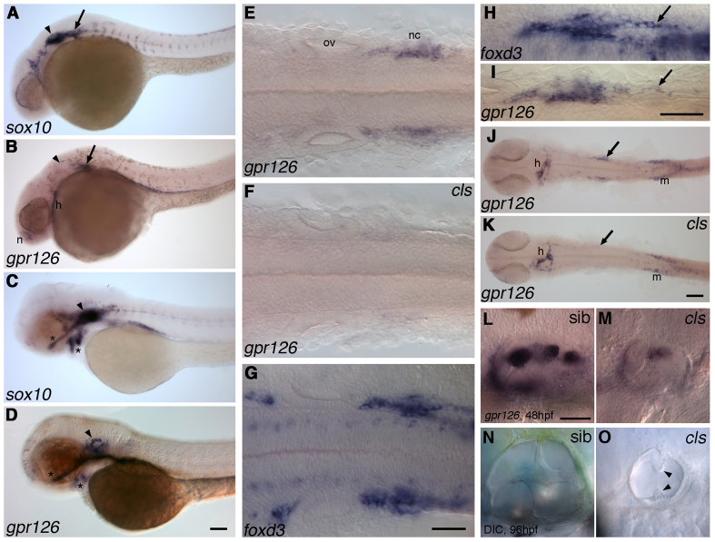
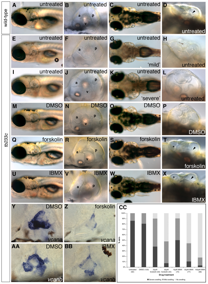
Morphogenesis of the semicircular canal ducts in the vertebrate inner ear is a dramatic example of epithelial remodelling in the embryo, and failure of normal canal development results in vestibular dysfunction. In zebrafish and Xenopus, semicircular canal ducts develop when projections of epithelium, driven by extracellular matrix production, push into the otic vesicle and fuse to form pillars. We show that in the zebrafish, extracellular matrix gene expression is high during projection outgrowth and then rapidly downregulated after fusion. Enzymatic disruption of hyaluronan in the projections leads to their collapse and a failure to form pillars: as a result, the ears swell. We have cloned a zebrafish mutant, lauscher (lau), identified by its swollen ear phenotype. The primary defect in the ear is abnormal projection outgrowth and a failure of fusion to form the semicircular canal pillars. Otic expression of extracellular matrix components is highly disrupted: several genes fail to become downregulated and remain expressed at abnormally high levels into late larval stages. The lau mutations disrupt gpr126, an adhesion class G protein-coupled receptor gene. Expression of gpr126 is similar to that of sox10, an ear and neural crest marker, and is partially dependent on sox10 activity. Fusion of canal projections and downregulation of otic versican expression in a hypomorphic lau allele can be restored by cAMP agonists. We propose that Gpr126 acts through a cAMP-mediated pathway to control the outgrowth and adhesion of canal projections in the zebrafish ear via the regulation of extracellular matrix gene expression.

Keywords: Adhesion GPCRs; Extracellular matrix; Inner ear; Semicircular canals; Zebrafish; gpr126.


Figures

Fig. 1.
Fig. 1.
Semicircular canal morphogenesis in the zebrafish ear. (A-F) Sketches of the developing semicircular canal system in the wild-type zebrafish ear, showing projection outgrowth, fusion and pillar formation. (G-L) Expression of vcana in the wild-type ear. (A,G) At 48 hpf, both anterior (A) and posterior (P) projections have begun to grow, and to express vcana at their tips. (B,H) The lateral projection, with its A and P bulges, is present by 50 hpf. The bulges and projections express vcana strongly at this time. The apparent downward growth of the A projection in H may be an artefact of fixation. (C,I) From 57-68 hpf, the A and P projections and bulges fuse to form the A and P pillars. The lateral projection now forms a ventral (V) bulge, and a V projection develops. Expression of vcana is downregulated in the A and P pillars, but is now strongly expressed in the V bulge and projection. (D,J) As fusion is completed at ∼70 hpf, expression of vcana is downregulated in all pillars. Expression remains in the dorsolateral septum (DLS) at 84 hpf. (E,K) At 72 hpf, all three pillars are fused [timing of fusion was slightly later than previously reported (Waterman and Bell, 1984)]. (F,L) At 4-5 dpf, only a trace of vcana expression remains in the DLS. Grey shading indicates the canal lumens. The positions of the cristae are shown. Abbreviations: A, anterior; DLS, dorsolateral septum; P, posterior; proj., projection; ssc, semicircular canal; V, ventral. Scale bar: 50 μm.
Fig. 2.
Fig. 2.
lauscher mutant zebrafish have swollen ears at 5 dpf and defects in semicircular canal formation. (A-F) Live images of wild-type sibling (A,B) and homozygous mutant (C-F) embryos at 5 dpf. The ears (arrowheads) of the mutants are swollen at 5 dpf, but otoliths appear normal. (G-J) Live images of wild-type sibling (G,H) and tb233c mutant (I,J) ears at 5 dpf. (K) Injection of hyaluronidase into the lateral projection of the left ear of a wild-type embryo results in the collapse of the projection, failure of fusion and a swollen ear. (L) Injection of PBS into the lateral projection of the left ear has no effect on ear development. Arrowheads mark the injected ears. (M,N) Comparison of ear swelling and projection fusion in the tb233c allele (M) with an ear in which the lateral projection has been injected with hyaluronidase (N) (dorsal views). Black arrows mark unfused projections; white arrows mark fused pillars. Asterisk in J marks projections that have touched but not fused correctly. Abbreviations: a and p, lumens of anterior and posterior semicircular canals; lp, enlarged lateral projection. A,C,E,F,H,J-N are dorsal views. Scale bars: in A, 200 μm for A,C,E,F,K,L; in B, 200 μm for B,D; in G, 50 μm for G-J.
Fig. 3.
Fig. 3.
Expression of genes involved in endolymph homeostasis in the lau mutant ear. (A-D) The endolymphatic duct, marked by expression of bmp4 (arrowheads, A,B) and foxi1 (C,D), appears to develop normally in lau mutants. The three cristae also express bmp4 normally (asterisks, A,B). (E-J) Expression of ion transporter genes in the lau mutant ear. (E,F) Expression of the atp1a1a.4 subunit of Na+/K+-ATPase1 is more diffuse and lacking from the ventral pillar (vp). (G-J) There is a substantial reduction in both kcnq1 and nkcc1 expression. Abbreviation: vp, ventral pillar (E) or ventral projection (F). Alleles: B,D,H,J, tk256a; F, tb233c. Scale bars: 50 μm.
Fig. 4.
Fig. 4.
Expression of extracellular matrix (ECM) genes and other semicircular canal markers is altered in the lauscher mutant ear. (A-JJ) Expression of ECM structural and enzyme genes in lau mutant ears at 76 hpf, 4 dpf and 5 dpf. Expression of the HA binding hapln1a (A-F) and the versican genes vcana and vcanb (M-X) is highly upregulated in mutant canal tissue. Expression of hapln3 is normally upregulated in wild-type ears on canal projection fusion (G,I,K); in the mutant, fusion fails, and transcript levels remain low (H,J,L). Antibody staining for type II Collagen (Y-DD) shows precocious protein accumulation at 76 hpf in mutants (Y,Z), persisting at 4-5 dpf (AA-DD); aberrant canal tissue is visible. Genes coding for enzymes for chondroitin synthesis (chsy1, EE,FF) and HA production (has3, GG,HH; ugdh, II,JJ) are upregulated in mutants at 76 hpf. (KK-PP) The canal markers aldh1a3, bmp7b and sox9b are upregulated in the unfused projections in the lau mutant ear. wt, wild type; sib, phenotypically wild-type sibling. Mutant alleles are shown on the panels. Scale bars: in A, 50 μm (applies to columns 1 and 2); in C, 50 μm (applies to all other panels).
Fig. 5.
Fig. 5.
Positional cloning, identification and confirmation of mutations in gpr126. (A) Genetic map of the lau locus. Numbers of meiotic recombinants for the flanking SSLP markers and SNP markers in slc25a27, schnurri2 and galnt14 are shown. (B) Sequence analysis of gpr126 cDNA in wild type (upper panels) and lau mutants (lower panels), with predicted changes to the coding sequence. (C) Schematic diagram of the Gpr126 protein, with its conserved domains: CUB (Complement C1r/C1s, Uegf, BMP1), PTX (Pentraxin), HBD (hormone binding domain), GAIN (GPCR autoproteolysis inducing) domain, GPS (GPCR proteolytic site) motif and 7TM (7-transmembrane) domain (not to scale). Positions of the mutations are shown. (D) Amino acid comparison of the fourth transmembrane (TMIV) region from eight zebrafish adhesion class GPCRs, showing the conserved hydrophobic residue at position 963 (mutated in tb233c), and the highly conserved proline (P) at position 969 (mutated in tk256a) (asterisks). (E) Genotyping of lau mutant fish by restriction digest of PCR-amplified genomic DNA. In tb233c, the mutation eliminated an SfaN1 site; in tk256a, the mutation eliminated a BsmF1 site; in the fr24 allele, a Bfa1 site was gained. (F,G) gpr126 morpholino injection recapitulates the lau mutant ear phenotype. (F) Wild-type (nac) embryos (left hand panels) injected with 5 ng control morpholino exhibit normal ear morphology at 5 dpf (a,c), and low vcanb expression at 5 dpf (e). Wild-type (nac) embryos injected with 5 ng gpr126 morpholino (right hand panels) have abnormal projection outgrowth (b,d). The lateral projection (lp) is enlarged, and the posterior projection (pp) in this ear has grown past the lateral projection without fusing. Expression of vcanb is upregulated (f). (G) RT-PCR analysis of gpr126 mRNA processing of the 19th exon in gpr126 morpholino-injected, or in control (mismatched) morpholino-injected, embryos at 5 dpf. Sequencing confirmed two aberrant splice variants. Panels c,d are dorsal views, anterior towards the top. Scale bars: 200 μm in a,b; 50 μm in c,d; 50 μm in e,f.
Fig. 6.
Fig. 6.
Expression of gpr126 in wild-type and lauscher mutant ears. (A-H) Expression of gpr126 mRNA in the ear at 24-96 hpf in wild-type (A-D) and fr24 mutant embryos (E-H). Strongest expression is in canal projections prior to fusion (48-72 hpf), with some expression remaining at 96 hpf. Inset in D: higher magnification showing expression in the anterior macula supporting cell layer. (I,M) Wild-type expression of gpr126 at 26 hpf in the anterior macula (I, dorsal view; M, transverse section). (J,N) Expression at 48 hpf shows stronger staining in the projections and sensory patches (J, dorsal view; N, transverse section). (K) Expression in sensory patches at 72 hpf is restricted to supporting cells (arrowhead) (transverse section; see also inset in D). There is strong expression in the projections. (L) Alternative focus view of D, showing residual expression in the lateral projection at 96 hpf. (O,P) Expression of gpr126 (blue) and vcanb (red) in the projections of wild-type and fr24 mutant embryos at 65 hpf. In wild-type embryos (O), there is co-expression (purple) in the recently fused ventral pillar; vcanb is downregulated in the lateral projection and in the anterior and posterior pillars (out of focus), whereas gpr126 is expressed at reduced levels. In fr24 mutants (P), vcanb and gpr126 are co-expressed in the unfused projections. Expression of vcanb persists in the dorsolateral septum, which does not express gpr126, in both wild-type and mutant embryos (O, arrow). Scale bars: in A, 50 μm for A-C,E-G,M,O,P; 50 μm in D,H,L; 25 μm in K; 100 μm in I,J,N.
Fig. 7.
Fig. 7.
Comparison of expression of gpr126 and sox10 in wild-type embryos, and expression of gpr126 in colourless (sox10-/-) mutant embryos. (A-D) Expression of gpr126 and sox10 in wild-type embryos at 24 hpf (A,B) and 48 hpf (C,D). Both genes are expressed in the otic vesicle (arrowhead), post-otic region (arrow), olfactory epithelium (nose, n) and head chondrocytes (asterisks). (E-G) Dorsal views of flat-mounted 24 hpf embryos: post-otic expression of gpr126 in the wild type (E) is lost in the cls mutant (F). Comparison with foxd3 expression identifies these cells as neural crest (G). (H,I) Dorsal view of the post-otic region showing foxd3-expressing Schwann cells extending posteriorly (H, arrow) and expression of gpr126 in the same location (I, arrow). (J,K) gpr126 expression in heart (h) and posterior mesoderm (m) persists in cls mutants, whereas neural crest expression is lost (arrows). (L,M) Expression of gpr126 is reduced in the cls mutant ear at 48 hpf, with weak expression in rudimentary projections (M). (N,O) DIC images of live ears at 96 hpf, showing a representative cls mutant ear (O). Note the small overall size and rudimentary unfused canal projections (arrowheads). Abbreviations: h, heart; m, posterior mesoderm, n, nose (olfactory epithelium); nc, neural crest; ov, otic vesicle; sib, phenotypically wild-type sibling embryo. Scale bars: in D, 100 μm for A-D; in G, 50 μm for to E-G; in I, 50 μm for H,I; in K, 100 μm for J,K; in L, 50 μm for L-O.
Fig. 8.
Fig. 8.
Treatment with cAMP agonists rescues the lauscher ear phenotype. (A-X) Live images at 5 dpf in control and drug-treated embryos. Ear swelling in tb233c mutants was variable, and categorized as ‘mild’ (E-H) or ‘severe’ (I-L) compared with wild type (A-D). Arrowhead in D indicates fused pillar in the untreated wild-type ear. Treatment between 60 and 90 hpf with cAMP agonists forskolin (25 μM; Q-T) and IBMX (100 μM; U-X) rescues the swollen ear phenotype; DMSO has no effect (M-P). Fusion of the anterior (a), posterior (p) and ventral (v) projections to form pillars is also restored: rescued pillars are present in drug-treated embryos (R,V; arrowheads in T,X) but not in DMSO-treated samples (N,P). However, small, ectopic tissue protrusions are present on the rescued pillars (asterisks, T,X). (Y-BB) Both drugs (forskolin, 50 μM; IBMX, 100 μM) can reduce expression of vcan genes in the ear (Y-BB). (CC) Graphical representation of the ear swelling data, analysed with a 3×3 (DMSO, forskolin) or 3×4 (DMSO, IBMX) chi-square contingency table; P<0.001 (both drugs). n values are in parentheses. (C,G,K,O,S,W) Dorsal views.

References

    1. Abbas L., Whitfield T. T. (2009). Nkcc1 (Slc12a2) is required for the regulation of endolymph volume in the otic vesicle and swim bladder volume in the zebrafish larva. Development 136, 2837–2848 - PMC - PubMed
    1. Abraira V. E., Del Rio T., Tucker A. F., Slonimsky J., Keirnes H. L., Goodrich L. V. (2008). Cross-repressive interactions between Lrig3 and netrin 1 shape the architecture of the inner ear. Development 135, 4091–4099 - PMC - PubMed
    1. Acampora D., Merlo G. R., Paleari L., Zerega B., Postiglione M. P., Mantero S., Bober E., Barbieri O., Simeone A., Levi G. (1999). Craniofacial, vestibular and bone defects in mice lacking the Distal-less-related gene Dlx5. Development 126, 3795–3809 - PubMed
    1. Araç D., Boucard A. A., Bolliger M. F., Nguyen J., Soltis S. M., Südhof T. C., Brunger A. T. (2012). A novel evolutionarily conserved domain of cell-adhesion GPCRs mediates autoproteolysis. EMBO J. 31, 1364–1378 - PMC - PubMed
    1. Asai Y., Chan D. K., Starr C. J., Kappler J. A., Kollmar R., Hudspeth A. J. (2006). Mutation of the atrophin2 gene in the zebrafish disrupts signaling by fibroblast growth factor during development of the inner ear. Proc. Natl. Acad. Sci. USA 103, 9069–9074 - PMC - PubMed

Publication types

MeSH terms