Review
. 2014 Apr;85(4):768-78.
doi: 10.1038/ki.2013.361. Epub 2013 Sep 25.

New magnetic resonance imaging methods in nephrology

Affiliations
Review

New magnetic resonance imaging methods in nephrology

Jeff L Zhang et al. Kidney Int. 2014 Apr.

Abstract

Established as a method to study anatomic changes, such as renal tumors or atherosclerotic vascular disease, magnetic resonance imaging (MRI) to interrogate renal function has only recently begun to come of age. In this review, we briefly introduce some of the most important MRI techniques for renal functional imaging, and then review current findings on their use for diagnosis and monitoring of major kidney diseases. Specific applications include renovascular disease, diabetic nephropathy, renal transplants, renal masses, acute kidney injury, and pediatric anomalies. With this review, we hope to encourage more collaboration between nephrologists and radiologists to accelerate the development and application of modern MRI tools in nephrology clinics.


Figures

Figure 1
Figure 1
Conventional anatomic MRI of kidney. The cyst, with long T1, is dark on T1-weighted image (A) and, with long T2, bright on T2-weighted image (B).
Figure 2
Figure 2
Kidney T2* maps from BOLD imaging. The scale on the right side is T2* value, with unit of milliseconds. Higher T2* corresponds to lower deoxyhemoglobin concentration. A) Typical T2* map with conventional BOLD scan (20-sec breath-hold, matrix size 256 × 256, FOV 32 × 32 cm). B) T2* map with free-breathing prospectively navigated sequence (10-minute imaging time, matrix size 512 × 512, FOV 50 × 50 cm). The free-breathing images offer greater image quality.
Figure 3
Figure 3
Difference images obtained from renal ASL scans. A) Acquired at 800 ms after arterial blood labeling, when the labeled blood is mostly in renal cortex; B) 1000 ms after the labeling, and some labeled blood reaches renal medulla. The images were acquired by a modified TrueFISP FAIR ASL sequence (8 averages, acquisition time ~24 sec, with breath hold).
Figure 4
Figure 4
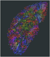
Kidney diffusion-weighted imaging using DTI methods. Following imaging processing, color-coded primary diffusion eigenvectors display radial pattern of medullary tubules.
Figure 5
Figure 5
Primitive right megaureter on a bifid ureter in a 6-month old boy. (A) T2-weighted image with fat saturation. (B) Coronal view of volume-rendered T2-weighted images. (C) Oblique view of volume-rendered T2-weighted images. (D) Maximum intensity projection of T1-weighted images at excretory phase. (E) Renography before contrast arrival. (F) Renography at arterial phase. (G) Renography at tubular phase. (H) Renography at excretory phase. Symmetric enhancement and excretion of contrast bilaterally suggests that the marked dilatation of the right collecting system and ureter is not a functional obstruction.

Comment in

References

    1. Lee VS, Rusinek H, Johnson G, et al. MR renography with low-dose gadopentetate dimeglumine: feasibility. Radiology. 2001;221:371–379. - PubMed
    1. Lee VS, Rusinek H, Bokacheva L, et al. Renal function measurements from MR renography and a simplified multicompartmental model. American journal of physiology. 2007;292:F1548–F1559. - PubMed
    1. Prasad PV, Edelman RR, Epstein FH. Noninvasive evaluation of intrarenal oxygenation with BOLD MRI. Circulation. 1996;94:3271–3275. - PubMed
    1. Textor SC, Glockner JF, Lerman LO, et al. The use of magnetic resonance to evaluate tissue oxygenation in renal artery stenosis. J Am Soc Nephrol. 2008;19:780–788. - PMC - PubMed
    1. Prasad PV. Functional MRI of the kidney: tools for translational studies of pathophysiology of renal disease. American journal of physiology. 2006;290:F958–F974. - PMC - PubMed