. 2013 Oct 15;110(42):17071-6.
doi: 10.1073/pnas.1315110110. Epub 2013 Sep 25.

Adaptor complex AP2/PICALM, through interaction with LC3, targets Alzheimer's APP-CTF for terminal degradation via autophagy

Affiliations

Adaptor complex AP2/PICALM, through interaction with LC3, targets Alzheimer's APP-CTF for terminal degradation via autophagy

Yuan Tian et al. Proc Natl Acad Sci U S A. .

Abstract

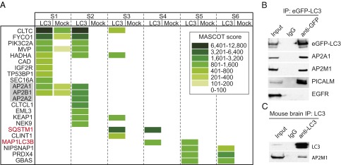
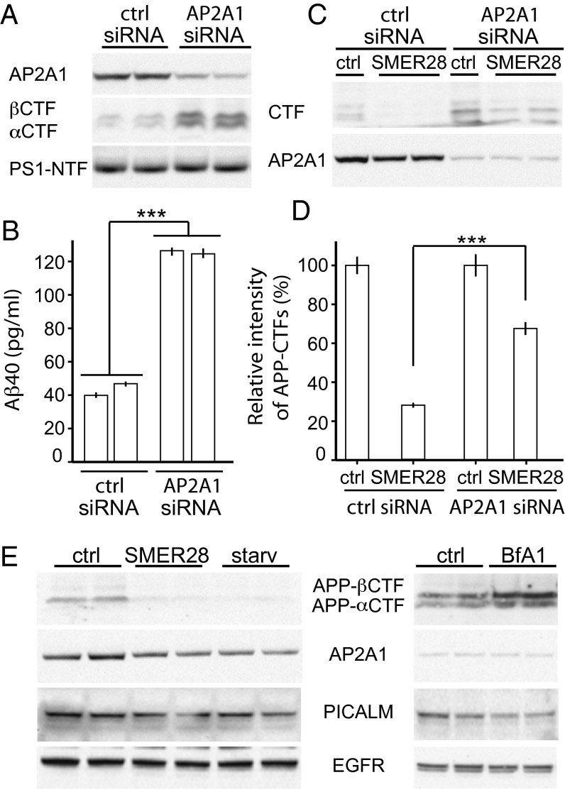
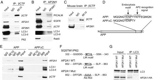
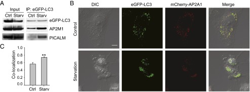
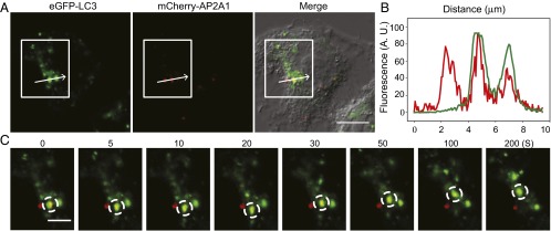
The hallmarks of Alzheimer's disease (AD) are the aggregates of amyloid-β (Aβ) peptides and tau protein. Autophagy is a major cellular pathway leading to the removal of aggregated proteins. We have reported recently that autophagy was responsible for amyloid precursor protein cleaved C-terminal fragment (APP-CTF) degradation and amyloid β clearance in an Atg5-dependent manner. Here we aimed to elucidate the molecular mechanism by which autophagy mediates the degradation of APP-CTF and the clearance of amyloid β. Through affinity purification followed by mass spectrum analysis, we identified adaptor protein (AP) 2 together with phosphatidylinositol clathrin assembly lymphoid-myeloid leukemia (PICALM) as binding proteins of microtubule-associated protein 1 light chain 3 (LC3). Further analysis showed that AP2 regulated the cellular levels of APP-CTF. Knockdown of AP2 reduced autophagy-mediated APP-CTF degradation. Immunoprecipitation and live imaging analysis demonstrated that AP2 and PICALM cross-link LC3 with APP-CTF. These data suggest that the AP-2/PICALM complex functions as an autophagic cargo receptor for the recognition and shipment of APP-CTF from the endocytic pathway to the LC3-marked autophagic degradation pathway. This molecular mechanism linking AP2/PICALM and AD is consistent with genetic evidence indicating a role for PICALM as a risk factor for AD.

Keywords: aggregate removal; endocytosis; trafficking.


Conflict of interest statement

The authors declare no conflict of interest.

Figures

Fig. 1.
Fig. 1.
Identification of AP2 as an LC3 binding protein. (A) HeLa cells stably expressing eGFP-LC3 were lysed and immunoprecipitated (IP) with mouse IgG or monoclonal GFP antibody. Eluates were resolved by SDS/PAGE and stained by colonial blue. Six bands (S1 to S6) from top to bottom of each IP were subjected to liquid chromatography-mass spectrometry. Proteins associated with LC3 were displayed as a heatmap based on their MASCOT score. The presence of the same polypeptides found in mock IP is also shown. (B) IP experiments with monoclonal GFP antibody were carried out as in A. (C) IP experiments with LC3 antibody using brain extracts from AD double transgenic mice, followed by immunoblotting using antibodies as indicated.
Fig. 2.
Fig. 2.
AP2 regulates protein levels of APP-CTF. N2a cells stably expressing APP were used and transfected with either control or AP2A1 siRNA for 48 h. (A) Cell lysates were analyzed by SDS/PAGE and Western blotting using antibodies as indicated. APP-CTFs were detected by using RU-369 antibody (C terminus of APP). (B) Aβ40 production was measured by ELISA using the supernatant. (C) Cells were then incubated with SMER28 (50 µM) for 16 h. Cell lysates were then analyzed by immunoblotting using RU-396 and AP2A1 antibodies. (D) Quantification of APP-CTF levels from repeated experiments as in C (n ≥ 3; ***P < 0.001; error bars represent SEM). (E) Cells were starved for 2 h or treated with SMER28 (50 μM) for 16, or treated with Bafilomycin (BfA1) (200 ng/mL) for 2 h. Cell lysates were analyzed by SDS/PAGE and Western blotting using antibodies as indicated.
Fig. 3.
Fig. 3.
AP2 interacts with APP-βCTF and LC3. (A) N2a cells stably expressing βCTF were lysed and immunoprecipitated with mouse IgG or 6E10 antibody and then analyzed by immunoblotting with corresponding antibodies. (B) IP was performed on the same cell lysates by using AP2M1 antibody. (C) IP experiments using brain extracts from AD double transgenic mice, using 6E10 antibody (N terminus of βCTF). (D) Schematic representation of the YxxФ motif deletion in APP C-terminal region. (E) N2a cells transiently transfected with APP or APP∆C were lysed and immunoprecipitations carried out with 6E10 or RU-369 antibodies, followed by SDS/PAGE and immunoblotting using AP2A1, LC3, and 6E10 antibodies. (F) Alignment of the LIR motifs of SQSTM1/P62 and AP2A1 is presented. The putative LIR motif of AP2A1 was mutated to alanine repeats in the AP2A1 Mut construct. (G) N2a cells were transiently transfected with HA-tagged AP2A1-WT or AP2A1 Mut constructs. Cell lysates were immunoprecipitated with LC3 antibody and immunoblotted with HA antibody.
Fig. 4.
Fig. 4.
Starvation enhances LC3 and AP2 interaction (A) and colocalization (B and C). (A) HeLa cells stably expressing eGFP-LC3 were used. Cells were starved or not for 2 h, and lysates were then used for IP (eGFP antibody). Immunoblots are presented as indicated. (B) Cells were transiently transfected with mCherry-AP2A1 and treated with complete medium or starvation medium for 2 h. Images were taken during the second hour of starvation by live-cell confocal microscopy. The micrographs presented are representative from three independent experiments. Two additional sets of images are shown in Fig. S4. (Scale bar: 10 μm.) (C) Quantification of Pearson's colocalization coefficient showed statistically significant difference in GFP-LC3 and mCherry-AP2A1 colocalization with or without serum starvation (n = 15–18; error bars represent SEM; **P < 0.001, Mann–Whitney u test).
Fig. 5.
Fig. 5.
Starvation-induced autophagy mediates LC3 and AP2 colocalization with time-scale on the order of a few hundred seconds. (A) Representative images of live HeLa cells expressing eGFP-LC3 and mCherry-AP2A1 upon 1-h serum starvation treatment. The white box highlights the colocalization of eGFP-LC3 and mCherry-AP2A1. (Scale bar: 10 μm.) (B) Fluorescent intensity profiles of the arrow in the white box indicate the position of a line scan, where two of three mCherry-AP2A1 (red line) punctate structures displayed high colocalization with eGFP-LC3 (green line). (C) Time-lapse images of the white box (A) showing that eGFP-LC3 and mCherry-AP2A1 present an increased colocalization following starvation. Note that the synchronized movement of eGFP-LC3 and mCherry-AP2A1 (dashed circles) lasted for at least 200 s. (Scale bar: 5 μm.)

References

    1. Haass C, Kaether C, Thinakaran G, Sisodia S. Trafficking and proteolytic processing of APP. Cold Spring Harb Perspect Med. 2012;2(5):a006270. - PMC - PubMed
    1. Perez RG, et al. Mutagenesis identifies new signals for beta-amyloid precursor protein endocytosis, turnover, and the generation of secreted fragments, including Abeta42. J Biol Chem. 1999;274(27):18851–18856. - PubMed
    1. Koo EH, Squazzo SL, Selkoe DJ, Koo CH. Trafficking of cell-surface amyloid beta-protein precursor. I. Secretion, endocytosis and recycling as detected by labeled monoclonal antibody. J Cell Sci. 1996;109(Pt 5):991–998. - PubMed
    1. Canfield WM, Johnson KF, Ye RD, Gregory W, Kornfeld S. Localization of the signal for rapid internalization of the bovine cation-independent mannose 6-phosphate/insulin-like growth factor-II receptor to amino acids 24-29 of the cytoplasmic tail. J Biol Chem. 1991;266(9):5682–5688. - PubMed
    1. Jadot M, Canfield WM, Gregory W, Kornfeld S. Characterization of the signal for rapid internalization of the bovine mannose 6-phosphate/insulin-like growth factor-II receptor. J Biol Chem. 1992;267(16):11069–11077. - PubMed

Publication types

MeSH terms

Substances