. 2013 Dec 1;305(11):C1098-113.
doi: 10.1152/ajpcell.00171.2013. Epub 2013 Sep 25.

Type-1 pericytes participate in fibrous tissue deposition in aged skeletal muscle

Affiliations

Type-1 pericytes participate in fibrous tissue deposition in aged skeletal muscle

Alexander Birbrair et al. Am J Physiol Cell Physiol. .

Abstract

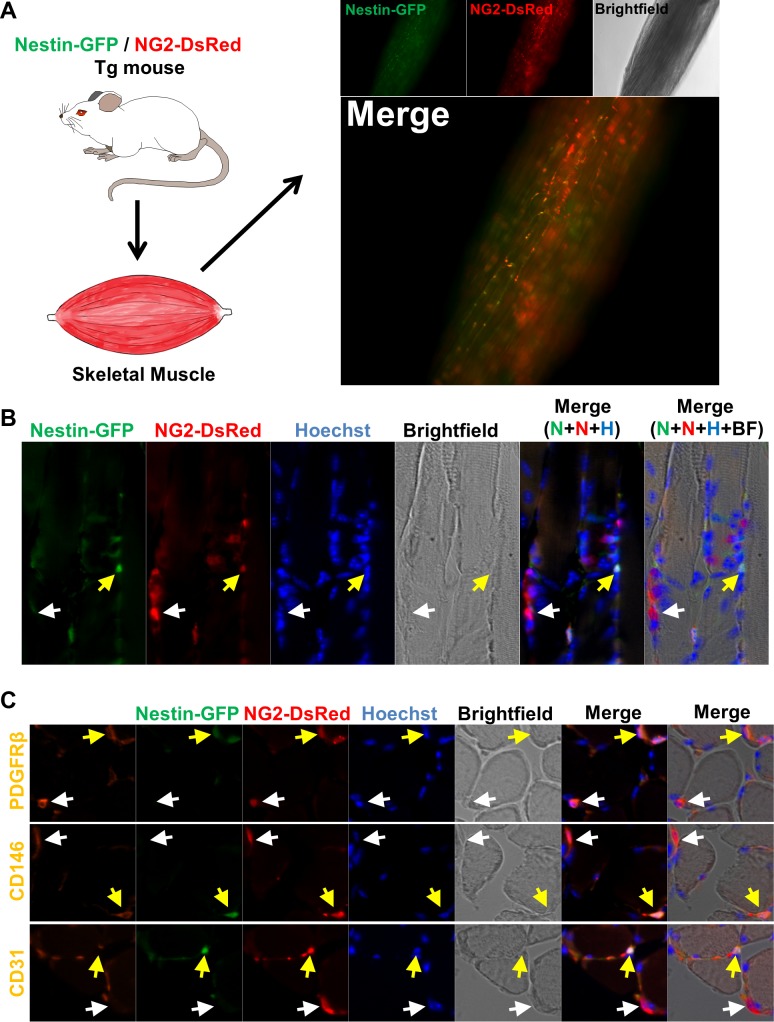
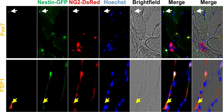
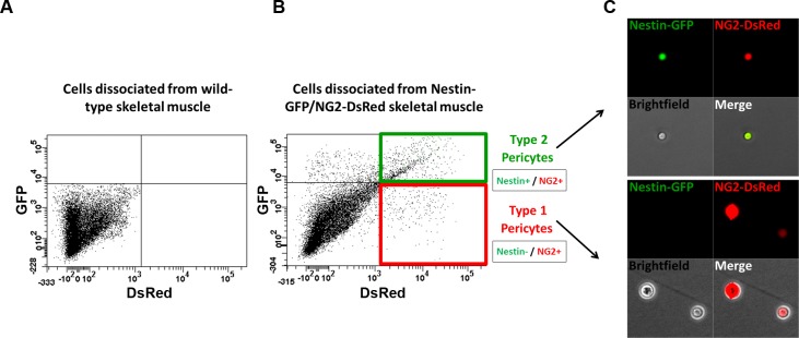
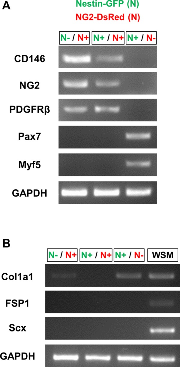
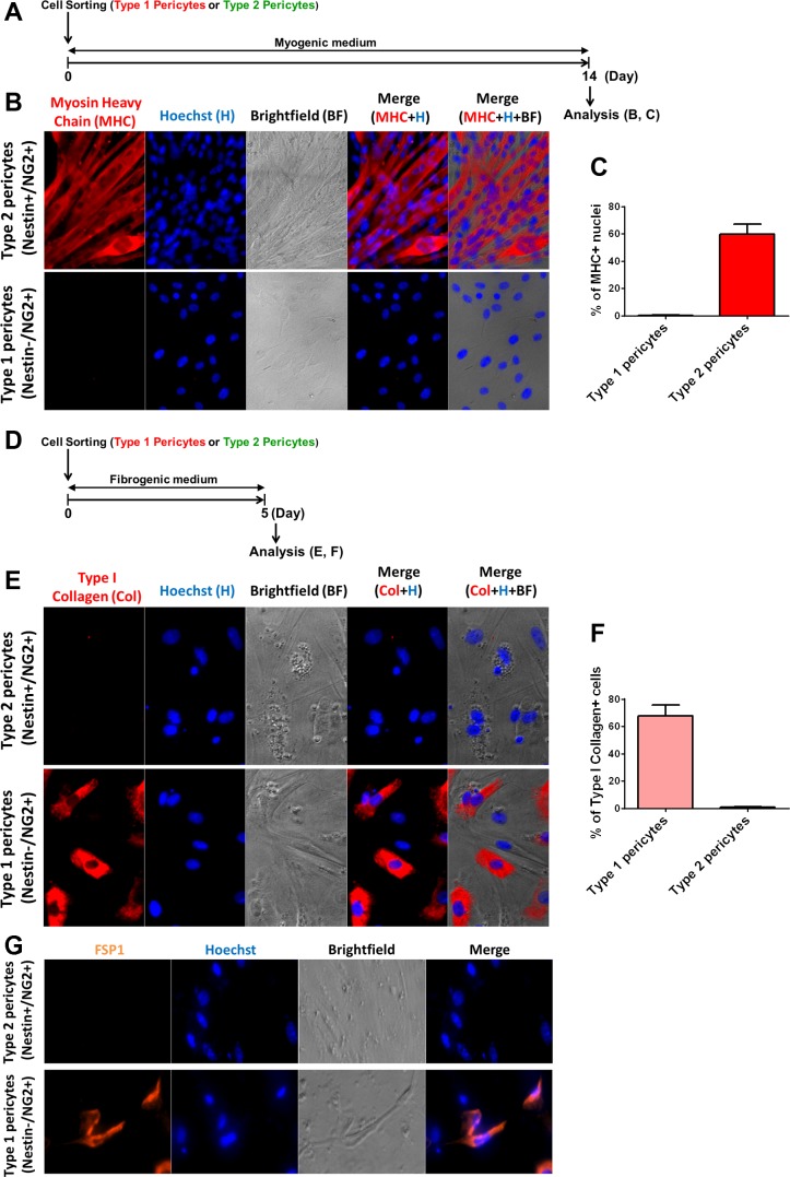
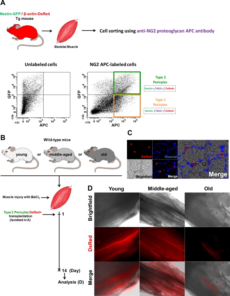
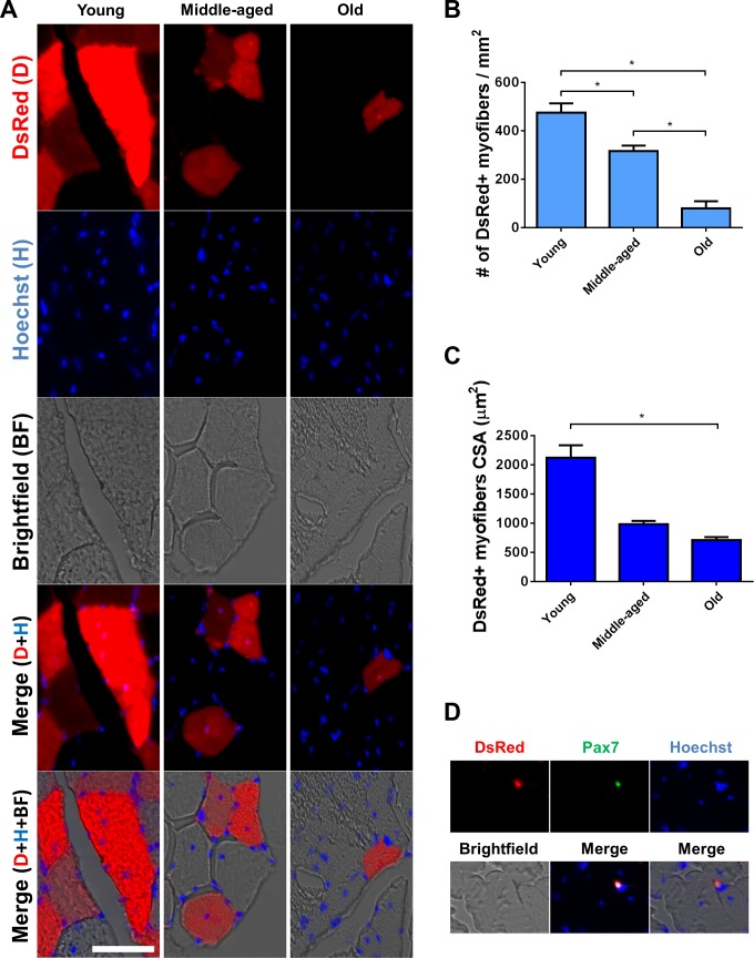
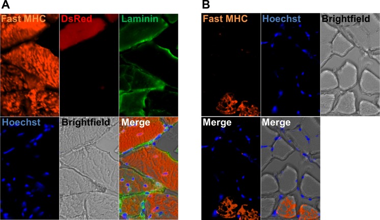
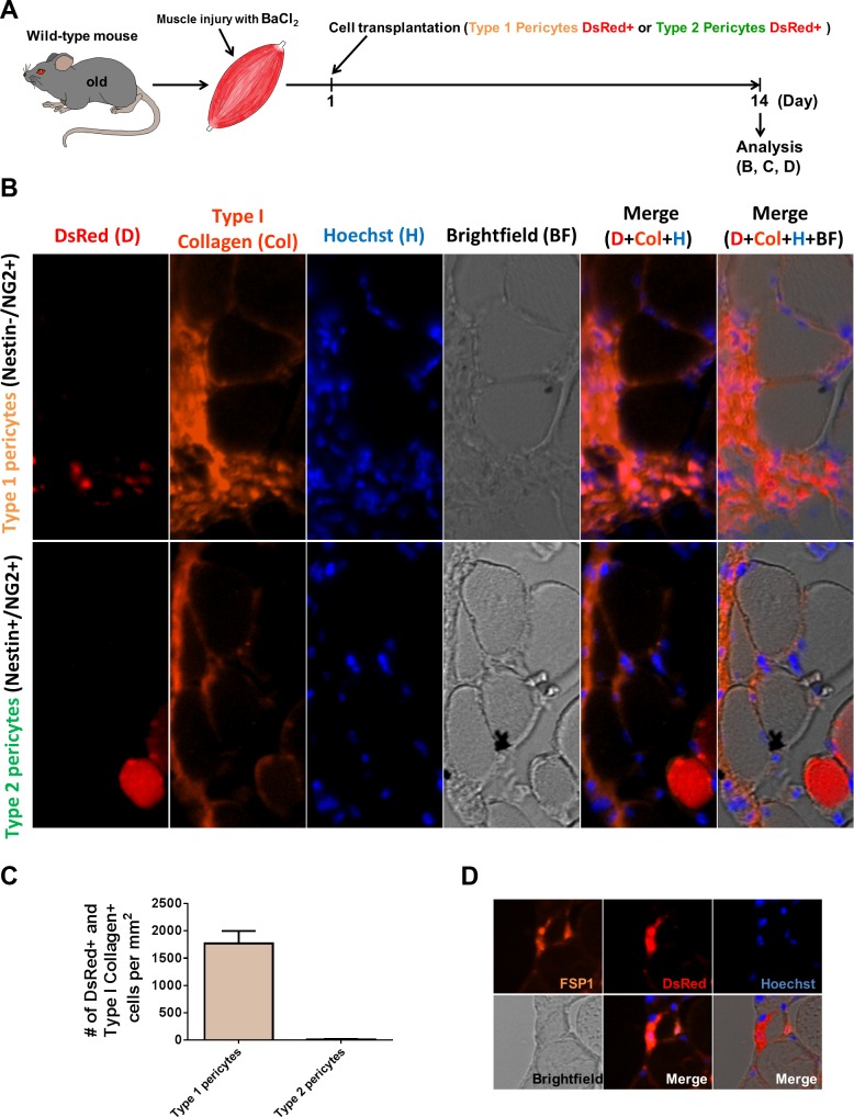
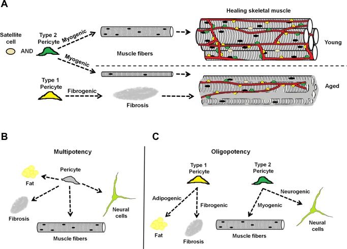
In older adults, changes in skeletal muscle composition are associated with increased fibrosis, loss of mass, and decreased force, which can lead to dependency, morbidity, and mortality. Understanding the biological mechanisms responsible is essential to sustaining and improving their quality of life. Compared with young mice, aged mice take longer to recover from muscle injury; their tissue fibrosis is more extensive, and regenerated myofibers are smaller. Strong evidence indicates that cells called pericytes, embedded in the basement membrane of capillaries, contribute to the satellite-cell pool and muscle growth. In addition to their role in skeletal muscle repair, after tissue damage, they detach from capillaries and migrate to the interstitial space to participate in fibrosis formation. Here we distinguish two bona fide pericyte subtypes in the skeletal muscle interstitium, type-1 (Nestin-GFP(-)/NG2-DsRed(+)) and type-2 (Nestin-GFP(+)/NG2-DsRed(+)), and characterize their heretofore unknown specific roles in the aging environment. Our in vitro results show that type-1 and type-2 pericytes are either fibrogenic or myogenic, respectively. Transplantation studies in young animals indicate that type-2 pericytes are myogenic, while type-1 pericytes remain in the interstitial space. In older mice, however, the muscular regenerative capacity of type-2 pericytes is limited, and type-1 pericytes produce collagen, contributing to fibrous tissue deposition. We conclude that in injured muscles from aging mice, the pericytes involved in skeletal muscle repair differ from those associated with scar formation.

Keywords: aging; fibrous tissue; pericytes; skeletal muscle.


Figures

Fig. 1.
Fig. 1.
Two pericyte subtypes are associated with skeletal muscle microvessels. A: histological analysis of pericyte subtypes in the skeletal muscle of double transgenic Nestin-GFP/NG2-DsRed mice. Whole lumbricalis muscle examined immediately after dissection. NG2-DsRed, Nestin-GFP, brightfield, and merged images are shown. NG2-DsRed+ cells can be detected; some overlap with Nestin-GFP fluorescence. B: longitudinal section of EDL muscle from Nestin-GFP/NG2-DsRed mice illustrating the 2 pericyte subtypes, type-1 (Nestin-GFP/NG2-DsREd+; white arrow) and type-2 (Nestin-GFP+/NG2-DsRed+; yellow arrow). AC show the same muscle area for different channels [Nestin-GFP (N), NG2-DsRed (N), Hoechst (H), brightfield (BF), merged fluorescence, and merged fluorescence and brightfield images]. C: immunofluorescence on transverse sections from the same muscle shown in B, stained with anti- platelet-derived growth factor receptor-β (PDGFR-β), anti-CD146, and anti-CD31 antibodies, confirms the presence of 2 types of pericytes, both PDGFR-β+/CD146+ and associated with microvessels (CD31+). Panels show identical muscle areas from left to right: PDGFR-β (1st line), CD146 (2nd line), or CD31 (3rd line, orange), Nestin-GFP+ (green), NG2-DsRed (red), Hoechst (blue), brightfield, and merged images. White arrows indicate type-1 pericytes (Nestin-GFP/NG2-DsRed+), and the yellow arrows indicate type-2 (Nestin-GFP+/NG2-DsRed+).
Fig. 2.
Fig. 2.
Satellite cells and fibroblasts do not express NG2-DsRed. Skeletal muscle from Nestin-GFP/NG2-DsRed transgenic mice shows Pax7 (satellite cells marker), fibroblast-specific protein 1 (FSP1; a fibroblast marker), Nestin-GFP, NG2-DsRed, and Hoechst staining in the same region. Brightfield and merged images are also shown. White arrow shows a satellite cell (Nestin-GFP+/Pax7+) that does not express NG2-DsRed; the yellow arrow indicates a Nestin-GFP/FSP1+ but NG2-DsRed fibroblast.
Fig. 3.
Fig. 3.
Isolation of skeletal muscle pericyte subtypes from Nestin-GFP/NG2-DsRed mice by sorting. Representative flow cytometry dot plot showing GFP vs. DsRed fluorescence with the gate set using cells isolated from skeletal muscle of wild-type mice (A). B: Nestin-GFP/NG2-DsRed skeletal muscle-derived cells were divided into 4 populations: Nestin-GFP+/NG2-DsRed-, Nestin-GFP/NG2-DsRed+ (type-1 pericytes), Nestin-GFP+/NG2-DsRed+ (type-2 pericytes), and Nestin-GFP/NG2-DsRed- cells. C: single cells in culture dishes immediately after sorting. Localization of Nestin-GFP+ (green) and NG2-DsRed+ (red) cells in freshly sorted fractions. All cells in the fraction of type-1 pericytes (Nestin/NG2+) were Nestin-GFP negative and NG2-DsRed positive and Nestin-GFP and NG2-DsRed positive in the fraction of type-2 pericytes (Nestin+/NG2+).
Fig. 4.
Fig. 4.
Nestin-GFP and NG2-DsRed cells gene expression. A: representative RT-PCR agarose gel from 3 experiments showing CD146, NG2, PDGFR-β, Pax7, Myf5 expression, and control GAPDH. The pericyte markers CD146, NG2, and PDGFR-β were present in Nestin-GFP/NG2-DsRed+ and Nestin-GFP+/NG2-DsRed+ cells but absent in Nestin-GFP+/NG2-DsRed- cells. The satellite cell markers Pax7 and Myf5 were detected only in Nestin-GFP+/NG2-DsRed- cells. B: RT-PCR agarose gel representative of 3 experiments, showing type I collagen (Col1a1), FSP1, Scleraxis (Scx) expression, and control GAPDH. Type I collagen gene was expressed in Nestin-GFP/NG2-DsRed+ and Nestin-GFP+/NG2-DsRed- cells, but absent in Nestin-GFP+/NG2-DsRed+ cells. The fibroblast markers were not present in Nestin-GFP/NG2-DsRed+, Nestin-GFP+/NG2-DsRed+, or Nestin-GFP+/NG2-DsRed cells. cDNA from whole skeletal muscle dissociated cells presorting (WSM) was used as a positive control.
Fig. 5.
Fig. 5.
Type-1 pericytes produce type I collagen, while type-2 pericytes differentiate into myotubes in vitro. Myogenic and fibrogenic induction of freshly isolated pericyte subtypes from Nestin-GFP/NG2-DsRed mouse muscle. A: time frame of myogenic differentiation in vitro: freshly isolated pericytes (Fig. 3) were cultured for 2 wk in myogenic differentiation medium. B: after 14 days in differentiation medium, Nestin-GFP/NG2-DsRed+ and Nestin-GFP+/NG2-DsRed+ cells were stained with anti-myosin heavy chain (MHC) antibody. C: percentage of MHC+ nuclei derived from each pericyte subtype was counted and normalized to the total number of nuclei (n = 3). Data are means ± SE. D: time frame for fibrogenic in vitro differentiation: freshly isolated pericytes (Fig. 3) were cultured for 5 days in fibrogenic medium containing TGF-β. E: after 5 days in fibrogenic medium, Nestin-GFP/NG2-DsRed+ and Nestin-GFP+/NG2-DsRed+ cells were stained with anti-type I collagen antibody. F: percentage of type I collagen+ cells derived from each pericyte subpopulation was counted and normalized to the total cell number (n = 3 preparations). G: Nestin-GFP/NG2-DsRed+ and Nestin-GFP+/NG2-DsRed+ cells were stained with FSP1 antibody at day 5 in fibrogenic conditions.
Fig. 6.
Fig. 6.
Skeletal muscle type-2 pericyte fate in vivo. A: pericytes subtypes from Nestin-GFP/β-actin-DsRed double-transgenic mice were isolated and sorted using an anti-NG2 proteoglycan APC antibody. All cells are DsRed+ and can be tracked in vivo. Representative dot plots showing GFP vs. APC fluorescence with the gate set using unlabeled cells before and after labeling with NG2 APC antibody. B: transplantation of isolated type-2 DsRed+ pericytes into injured skeletal muscles. C: representative tibialis anterior (TA) muscle section from a young mouse showing transplanted type-1 pericytes (DsRed+) remaining in the interstital space. Panels show identical muscle areas. Nuclei were stained with Hoechst 33342. D: whole TA muscle visualized immediately after dissection from young, middle-aged, and old mice 2 wk after type-2 DsRed+ pericytes transplantation. Brightfield, DsRed fluorescence, and merged images are shown. Notice the scarcity of DsRed+ cells in the muscle from old animals.
Fig. 7.
Fig. 7.
Type-2 pericytes participate in the formation of fewer and smaller myofibers in injured old mice. A: 2 wk after transplantation, DsRed+ myofibers (red) are present in the muscle of mice injected with type-2 pericytes. Panels show identical muscle areas from top to bottom: DsRed fluorescence (first line), Hoechst (blue), brightfield, merged fluorescence, and merged fluorescence and brightfield images. B and C: quantification of the data illustrated in A. B: number of DsRed+/type I collagen+ cells normalized to the cross-sectional area at day 14 of cell transplantation into injured skeletal muscle from old mice (n = 3 muscles). For data analysis, we used one-way ANOVA followed by Holm-Sidak test. C: cross-sectional area of DsRed+ myofibers in young, middle-aged, and old mice (n = 3 muscles). For data analysis, we used one-way ANOVA followed by Tukey test. D: representative TA muscle section from a transplanted mouse (as in A), showing a rare type-2 pericyte (DsRed+) expressing the satellite cell marker Pax7. Panels show identical muscle areas. Nuclei were stained with Hoechst 33342. *P < 0.05.
Fig. 8.
Fig. 8.
DsRed+ muscle fibers in muscles transplanted with DsRed+ type-2 pericytes are fast (type II). A: all fibers express fast myosin heavy chain in TA muscle injected with DsRed+ type-2 pericytes. B: soleus muscle, used as a staining control, also shows some fibers expressing fast myosin heavy chain. A and B show the same area for different channels: Fast MHC staining (orange), DsRed (red), Laminin (green), Hoechst (blue), brightfield, merged fluorescence images, and fluorescence and brightfield merge image; n = 3.
Fig. 9.
Fig. 9.
Type-1 pericytes are fibrogenic in vivo in old mouse skeletal muscle. A: time frame for type-1 or type-2 DsRed+ pericyte isolation and injection into injured skeletal muscles from old wild-type mice. B: TA muscle cross sections from old mice injected with type-1 or type-2 DsRed pericytes 2 wk after transplantation. Panels show identical muscle areas from left to right: DsRed, type I collagen, Hoechst, brightfield, merged fluorescence, and merged fluorescence and brightfield images. Anti-type I collagen staining confirms that type-1 pericytes stay in the interstitium and do not differentiate into muscle cells; type-2 pericytes do not express type I collagen and fuse in DsRed+ myofibers. C: quantification of the data illustrated in B. Number of DsRed+/type I collagen+ cells normalized to the cross-sectional area at day 14 of cell transplantation into injured skeletal muscle from old mice (n = 3 muscles). D: TA muscle cross-sections from old mice injected with type-1 DsRed+ pericytes 2 wk after transplantation. All panels show the same muscle area for different channels (FSP1 staining, DsRed, Hoechst, brightfield, merged fluorescence, and fluorescence and brightfield merged images). Type-1 pericytes are located in the interstitium and express the fibroblast marker FSP1.
Fig. 10.
Fig. 10.
Diagram of the role of pericyte subtypes in healing young and old skeletal muscle. Two subpopulations of pericytes are associated with blood vessels in the skeletal muscle: type-1 (yellow) and type-2 (green). A: we propose that during muscle repair, type-2 pericytes together with satellite cells contribute to myogenesis, producing larger myofibers in young than in old mice, while type-1 pericytes participate in the fibrous tissue accumulation observed in aged mice. B: classical model of the pericyte as a multipotent stem cell able to differentiate into fat, fibrous tissue, muscle, or neural cells. C: our model proposes that pericytes are heterogeneous and multipotent, but their subtypes are oligopotent and their ability to differentiate more restricted.

References

    1. Alnaqeeb MA, Al Zaid NS, Goldspink G. Connective tissue changes and physical properties of developing and ageing skeletal muscle. J Anat 139: 677–689, 1984 - PMC - PubMed
    1. Andrae J, Gallini R, Betsholtz C. Role of platelet-derived growth factors in physiology and medicine. Genes Dev 22: 1276–1312, 2008 - PMC - PubMed
    1. Barnes JL, Glass WF., 2nd Renal interstitial fibrosis: a critical evaluation of the origin of myofibroblasts. Contrib Nephrol 169: 73–93, 2011 - PubMed
    1. Barton-Davis ER, Shoturma DI, Musaro A, Rosenthal N, Sweeney HL. Viral mediated expression of insulin-like growth factor I blocks the aging-related loss of skeletal muscle function. Proc Natl Acad Sci USA 95: 15603–15607, 1998 - PMC - PubMed
    1. Beauchamp JR, Heslop L, Yu DS, Tajbakhsh S, Kelly RG, Wernig A, Buckingham ME, Partridge TA, Zammit PS. Expression of CD34 and Myf5 defines the majority of quiescent adult skeletal muscle satellite cells. J Cell Biol 151: 1221–1234, 2000 - PMC - PubMed

Publication types

LinkOut - more resources