. 2013 Nov;24(22):3545-56.
doi: 10.1091/mbc.E13-06-0332. Epub 2013 Sep 25.

A deubiquitinase negatively regulates retro-translocation of nonubiquitinated substrates

Affiliations

A deubiquitinase negatively regulates retro-translocation of nonubiquitinated substrates

Kaleena M Bernardi et al. Mol Biol Cell. 2013 Nov.

Abstract

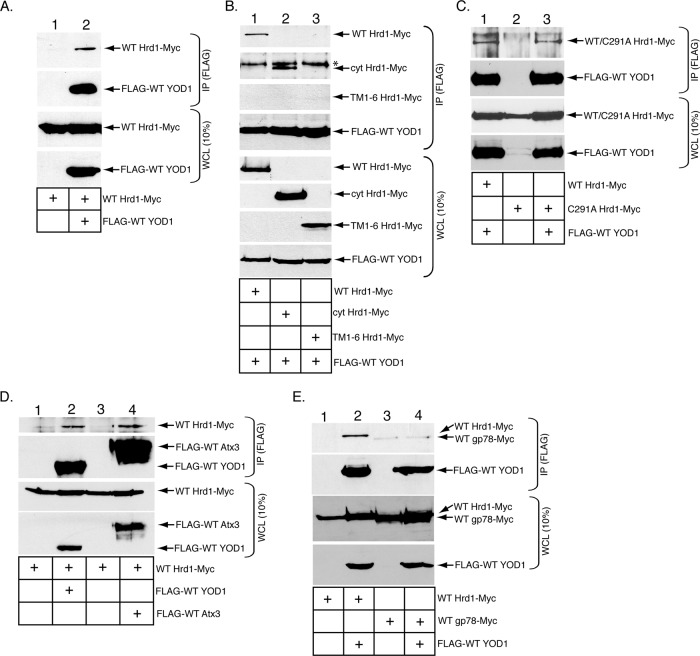
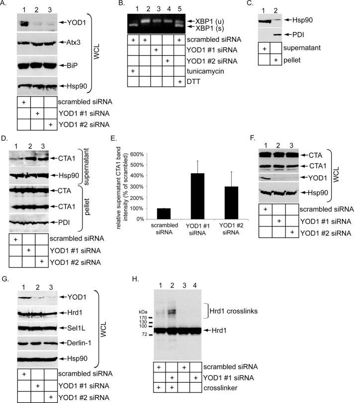
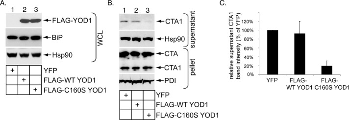
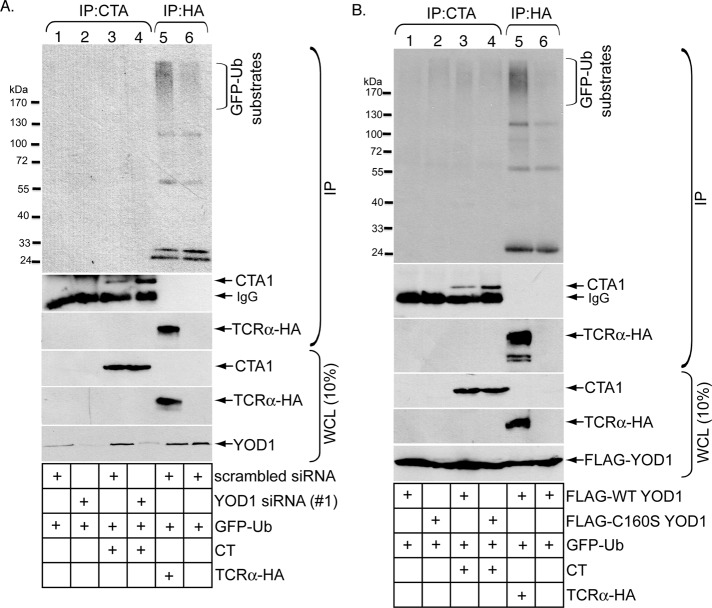
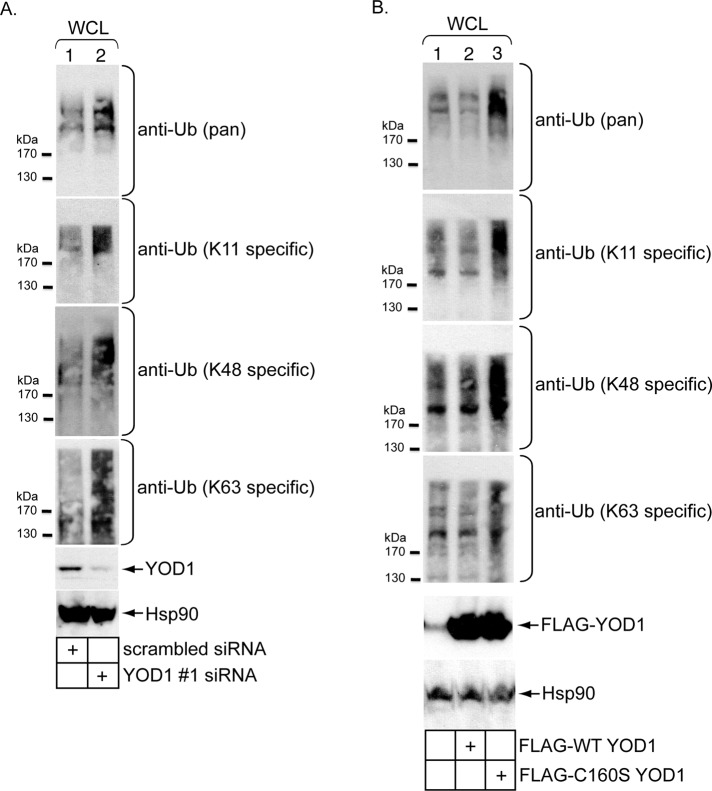
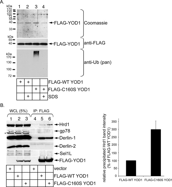
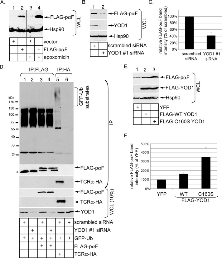
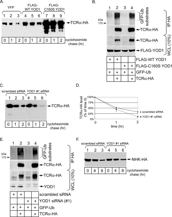
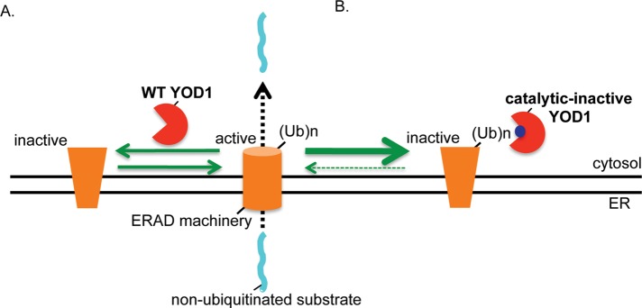
Endoplasmic reticulum (ER) membrane-bound E3 ubiquitin ligases promote ER-associated degradation (ERAD) by ubiquitinating a retro-translocated substrate that reaches the cytosol from the ER, targeting it to the proteasome for destruction. Recent findings implicate ERAD-associated deubiquitinases (DUBs) as positive and negative regulators during ERAD, reflecting the different consequences of deubiquitinating a substrate prior to proteasomal degradation. These observations raise the question of whether a DUB can control the fate of a nonubiquitinated ERAD substrate. In this study, we probed the role of the ERAD-associated DUB, YOD1, during retro-translocation of the nonubiquitinated cholera toxin A1 (CTA1) peptide, a critical intoxication step. Through combining knockdown, overexpression, and binding studies, we demonstrated that YOD1 negatively controls CTA1 retro-translocation, likely by deubiquitinating and inactivating ubiquitinated ERAD components that normally promote toxin retro-translocation. YOD1 also antagonizes the proteasomal degradation of nonglycosylated pro-α factor, a postulated nonubiquitinated yeast ERAD substrate, in mammalian cells. Our findings reveal that a cytosolic DUB exerts a negative function during retro-translocation of nonubiquitinated substrates, potentially by acting on elements of the ERAD machinery.


Figures

FIGURE 1:
FIGURE 1:
The YOD1 deubiquitinase binds to the Hrd1 E3 ubiquitin ligase. (A) 293T cells were transfected with WT Hrd1-Myc alone or with FLAG-WT YOD1 and lysed in a buffer containing 1% NP-40. The resulting WCLs were incubated with FLAG antibody–conjugated beads, and the immunoprecipitates were subjected to SDS–PAGE followed by immunoblotting with the indicated antibodies. The corresponding WCLs were analyzed by SDS–PAGE and immunoblotting with the indicated antibodies. (B) Cells were transfected with FLAG-WT YOD1 and either WT Hrd1-Myc, cyt Hrd1-Myc, or TM1-6 Hrd1-Myc, and processed as in (A). *, An unidentified band that cross-reacts with the Myc antibody. (C) Cells were transfected with FLAG-WT YOD1 and WT Hrd1-Myc, C291A Hrd1-Myc alone, or C291A Hrd1-Myc and FLAG-WT YOD1, and processed as in (A). (D) Cells were transfected with WT Hrd1-Myc alone or with FLAG-WT YOD1 or FLAG-WT Atx3, and processed as in (A). (E) Cells were transfected with WT Hrd1-Myc alone, WT Hrd1-Myc and FLAG-WT YOD1, WT gp78-Myc alone, or WT gp78-Myc and FLAG-WT YOD1, and processed as in (A). At least three independent experiments were performed in each condition.
FIGURE 2:
FIGURE 2:
YOD1 knockdown stimulates CTA1 retro-translocation. (A) WCLs from 293T cells transfected with a scrambled, YOD1 #1, or YOD1 #2 siRNA were analyzed by SDS–PAGE and immunoblotting with the indicated antibodies. (B) Reverse transcription–PCR analysis of the unspliced (u) and spliced (s) forms of the XBP1 mRNA derived from cells transfected with a scrambled siRNA treated with or without tunicamycin or DTT, or from cells transfected with YOD1 #1 or #2 siRNA. (C) Cells were incubated with digitonin and centrifuged. The resulting supernatant and pellet fractions were analyzed for the presence of the cytosolic Hsp90 and ER-resident PDI markers. This protocol is the fractionation procedure utilized in the retro-translocation assay. (D) Cells transfected with a scrambled, YOD1 #1, or YOD1 #2 siRNA were incubated with CT (10 nM) for 90 min and subjected to the retro-translocation assay as in (C). The pellet and supernatant fractions were subjected to SDS–PAGE and were analyzed by immunoblotting with the indicated antibodies. (E) The supernatant CTA1 band intensity generated in (D) was quantified with ImageJ. Data represent the mean of at least three independent experiments. Error bars: ±SD. (F) Cells transfected with a scrambled, YOD1 #1, or YOD1 #2 siRNA were intoxicated with CT (10 nM) for 90 min and harvested, and the resulting WCLs were analyzed with nonreducing SDS–PAGE followed by immunoblotting with the indicated antibodies. (G) WCLs derived from cells transfected with a scrambled, YOD1 #1, or YOD1 #2 siRNA were analyzed by immunoblotting using the indicated antibodies. (H) WCLs derived from cells transfected with a scrambled or YOD1 #1 siRNA and incubated with or without the EDAC cross-linker were analyzed by immunoblotting with the Hrd1 antibody.
FIGURE 3:
FIGURE 3:
Catalytically inactive YOD1 overexpression decreases CTA1 retro-translocation. (A) Cells were transfected with YFP, FLAG-WT YOD1, or catalytically inactive FLAG-C160S YOD1. The resulting WCLs were subjected to SDS–PAGE followed by immunoblotting with the indicated antibodies. (B) Cells transfected with YFP, FLAG-WT YOD1, and FLAG-C160S YOD1 were incubated with CT (10 nM) for 90 min and subjected to the retro-translocation assay as in Figure 2C. (C) The supernatant CTA1 band intensity in (B) was analyzed as in Figure 2D. Mean of at least three independent experiments. Error bars: ±SD.
FIGURE 4:
FIGURE 4:
Disrupting YOD1 activity does not promote CT polyubiquitination. (A) Cells transfected with scrambled siRNA, YOD1 #1 siRNA, or TCRα-HA were intoxicated with or without CT (10 nM) for 90 min and lysed in a RIPA buffer containing 0.1% SDS. The resulting WCLs were incubated with a CTA antibody (lanes 1–4) or HA antibody–conjugated beads (lanes 5–6). The immunoprecipitates were subjected to reducing SDS–PAGE followed by immunoblotting with the indicated antibodies. WCLs were also analyzed by immunoblotting with the appropriate antibodies. All cells were transfected with GFP-Ub and incubated with epoxomicin. (B) As in (A), except cells were transfected with FLAG-WT YOD1 or FLAG-C160S YOD1 instead of the siRNAs.
FIGURE 5:
FIGURE 5:
Perturbing YOD1 increases cellular K11-, K48-, and K63-linked polyubiquitinated proteins. (A) WCLs derived from cells transfected with scrambled or YOD1 #1 siRNA were subjected to SDS–PAGE and analyzed by immunoblotting using the indicated antibodies. (B) As in (A), except cells were untransfected or transfected with either FLAG-WT YOD1 or FLAG-C160S YOD1.
FIGURE 6:
FIGURE 6:
Catalytically inactive YOD1 traps polyubiquitinated proteins. (A) For purification of YOD1, cells transfected with either FLAG-WT YOD1 or FLAG-C160S YOD1 were lysed in a buffer containing 1% NP-40 with or without 1% SDS. The resulting WCLs were diluted 10-fold and incubated with FLAG antibody–conjugated beads; the precipitated material was eluted with a FLAG peptide and subjected to SDS–PAGE followed by Coomassie staining or immunoblotting with the indicated antibodies. (B) Transfected FLAG-WT YOD1 or FLAG-C160S YOD1 were precipitated, and the samples were immunoblotted with the indicated antibodies. Quantification of the Hrd1 band intensity is as in Figure 2E. Data represent the mean of three independent experiments. Error bars: ±SD.
FIGURE 7:
FIGURE 7:
Perturbing YOD1 activity disrupts ERAD of the nonubiquitinated yeast pro-α factor. (A) Cells pretreated with or without epoxomicin were transfected with a pcDNA3.1(−) vector or FLAG-pαF. The resulting WCLs were subjected to SDS–PAGE and immunoblotting with the indicated antibodies. (B) WCLs derived from cells transfected with FLAG-pαF and either scrambled siRNA or YOD1 #1 siRNA were immunoblotted with the indicated antibodies. (C) The FLAG-pαF band intensity in (B) was quantified as in Figure 2D. Mean of at least three independent experiments. Error bars: ±SD. (D) Cells transfected with scrambled siRNA, YOD1 #1 siRNA, FLAG-pαF, and/or TCRα-HA were lysed in a RIPA buffer containing 0.1% SDS. The resulting WCLs were incubated with a FLAG antibody (lanes 1–4) or HA antibody–conjugated beads (lanes 5–6). The immunoprecipitates were subjected to reducing SDS–PAGE followed by immunoblotting with the indicated antibodies. WCLs were also analyzed by immunoblotting with the appropriate antibodies. All cells were transfected with GFP-Ub and incubated with epoxomicin. (E) As in (B), except cells were transfected with YFP, FLAG-WT YOD1, or FLAG-C160S YOD1. (F) The FLAG-pαF band intensity in (E) was quantified as in Figure 2D. Mean of at least three independent experiments. Error bars: ±SD.
FIGURE 8:
FIGURE 8:
Disrupting YOD1 activity affects TCRα degradation without altering its ubiquitination level. (A) Cells transfected with TCRα-HA and cotransfected with YFP, FLAG-WT YOD1, or FLAG-C160S YOD1 were incubated with cycloheximide for the indicated time. The resulting WCLs were immunoblotted with an HA antibody. (B) As in Figure 5B. (C) As in (A), except cells were transfected with scrambled siRNA or YOD1 #1 siRNA. (D) The TCRα-HA band intensity in (C) was quantified as in Figure 2C. Mean of at least three independent experiments. Error bars: ±SD. (E) As in (B), except cells were transfected with scrambled siRNA or YOD1 #1 siRNA. (F) As in (C), except cells were transfected with NHK-HA.
FIGURE 9:
FIGURE 9:
A model of how YOD1 negatively regulates retro-translocation of nonubiquitinated substrates. (A) YOD1 deubiquitinates and inactivates a ubiquitinated component of the ERAD machinery normally important for promoting retro-translocation. Hence, YOD1 down-regulation results in accumulation of the ubiquitinated ERAD component and consequently enhances retro-translocation. (B) By contrast, the catalytically inactive C160S YOD1 mutant binds to and traps the ubiquitinated ERAD component. Trapping effectively inactivates the function of the ubiquitinated ERAD factor, leading to a block in retro-translocation. A potential candidate for the ERAD component trapped by mutant YOD1 is Hrd1 itself.

References

    1. Bernardi KM, Forster ML, Lencer WI, Tsai B. Derlin-1 facilitates the retro-translocation of cholera toxin. Mol Biol Cell. 2008;19:877–884. - PMC - PubMed
    1. Bernardi KM, Williams JM, Kikkert M, van Voorden S, Wiertz EJ, Ye Y, Tsai B. The E3 ubiquitin ligases Hrd1 and gp78 bind to and promote cholera toxin retro-translocation. Mol Biol Cell. 2010;21:140–151. - PMC - PubMed
    1. Blount JR, Burr AA, Denuc A, Marfany G, Todi SV. Ubiquitin-specific protease 25 functions in endoplasmic reticulum-associated degradation. PLoS One. 2012;7:e36542. - PMC - PubMed
    1. Brodsky JL. Cleaning up: ER-associated degradation to the rescue. Cell. 2012;151:1163–1167. - PMC - PubMed
    1. Carvalho P, Stanley AM, Rapoport TA. Retrotranslocation of a misfolded luminal ER protein by the ubiquitin-ligase Hrd1p. Cell. 2010;143:579–591. - PMC - PubMed

Publication types

MeSH terms

LinkOut - more resources