Review
. 2014 Jan;42(1):20-44.
doi: 10.1093/nar/gkt847. Epub 2013 Sep 24.

Type I restriction enzymes and their relatives

Affiliations
Review

Type I restriction enzymes and their relatives

Wil A M Loenen et al. Nucleic Acids Res. 2014 Jan.

Abstract

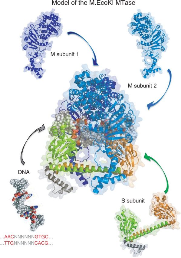
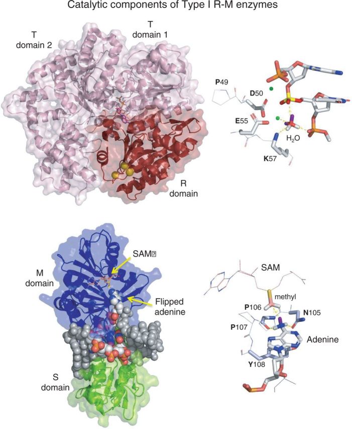
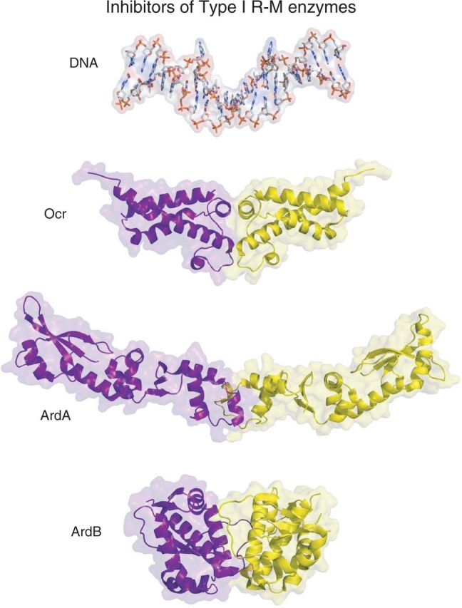
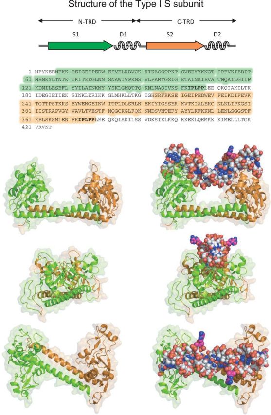
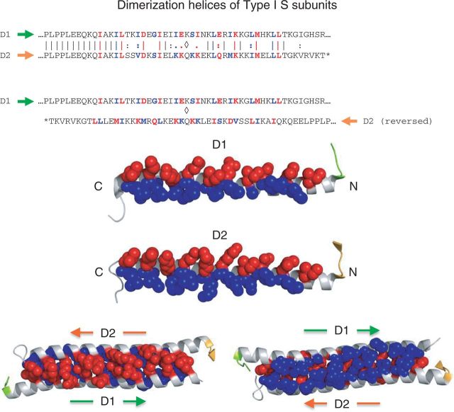
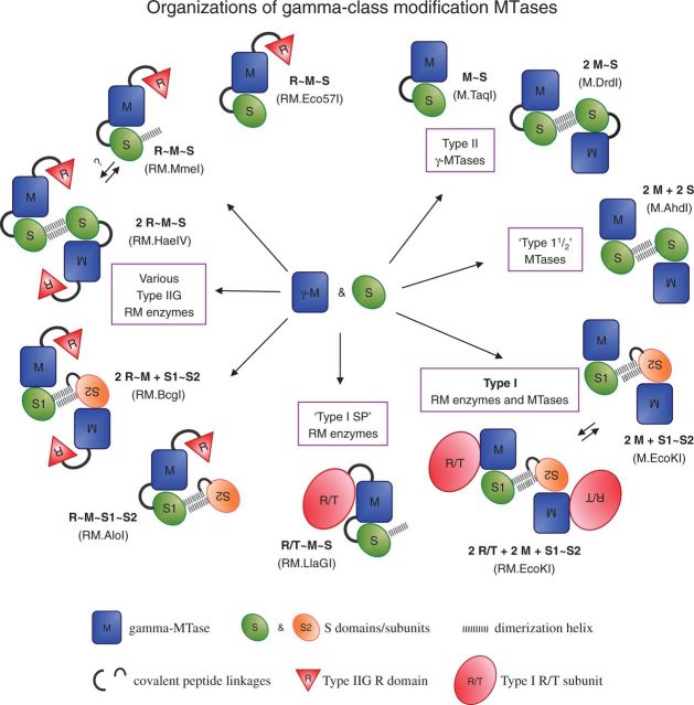
Type I restriction enzymes (REases) are large pentameric proteins with separate restriction (R), methylation (M) and DNA sequence-recognition (S) subunits. They were the first REases to be discovered and purified, but unlike the enormously useful Type II REases, they have yet to find a place in the enzymatic toolbox of molecular biologists. Type I enzymes have been difficult to characterize, but this is changing as genome analysis reveals their genes, and methylome analysis reveals their recognition sequences. Several Type I REases have been studied in detail and what has been learned about them invites greater attention. In this article, we discuss aspects of the biochemistry, biology and regulation of Type I REases, and of the mechanisms that bacteriophages and plasmids have evolved to evade them. Type I REases have a remarkable ability to change sequence specificity by domain shuffling and rearrangements. We summarize the classic experiments and observations that led to this discovery, and we discuss how this ability depends on the modular organizations of the enzymes and of their S subunits. Finally, we describe examples of Type II restriction-modification systems that have features in common with Type I enzymes, with emphasis on the varied Type IIG enzymes.


Figures

Figure 1.
Figure 1.
Model of the M.EcoKI MTase (pdb file 2Y7H). The S subunit is composed of two TRDs in inverted orientations. Each TRD comprises a globular DNA-binding domain and an alpha helical dimerization domain. The N-TRD (green) in this protein is specific for the sequence AAC (the 5-half-sequence), and the C-terminal domain (orange) is specific for GCAC (the 3 half-sequence). Zipper-like association of the helices separates the globular domains by a fixed distance and reverses the orientation of the C-TRD, resulting in the composite recognition sequence that is bipartite: AAC N6 GTGC. Each TRD also associates with one M subunit (identical, but shown here in different shades of blue for clarity) to form an M2S trimer. Neither S nor M subunits bind to DNA alone, but the trimer binds specifically at the recognition sequence and catalyzes methylation of one adenine in each half-sequence. Because the TRDs are inverted, the two M subunits have opposite orientations. Consequently, both strands of the recognition sequence become methylated, the ‘top’ strand of the 5 half-sequence (Am6AC) and the ‘bottom’ strand of the 3 half-sequence (GCm6AC). This enables the DNA of the host cell to be distinguished from infecting DNA during DNA replication.
Figure 2.
Figure 2.
Catalytic components of Type I R–M enzymes. Upper figure, left: The R/T subunit of EcoR124I (pdb: 2W00). The R domain (red), responsible for DNA cleavage, comprises the N-terminal ∼260 aa. It contains the common PD-D/EXK endonuclease catalytic site, composed of D151, E165 and K167 (yellow spheres). Upper figure, right. The PD-D/EXK catalytic site of the Type II REase, MvaI (pdb:2OAA). D50 and E55 coordinate divalent metal ions (in this case two Ca2+ ions, shown as green spheres at reduced scale for clarity). The hydrolytic water molecule is oriented by interaction with a metal ion, the general base K57, and a phosphate oxygen from the adjacent base. These interactions position a lone pair electron orbital (purple sticks) of the water molecule for in-line nucleophilic attack on the phosphorus atom (bright yellow), initiating the DNA cleavage reaction. Catalysis occurs in the presence of Mg2+, but not in the presence of Ca2+; hence, this structure represents the pre-cleavage complex. Lower figure, left: The monomeric γ-MTase, M.TaqI, with bound DNA (pdb: 1G38). SAM was absent in this complex, which represents the pre-methylation complex. SAM has been added here through modeling by structural alignment with pdb:2ADM. Lower figure, right: The catalytic NPPY of M.TaqI is composed of N105, P106, P107 and Y108. When the target adenine is flipped into the catalytic site, the hydrogens of the 6-amino group form hydrogen bonds with the side chain amide carbonyl of N104 and the main chain carbonyl of P105. These lie below the plane of the base and likely induce the nitrogen to switch from the planar sp2 orbital configuration it normally possesses, to the tetrahedral sp3 configuration (105). In this latter configuration, the lone pair orbital of nitrogen (purple stick) is appropriately positioned for in-line nucleophilic attack on the carbon thiol (pink) of SAM, initiating the DNA methylation reaction (105,106).
Figure 3.
Figure 3.
Protein inhibitors of Type I R–M enzymes. Top panel: DNA model (hydrogen atoms omitted) from pdb:2Y7H displayed on the same scale as the proteins for structural comparisons. Panel b: Ocr (pdb:1S7Z and 2Y7C) from bacteriophage T7; panel c: ArdA (pdb:2W82) from Tn916 of E. faecalis (117); panel d: ArdB (pdb:2WJ9) from a pathogenicity island of E. coli CFT073. All three proteins are homodimeric. Their subunits are identical, but are displayed here in different colors.
Figure 4.
Figure 4.
Structure of the Type I S subunit (pdb:1YF2). The recognition sequence of this protein, S-MjaXI, from Methanocaldococcus (formerly Methanococcus) jannaschii is not known. It is closely related to the EcoKI-family (Type IA) of enzymes depicted in Figure 1. The upper diagram shows the domain organization of the protein; arrows represent DNA-binding domains, and curly lines represent dimerization alpha helices. The aa sequence of the protein is shown below, with the domains in corresponding colors. Below this are three views of the structure, from three perpendicular directions, ‘sideways’, ‘end-on’, and ‘above’. The panels on the left depict the protein; those on the right depict the protein with modeled DNA positioned approximately as it is bound. The DNA was taken from pdb:2Y7H and transferred by structural alignment of the S subunits.
Figure 5.
Figure 5.
Dimerization helices of Type I S subunits. Upper diagram: aa sequence alignment of the dimerization helices of S-MjaXI (pdb:1YF2; D1 and D2 in Figure 3) show that they are similar but not identical (top). The proline-rich motif that precedes each helix (IPLPP) is a hallmark of Type I S subunits and likely plays a structural role establishing the correct architectural relationship between the S and D domains. The helices interact in opposite orientations to form an antiparallel coiled-coil (bottom). Adjacent pairs of aa (red and blue) form the dimerization interface and occur with the 4-3-4-3 … spacing characteristic of leucine zippers. Middle diagram: Exposed dimerization surfaces of two helices. Side chains of red aa point ‘up’ toward the bound DNA, and those of blue aa point ‘down’. To form the coiled coil, D1 must be rotated 180 degrees around the vertical axis and docked against the surface of D2 shown. Lower diagram: Within the coiled coil, red aa from one helix interdigitate with red aa from the other helix, forming the ‘upper’ surface of the coiled coil, the surface closest to the bound DNA (left). And blue aa from one helix inter-digitate with blue aa from the other helix to form the ‘lower’ surface on the other side (red). Within the coiled-coiled, red side chains from one helix stack on blue side chains from the other helix in an alternating pattern. At this interface, the helices have complimentary topologies such that a ridge or bump in one is accommodated by a valley or depression in the other, resulting in a close hydrophobic fit.
Figure 6.
Figure 6.
Organizations of gamma-class modification MTases. γ-MTases catalyze transfer of a methyl group from SAM to the exocyclic amino group of adenine (or in rare cases, cytosine) at specific target sequences in duplex DNA. Their organizations vary depending on whether they methylate both DNA strands, or only one, and whether they cleave the DNA instead if the sequence is completely unmodified. At minimum, γ-MTases comprise an M component for base flipping and methyl transfer (blue oblongs), and an S component for sequence-recognition (green and orange ovals). These can be separate subunits or discrete domains. S components can function individually, or as inverted pairs that associate through antiparallel dimerization helices. The members of such pairs can be identical, in which case the structure is homodimeric, or they can differ, in which case the structure is functionally heterodimeric albeit usually connected into a single protein chain. Type I and Type IIG R–M enzymes are γ-MTases that also restrict DNA. For Type I enzymes, a supplementary R/T subunit (large red ovals) catalyzes endonuclease and DNA-translocase activities. For Type IIG enzymes, an R domain (red triangle) is permanently present at the N-terminus. The latter combine with S components in configurations that mirror those of the monofunctional γ-MTases. The figure is not exhaustive; it depicts only the commonest variants of γ-MTases but reveals a close evolutionary connection between Type I R–M systems and certain Type II R–M systems. An example of each organization is given in parentheses.

References

    1. Dussoix D, Arber W. Host specificity of DNA produced by Escherichia coli. II. Control over acceptance of DNA from infecting phage lambda. J. Mol. Biol. 1962;5:37–49. - PubMed
    1. Arber W, Dussoix D. Host specificity of DNA produced by Escherichia coli. I. Host controlled modification of bacteriophage lambda. J. Mol. Biol. 1962;5:18–36. - PubMed
    1. Luria SE, Human ML. A nonhereditary, host-induced variation of bacterial viruses. J. Bacteriol. 1952;64:557–569. - PMC - PubMed
    1. Anderson ES, Felix A. Variation in Vi-phage II of Salmonella typhi. Nature. 1952;170:492–494. - PubMed
    1. Bertani G, Weigle JJ. Host controlled variation in bacterial viruses. J. Bacteriol. 1953;65:113–121. - PMC - PubMed

MeSH terms

Substances