. 2013 Sep 25;33(39):15626-41.
doi: 10.1523/JNEUROSCI.1483-13.2013.

Sodium-mediated plateau potentials in lumbar motoneurons of neonatal rats

Affiliations

Sodium-mediated plateau potentials in lumbar motoneurons of neonatal rats

Mouloud Bouhadfane et al. J Neurosci. .

Abstract

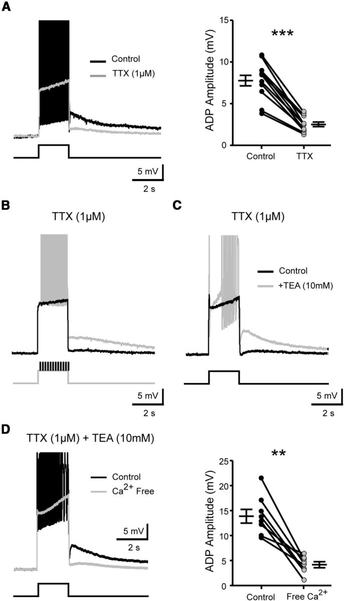
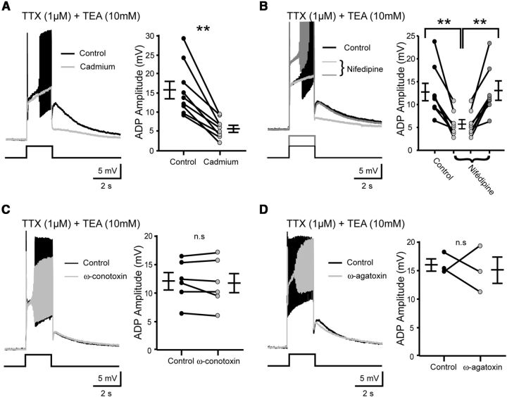
The development and the ionic nature of bistable behavior in lumbar motoneurons were investigated in rats. One week after birth, almost all (∼80%) ankle extensor motoneurons recorded in whole-cell configuration displayed self-sustained spiking in response to a brief depolarization that emerged when the temperature was raised >30°C. The effect of L-type Ca(2+) channel blockers on self-sustained spiking was variable, whereas blockade of the persistent sodium current (I(NaP)) abolished them. When hyperpolarized, bistable motoneurons displayed a characteristic slow afterdepolarization (sADP). The sADPs generated by repeated depolarizing pulses summed to promote a plateau potential. The sADP was tightly associated with the emergence of Ca(2+) spikes. Substitution of extracellular Na(+) or chelation of intracellular Ca(2+) abolished both sADP and the plateau potential without affecting Ca(2+) spikes. These data suggest a key role of a Ca(2+)-activated nonselective cation conductance ((CaN)) in generating the plateau potential. In line with this, the blockade of (CaN) by flufenamate abolished both sADP and plateau potentials. Furthermore, 2-aminoethoxydiphenyl borate (2-APB), a common activator of thermo-sensitive vanilloid transient receptor potential (TRPV) cation channels, promoted the sADP. Among TRPV channels, only the selective activation of TRPV2 channels by probenecid promoted the sADP to generate a plateau potential. To conclude, bistable behaviors are, to a large extent, determined by the interplay between three currents: L-type I(Ca), I(NaP), and a Na(+)-mediated I(CaN) flowing through putative TRPV2 channels.


Figures

Figure 1.
Figure 1.
Electrophysiological properties of plateau potentials. A, Confocal image of a transverse section of the lumbar spinal cord shows motoneurons retrogradely labeled with the fluorescein-conjugated cholera toxin B subunit tracer injected into the triceps surae muscle. Scale bar, 100 μm. B, Bistable motoneuron displaying a plateau potential (black traces) when the depolarizing current pulse was increased either in amplitude (B1) or in duration (B2) or when the holding membrane potential was depolarized (B3). Current pulses in B2 and B3 were given from a depolarized membrane potential using bias current. Gray traces show the sADP outlasting the current pulse when the voltage threshold to trigger the plateau potential was not reached. C, Firing properties of a non-bistable motoneuron unable to generate a plateau potential in response to a 2 s depolarizing current pulse. C, Inset, Illustrates with expanded time scale the typical pronounced afterhyperpolarization (AHP) that follows the current pulse (action potentials are truncated). B, C, Instantaneous frequency plots are shown on top of intracellular recordings. D, Proportions of bistable and non-bistable motoneurons at the beginning and the end of the first postnatal week. E, Plateau potential triggered by a brief depolarizing current pulse in the presence of CNQX (10 μm), AP5 (50 μm), strychnine (5 μm) and bicuculline (20 μm). F, Electrical stimulation of the dorsal root (10 Hz, 2 s) induced plateau potential, which was stopped by hyperpolarization. In this and the following figures, bottom traces are the injected current.
Figure 2.
Figure 2.
Electrophysiological properties of the sADP. A, Superimposed voltage traces from a motoneuron showing the sADP in response to current steps of increasing amplitude (A1) or duration (A2). A, Insets, The linear relationship between the area of the sADP and the number of spikes during the stimulus. B, Voltage traces illustrating the wind-up of the sADP in response to repetitive depolarizing current pulses of constant amplitude applied either at resting membrane potential (B1) or at a more depolarized potential producing a plateau potential (B2). C, Superimposed voltage traces from a bistable motoneuron recorded at resting membrane potential illustrating the switch of the sADP (gray trace) into a plateau potential (black trace) when the initial 2 s square pulse was followed by a sequence of suprathreshold depolarizing current pulses.
Figure 3.
Figure 3.
Pharmacological profile of plateau potentials. A1A3: Effect of blocking the L-type Ca2+ channels by nifedipine (20 μm) on plateau potentials. B, Effect of the progressive blockade of the persistent sodium current by 5 μm riluzole (B1) or 10 nm TTX (B2) on plateau potentials. C, Facilitation of plateau potentials by upregulating the persistent sodium current with veratridine (40 nm). Motoneurons (Mns) were recorded at a holding potential between −60 and −55 mV.
Figure 4.
Figure 4.
The sADP requires spiking-dependent Ca2+ influx. A, Superimposed voltage traces in response to a 2 s depolarizing pulse before (black trace) and after (gray trace) TTX application (1 μm). The graph shows the mean amplitude of the sADP before and after drug application. B, Superimposed voltage traces collected under TTX, in response to a 2 s depolarizing pulse on which additional brief depolarizing pulses that mimic action potentials were (black trace) or were not (gray trace) applied. C, Superimposed voltage traces in response to a 2 s depolarizing pulse, collected under TTX before and after adding TEA (10 mm). D, Superimposed voltage traces in response to a 2 s depolarizing pulse, collected under TTX and TEA before and after the removal of extracellular Ca2+. Graphs show the mean amplitude of the sADP before and after removing Ca2+. Note that action potentials were truncated for the visualization of the sADP. Holding potential, −60 mV. Error bars indicate SEM. ns, Not significant; **p < 0.01, ***p < 0.001 (Wilcoxon paired test).
Figure 5.
Figure 5.
The L-type voltage-gated Ca2+ channels are primarily responsible for the spiking-dependent Ca2+ influx necessary to generate the sADP. A–D, Left, Superimposed voltage traces in response to a 2 s depolarizing pulse before (black traces) and after (gray traces) the addition of: (A) cadmium, a broad spectrum voltage-gated Ca2+ channels blocker (200 μm), (B) nifedipine a L-type Ca2+ channels blocker (20 μm), (C) ω-conotoxin GVIA (1 μm) a N-type Ca2+ channels blocker, and (D) ω-agatoxin VIA (200 nm) a P/Q-type Ca2+ channels blocker. Note that in the presence of nifedipine large depolarizing current pulses allow the recovery of both action potentials and sADP (dark gray trace). Right, Graphs show the mean amplitude of the sADP before and after the drug application. All recordings were made under TTX (1 μm) and TEA (10 mm). Holding potential, −60 mV. Error bars indicate SEM. ns, Not significant; *p < 0.05, **p < 0.01 (Wilcoxon paired test in A, C, and D and Kruskal–Wallis test in B).
Figure 6.
Figure 6.
The sADP and plateau potentials are dependent on ICaN that uses Na+ as the main charge carrier, not a Na+/Ca2+ exchanger. A, Superimposed voltage traces in response to a 2 s depolarizing pulse before (black traces) and after (gray traces) chelating the intracellular Ca2+ with BAPTA (10 mm) in the pipette solution. B, Left, Superimposed current traces from a motoneuron recorded under TEA (10 mm), TTX (1 μm), and apamin (100 nm), held at −60 mV, step-depolarized to +10 mV and then returned to potentials between −60 and 0 mV. On the right, I–V relationship of the peak inward tail current plotted against the return potential before (black trace) and after (gray trace) lowering the extracellular concentration of Na+. C, D, Superimposed current (C) and voltage (D) traces in response to a 2 s depolarizing pulse before (black traces) and after (gray traces) lowering the extracellular concentration of Na+. E, Superimposed voltage traces from a bistable motoneuron recorded at resting membrane potential in response to a 2 s square pulse followed by a series of brief depolarizing current pulses before (black trace) and after (gray trace) lowering the concentration of Na+. F, Superimposed voltage traces in response to a 2 s depolarizing pulse before (black traces) and after (gray traces) the blockade of the Na+/Ca2+ exchanger by lithium. A, C, D, F, Right, Graphs show the mean amplitude of the sADP before and after the drug application. Recordings were made under TTX (1 μm) and TEA (10 mm). The apamin (100 nm) was superfused for voltage-clamp recordings. Holding potential, −60 mV. Error bars indicate SEM. ns, Not significant; *p < 0.05, **p < 0.01 (Wilcoxon paired test).
Figure 7.
Figure 7.
The sADP is upregulated by thermosensitive TRPV cation channels. A–D, Superimposed voltage traces in response to a 2 s depolarizing pulse before (black traces) and after (gray traces) the addition of: (A) flufenamate (75 μm) or (B) lanthanum (100 μm), two broad spectrum TRP channel blockers; (C) SKF 96365, a nonselective TRPC channel blocker (20 μm); and (D) 2-APB, a common activator of TRPV channels. Right, Graphs show the mean amplitude of the sADP before and after the drug application. Recordings were made under TTX and TEA. Holding potential, −60 mV. Error bars indicate SEM. ns, Not significant; *p < 0.05, **p < 0.01, ***p < 0.001 (Wilcoxon paired test).
Figure 8.
Figure 8.
Among TRPV cationic channels, TRPV2 upregulates the sADP. A–D, Superimposed voltage traces in response to a 2 s depolarizing pulse before (black traces) and after (gray traces) the addition of agonists (left in A1, B1, C1, and D1) or antagonists (right in A2, B2, C2, and D2) of specific TRPV1 (A), TRPV2 (B), TRPV3 (C), or TRPV4 (D) channels. B2, Left, Superimposed current traces in response to a 2 s depolarizing pulse before (black traces) and after (gray traces) applying probenecid (250 μm). Right, I–V relationship of the peak inward tail current plotted against the return potential. B4, Superimposed current traces in response to a 2 s depolarizing pulse recorded under mefloquine (50 μM) before (black traces) and after (gray traces) applying probenecid (250 μM). Recordings were made under TTX and TEA. Holding potential, −60 mV. Error bars indicate SEM. ns, Not significant; *p < 0.05, **p < 0.01 (Wilcoxon paired test).
Figure 9.
Figure 9.
Activation of TRPV2 channels generates self-sustained firing. A–D, Superimposed voltage traces recorded in normal saline (without TTX and TEA) in response to a 2 s depolarizing pulse before (black traces) and after (gray traces) bath applying probenecid (A), chelating intracellular Ca2+ with BAPTA (B), blocking IKCa with apamine (C), or reducing the temperature of the bath from 34°C to 28°C (D). C, Instantaneous frequency plots are shown on top of intracellular recordings.
Figure 10.
Figure 10.
A schematic ménage à trois relationship between currents underlying the different phases of the plateau firing mode. The Ca2+ entry via voltage-dependent Ca2+ current occurs during the train of action potential (step 1 and 2 in A and B). This increase in intracellular Ca2+ triggers a voltage-independent cation current that depolarizes the membrane and mediates the sADP (step 3 in A and B). The positive change in membrane potential opens voltage-dependent persistent Na+ current to maintain a train of action potential (step 4 in A and B). The repetitive spiking activity will then induce Ca2+ entry via voltage-dependent Ca2+ current and so forth.

References

    1. Altman J, Sudarshan K. Postnatal development of locomotion in the laboratory rat. Anim Behav. 1975;23:896–920. doi: 10.1016/0003-3472(75)90114-1. - DOI - PubMed
    1. Angstadt JD, Choo JJ. Sodium-dependent plateau potentials in cultured Retzius cells of the medicinal leech. J Neurophysiol. 1996;76:1491–1502. - PubMed
    1. Bang S, Kim KY, Yoo S, Lee SH, Hwang SW. Transient receptor potential V2 expressed in sensory neurons is activated by probenecid. Neurosci Lett. 2007;425:120–125. doi: 10.1016/j.neulet.2007.08.035. - DOI - PubMed
    1. Bang S, Yoo S, Yang TJ, Cho H, Hwang SW. Isopentenyl pyrophosphate is a novel antinociceptive substance that inhibits TRPV3 and TRPA1 ion channels. Pain. 2011;152:1156–1164. doi: 10.1016/j.pain.2011.01.044. - DOI - PubMed
    1. Benham CD, Gunthorpe MJ, Davis JB. TRPV channels as temperature sensors. Cell Calcium. 2003;33:479–487. doi: 10.1016/S0143-4160(03)00063-0. - DOI - PubMed

Publication types