Review
. 2013 Sep 23:7:612.
doi: 10.3389/fnhum.2013.00612.

Ephaptic communication in the vertebrate retina

Affiliations
Review

Ephaptic communication in the vertebrate retina

Rozan Vroman et al. Front Hum Neurosci. .

Abstract

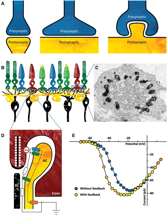
In the vertebrate retina, cones project to the horizontal cells (HCs) and bipolar cells (BCs). The communication between cones and HCs uses both chemical and ephaptic mechanisms. Cones release glutamate in a Ca(2+)-dependent manner, while HCs feed back to cones via an ephaptic mechanism. Hyperpolarization of HCs leads to an increased current through connexin hemichannels located on the tips of HC dendrites invaginating the cone synaptic terminals. Due to the high resistance of the extracellular synaptic space, this current makes the synaptic cleft slightly negative. The result is that the Ca(2+)-channels in the cone presynaptic membrane experience a slightly depolarized membrane potential and therefore more glutamate is released. This ephaptic mechanism forms a very fast and noise free negative feedback pathway. These characteristics are crucial, since the retina has to perform well in demanding conditions such as low light levels. In this mini-review we will discuss the critical components of such an ephaptic mechanism. Furthermore, we will address the question whether such communication appears in other systems as well and indicate some fundamental features to look for when attempting to identify an ephaptic mechanism.

Keywords: cones; ephaptic communication; horizontal cells; inhibition; vertebrate retina.


Figures

Figure 1
Figure 1
(A) Ephaptic interactions will occur in every synapse. The strength of the ephaptic interaction will depend on the organization of the synapse. It will only become significant if the extracellular resistance is high enough. Left: synapse without significant ephaptic interaction. Middle and right: synapse with potentially significant ephaptic interactions. (B) Schematic drawing of the vertebrate outer retina. The photoreceptors are the light sensitive neurons in the retina (top layer). They are contacted by horizontal cells (HCs) (yellow) and bipolar cells (BCs) (black). The dendrites of both the HCs and the BCs invaginate the photoreceptor synaptic terminal. (C) Electron micrograph of a cone synaptic terminal. In this zebrafish HCs express Green Fluorescent Protein (GFP), which is visible as a black label in this image. Note that every ribbon is flanked by HC dendrites. R: synaptic ribbons. (Taken from: Klaassen et al., 2012) (D) Schematic drawing of the cone/HC synapse. In the dark, glutamate release is high and thus the glutamate receptors are activated (green resistors). A constant inward current flows through the connexin hemichannels (blue resistor) and because the resistance of the synaptic cleft is relatively high (white resistors), the synaptic space is slightly negative compared to the extrasynaptic space. When the retina receives a full field light stimulus, cones and consequently HCs hyperpolarize. This causes an increased inward current through the connexin hemichannels, resulting in an increased negativity of the synaptic cleft. The voltage sensitive Ca-channels on the cone (ICa, red circle) detect this as a slight depolarization of the membrane potential, effectively shifting the Ca-current activation potential towards more negative potentials (see Panel E). The influx of calcium increases and consequently the glutamate release. The current entering the HCs via the connexin hemichannels leaves the HCs via the potassium channel on the HC somata (orange resistor). (E) Feedback from HCs to cones modulates the Ca-current of cones. Ca-current of a cone in control condition (blue) and when HCs are hyperpolarized and feedback is active (yellow). (Modified from: Verweij et al., 1996).

References

    1. Ala-Laurila P., Greschner M., Chichilnisky E. J., Rieke F. (2011). Cone photoreceptor contributions to noise and correlations in the retinal output. Nat. Neurosci. 14, 1309–1316 10.1038/nn.2927 - DOI - PMC - PubMed
    1. Berretta N., Rossokhin A. V., Kasyanov A. M., Sokolov M. V., Cherubini E., Voronin L. L. (2000). Postsynaptic hyperpolarization increases the strength of AMPA-mediated synaptic transmission at large synapses between mossy fibers and CA3 pyramidal cells. Neuropharmacology 39, 2288–2301 10.1016/s0028-3908(00)00076-9 - DOI - PubMed
    1. Bogdanik L., Framery B., Frolich A., Franco B., Mornet D., Bockaert J., et al. (2008). Muscle dystroglycan organizes the postsynapse and regulates presynaptic neurotransmitter release at the Drosophila neuromuscular junction. PLoS One 3:e2084 10.1371/journal.pone.0002084 - DOI - PMC - PubMed
    1. Byzov A. L., Golubtzov K. V., Trifonov J. A. (1977). “The model of mechanism of feedback between horizontal cells and photoreceptors in vertebrate retina,” in Vertebrate Photoreception, eds Barlow H. B. and Fatt P. (London: Academic Press; ), 265–274
    1. Byzov A. L., Shura-Bura T. M. (1986). Electrical feedback mechanism in the processing of signals in the outer plexiform layer of the retina. Vision Res. 26, 33–44 10.1016/0042-6989(86)90069-6 - DOI - PubMed

LinkOut - more resources