Brain morphometry shows effects of long-term musical practice in middle-aged keyboard players
- PMID: 24069009
- PMCID: PMC3779931
- DOI: 10.3389/fpsyg.2013.00636
Brain morphometry shows effects of long-term musical practice in middle-aged keyboard players
Abstract
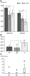
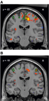
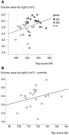
To what extent does musical practice change the structure of the brain? In order to understand how long-lasting musical training changes brain structure, 20 male right-handed, middle-aged professional musicians and 19 matched controls were investigated. Among the musicians, 13 were pianists or organists with intensive practice regimes. The others were either music teachers at schools or string instrumentalists, who had studied the piano at least as a subsidiary subject, and practiced less intensively. The study was based on T1-weighted MR images, which were analyzed using deformation-based morphometry. Cytoarchitectonic probabilistic maps of cortical areas and subcortical nuclei as well as myeloarchitectonic maps of fiber tracts were used as regions of interest to compare volume differences in the brains of musicians and controls. In addition, maps of voxel-wise volume differences were computed and analyzed. Musicians showed a significantly better symmetric motor performance as well as a greater capability of controlling hand independence than controls. Structural MRI-data revealed significant volumetric differences between the brains of keyboard players, who practiced intensively and controls in right sensorimotor areas and the corticospinal tract as well as in the entorhinal cortex and the left superior parietal lobule. Moreover, they showed also larger volumes in a comparable set of regions than the less intensively practicing musicians. The structural changes in the sensory and motor systems correspond well to the behavioral results, and can be interpreted in terms of plasticity as a result of intensive motor training. Areas of the superior parietal lobule and the entorhinal cortex might be enlarged in musicians due to their special skills in sight-playing and memorizing of scores. In conclusion, intensive and specific musical training seems to have an impact on brain structure, not only during the sensitive period of childhood but throughout life.
Keywords: DBM; MRI; brain plasticity; cerebral cortex; deformation-based morphometry; long-term musical practice; musicians.
Figures


Similar articles
-
Structural neuroplasticity in expert pianists depends on the age of musical training onset.Neuroimage. 2016 Feb 1;126:106-19. doi: 10.1016/j.neuroimage.2015.11.008. Epub 2015 Nov 14. Neuroimage. 2016. PMID: 26584868
-
Motor cortex and hand motor skills: structural compliance in the human brain.Hum Brain Mapp. 1997;5(3):206-15. doi: 10.1002/(SICI)1097-0193(1997)5:3<206::AID-HBM5>3.0.CO;2-7. Hum Brain Mapp. 1997. PMID: 20408216
-
Instrument specific use-dependent plasticity shapes the anatomical properties of the corpus callosum: a comparison between musicians and non-musicians.Front Behav Neurosci. 2014 Jul 16;8:245. doi: 10.3389/fnbeh.2014.00245. eCollection 2014. Front Behav Neurosci. 2014. PMID: 25076879 Free PMC article.
-
The brain of musicians. A model for functional and structural adaptation.Ann N Y Acad Sci. 2001 Jun;930:281-99. Ann N Y Acad Sci. 2001. PMID: 11458836 Review.
-
Brain Plasticity and the Concept of Metaplasticity in Skilled Musicians.Adv Exp Med Biol. 2016;957:197-208. doi: 10.1007/978-3-319-47313-0_11. Adv Exp Med Biol. 2016. PMID: 28035567 Review.
Cited by
-
Increased Callosal Thickness in Early Trained Opera Singers.Brain Topogr. 2025 Aug 7;38(5):56. doi: 10.1007/s10548-025-01134-x. Brain Topogr. 2025. PMID: 40772978 Free PMC article.
-
Enriching activities during childhood are associated with variations in functional connectivity patterns later in life.Neurobiol Aging. 2021 Aug;104:92-101. doi: 10.1016/j.neurobiolaging.2021.04.002. Epub 2021 Apr 20. Neurobiol Aging. 2021. PMID: 33984626 Free PMC article.
-
Musical Activity During Life Is Associated With Multi-Domain Cognitive and Brain Benefits in Older Adults.Front Psychol. 2022 Aug 25;13:945709. doi: 10.3389/fpsyg.2022.945709. eCollection 2022. Front Psychol. 2022. PMID: 36092026 Free PMC article.
-
Motor Cortical Plasticity to Training Started in Childhood: The Example of Piano Players.PLoS One. 2016 Jun 23;11(6):e0157952. doi: 10.1371/journal.pone.0157952. eCollection 2016. PLoS One. 2016. PMID: 27336584 Free PMC article.
-
Investigating Neuroanatomical Features in Top Athletes at the Single Subject Level.PLoS One. 2015 Jun 16;10(6):e0129508. doi: 10.1371/journal.pone.0129508. eCollection 2015. PLoS One. 2015. PMID: 26079870 Free PMC article.
References
LinkOut - more resources
Full Text Sources
Other Literature Sources

