doi: 10.5489/cuaj.411.
Malignant peripheral nerve sheath tumour of the renal parenchyma presenting as a fast growing atypical renal cyst
Affiliations
- PMID: 24069105
- PMCID: PMC3776038
- DOI: 10.5489/cuaj.411
Item in Clipboard
Malignant peripheral nerve sheath tumour of the renal parenchyma presenting as a fast growing atypical renal cyst
Can Urol Assoc J.
2013 Sep-Oct.
Abstract
Malignant peripheral nerve sheath tumours (MPNST) of the kidney are very rare, with only 3 cases reported in the English and French literature. However, we report the first case of fast growing atypical renal cyst where a magnetic resonance imaging was an interesting adjunct to the computed tomography scan in imaging this rare tumour.
Figures
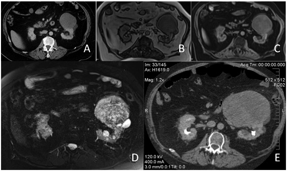
(A) Contrast enhanced computed tomography (CT) scan shows a 8-cm left renal lesion with a density of 13 Hounsfield units and slight peripheral rim enhancement. (B) T1-weighted magnetic resonance image reveals a slightly hypointense left renal lesion (C) which shows peripheral and heterogeneous internal enhancement following gadolinium administration. (D) On T2-weighted image, the left renal lesion appears heterogeneously hyperintense, with a small and very hyperintense (cystic) posterior component. (E) There is a significant growth to 14 cm of the left renal lesion on a follow-up contrast enhanced CT scan done 3 months later. The lesion shows heterogeneous internal enhancement and displaces the kidney posteriorly.
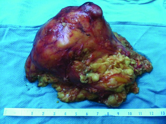
Gross pathology specimen removed by a transperitoneal laparoscopic approach. Ruler graduated in inches.
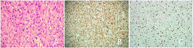
(A) Microscopic imaging (hematoxylin and eosin stain; 40×) of the tumoural proliferation composed of spindle cells with moderate cellularity and nuclear atypia (B) Immunohistochemical study demonstrating strong S-100 immunoreactivity consistent with the diagnosis of malignant peripheral nerve sheath tumours. (C) 40% to 50% of the tumour cells were positive for Ki-67. Tumour cells were negative for muscle markers.
References
LinkOut - more resources
Full Text Sources
Other Literature Sources