. 2013 Sep 12;8(9):e72995.
doi: 10.1371/journal.pone.0072995. eCollection 2013.

Bortezomib-induced painful peripheral neuropathy: an electrophysiological, behavioral, morphological and mechanistic study in the mouse

Affiliations

Bortezomib-induced painful peripheral neuropathy: an electrophysiological, behavioral, morphological and mechanistic study in the mouse

Valentina A Carozzi et al. PLoS One. .

Abstract

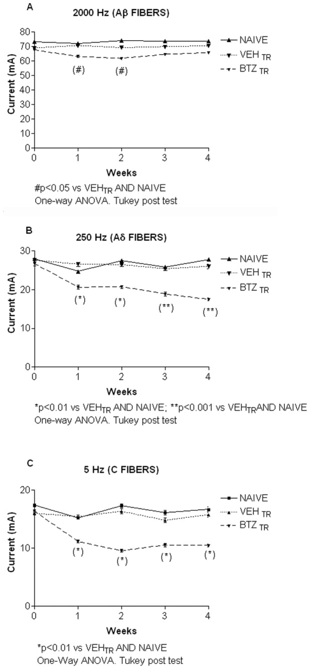
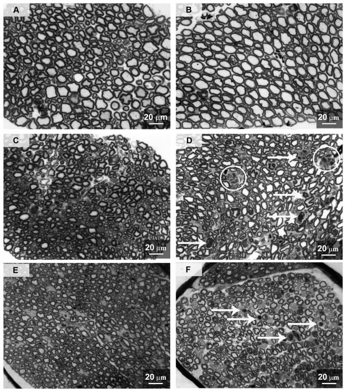
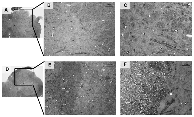
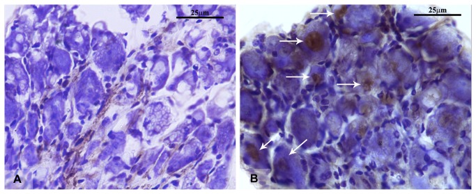
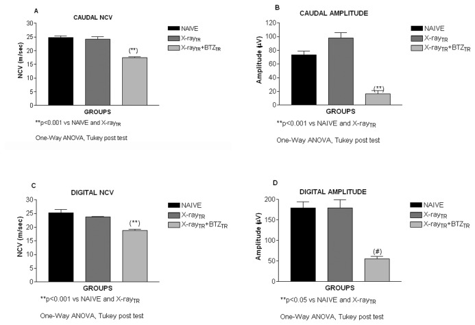
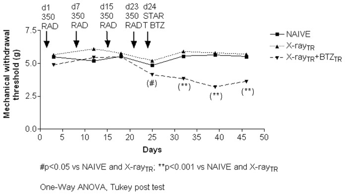
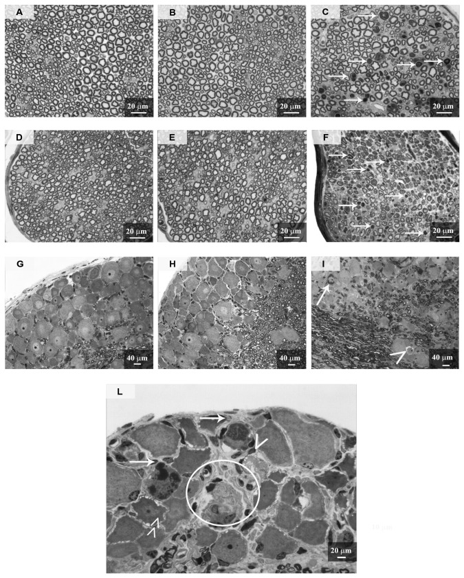
Bortezomib is the first proteasome inhibitor with significant antineoplastic activity for the treatment of relapsed/refractory multiple myeloma as well as other hematological and solid neoplasms. Peripheral neurological complications manifesting with paresthesias, burning sensations, dysesthesias, numbness, sensory loss, reduced proprioception and vibratory sensitivity are among the major limiting side effects associated with bortezomib therapy. Although bortezomib-induced painful peripheral neuropathy is clinically easy to diagnose and reliable models are available, its pathophysiology remains partly unclear. In this study we used well-characterized immune-competent and immune-compromised mouse models of bortezomib-induced painful peripheral neuropathy. To characterize the drug-induced pathological changes in the peripheral nervous system, we examined the involvement of spinal cord neuronal function in the development of neuropathic pain and investigated the relevance of the immune response in painful peripheral neuropathy induced by bortezomib. We found that bortezomib treatment induced morphological changes in the spinal cord, dorsal roots, dorsal root ganglia (DRG) and peripheral nerves. Neurophysiological abnormalities and specific functional alterations in Aδ and C fibers were also observed in peripheral nerve fibers. Mice developed mechanical allodynia and functional abnormalities of wide dynamic range neurons in the dorsal horn of spinal cord. Bortezomib induced increased expression of the neuronal stress marker activating transcription factor-3 in most DRG. Moreover, the immunodeficient animals treated with bortezomib developed a painful peripheral neuropathy with the same features observed in the immunocompetent mice. In conclusion, this study extends the knowledge of the sites of damage induced in the nervous system by bortezomib administration. Moreover, a selective functional vulnerability of peripheral nerve fiber subpopulations was found as well as a change in the electrical activity of wide dynamic range neurons of dorsal horn of spinal cord. Finally, the immune response is not a key factor in the development of morphological and functional damage induced by bortezomib in the peripheral nervous system.


Conflict of interest statement

Competing Interests: The authors have declared that no competing interests exist.

Figures

Figure 1
Figure 1. Flow chart of experiment 2.
In experiment 2, animals were exposed to X-Ray irradiation on day 1, 7, 15 and 23. Twenty-four hours after the last irradiation, animals started the 4-week period of bortezomib chemotherapy. The Dynamic Aesthesiometer Test was performed on day 3, 12, 18, 25, 32, 46 and the cytofluorimetric analysis of PB and BM CD45 positive cells on days 26, 40 and 46. On day 46 animals underwent the neurophysiologic analysis and, once euthanized, the sample collection for the neuropathological analysis.
Figure 2
Figure 2. Body weight, experiment 1.
Body weights were measured twice a week to monitor bortezomib general toxicity. Ten days after the first injection through the end of the experimental period, bortezomib caused a significant decrease in the body weight of drug-treated mice compared to the naïve and to the vehicle treated ones.
Figure 3
Figure 3. Neurophysiology, experiment 1.
Nerve Conduction Velocity (NCV) and action potential amplitudes were tested with an electromyographic apparatus in the caudal and digital nerves two days after the last bortezomib administration. Bortezomib induced a significant decrease in the caudal (A) and digital (C) NCV compared to the naïve and vehicle-treated mice. Bortezomib induced a statistically significant reduction in the caudal action potential amplitude (B). There was no difference between naïve/vehicle and bortezomib-treated animals in the digital action potential amplitude (D).
Figure 4
Figure 4. Mechanical and thermal thresholds, experiment 1.
The mice were tested weekly using the Dynamic Aesthesiometer Test and the incremental hot/cold plate. Bortezomib induced a significant decrease in the mechanical threshold to induce a paw withdrawal at one week, which persisted through week 4 (A). There was no difference between naïve, vehicle and bortezomib-treated mice in the response threshold to a heat (B) and cold (D) stimulus.
Figure 5
Figure 5. Electrophysiology of the spinal cord, experiment 1.
Bortezomib increased the activity of wide dynamic range neurons in the spinal dorsal horn. Mice were tested for neuronal excitability during stimulation of the left hind paw with a brush and von Frey filaments (0.4, 1 and 4 grams). Bortezomib induced an increase of the number of spikes per second (spikes/s) compared to naïve and vehicle-treated mice in response to innocuous stimulation with a brush (A) and 0.4g filament (B), to moderate stimulation with the 1 g filament (C) and noxious stimulation with the 4 g (D) filament.
Figure 6
Figure 6. Neurometer, experiment 1.
Bortezomib decreased the current perception threshold. The mice were treated with bortezomib or vehicle and tested weekly using the Neurometer to determine the amount of current needed at each frequency (5 Hz, C fibers; 250 Hz, Aδ fibers; 2000 Hz, Aβ fibers) to elicit a paw withdrawal. There was a small but significant decrease in the amount of current needed at 2000 Hz to elicit a paw withdrawal at weeks 1 and 2 of treatment, which resolved back to baseline in weeks 3-4 (A). Bortezomib induced a significant decrease in the amount of current needed at 250 Hz to elicit a paw withdrawal at one week, which persisted through week 4 (B). Bortezomib induced a significant decrease in the amount of current needed at 5 Hz to elicit a paw withdrawal at one week, which persisted through week 4 (C).
Figure 7
Figure 7. Morphological analysis of ventral/dorsal roots and caudal nerve, experiment 1.
Dorsal and ventral roots and caudal nerves were collected at sacrifice and processed for light microscopy. A: vehicle ventral root, B: bortezomib ventral root, C: vehicle dorsal root, D: bortezomib dorsal root, E: vehicle caudal nerve, F: bortezomib caudal nerve. Bortezomib did not alter the morphology of the nerve fibers in the ventral roots (B). Bortezomib induced some morphological alterations in the axoplasm and myelin of the nerve fibers in dorsal roots (arrows in D), sometimes leading to advanced axonal degeneration (circles in D). Moderate to severe axonal degeneration of the myelinated and unmyelinated fibers was evident in the caudal nerves of bortezomib-treated animals (arrows in F). Ventral root, dorsal root and caudal nerve of vehicle-treated mice did not manifest any morphological alterations.
Figure 8
Figure 8. Morphological analysis of lumbar spinal cord, experiment 1.
Lumbar spinal cords from vehicle- (A, B, C) and bortezomib-treated (D-E-F) mice were collected at the time of sacrifice and processed for light microscopy. No evident alterations of dorsal horn neurons were found while sporadic axonal degeneration was present in the myelinated fibers of the dorsal column of bortezomib-treated mice (arrows in E and F) compared to the vehicle-treated ones (B-C).
Figure 9
Figure 9. ATF3 immunolabeling, experiment 1.
DRG were collected at the time of sacrifice for the immunohistochemical detection of the nuclear transcription factor ATF3. Thick (15 µm) cryostat sections of DRG were collected from vehicle (A) and bortezomib-treated (B) animals. Bortezomib treatment induced a marked nuclear ATF3 expression in DRG sensory neurons (arrows in B). No ATF3-positive nuclei were present in DRG of vehicle-treated mice.
Figure 10
Figure 10. Body weight, experiment 2.
Body weights were measured twice a week to monitor X-Ray irradiation and bortezomib-induced toxicity. X-Ray-treated animals showed an initial slight body weight decrease on day 4 and 8 then resumed a normal growth trend similar to naïve animals. Starting from the second bortezomib administration (day 32) animals showed a marked and statistically significant decrease of body weight that persisted till the end of the experimental period.
Figure 11
Figure 11. Bone marrow and peripheral blood and bone marrow white blood cells (WBC) count and FACS analysis, experiment 2.
Black arrows indicate the treatment given to mice, such as the X-ray irradiation doses (350 RAD induction and 100 RAD maintenance), and bortezomib administrations. For each group of animals, the WBC concentration within the BM (A) or peripheral blood (B) is indicated for the corresponding analysis time point. The number of hematopoietic cells in the peripheral blood expressing the pan-leukocyte antigen CD45 is also reported (C). A comparable immune-suppression was observed in both X-Ray and X-Ray-bortezomib-treated animals after the irradiation induction and maintenance doses, as shown by the decreased cellularity and leukopenia in the BM and spleen of irradiated mice, regardless of bortezomib treatment.
Figure 12
Figure 12. Neurophysiology, experiment 2.
Nerve Conduction Velocity (NCV) and action potential amplitudes were tested with an electromyographic instrument in the caudal and digital nerves two days after last bortezomib administration. X-Ray irradiation alone did not alter the NCV while bortezomib induced a significant decrease of the caudal (A) and digital (C) NCV compared to naïve and X-Ray animals. X-Ray irradiation alone did not alter the action potential amplitude while bortezomib induced a statistically significant reduction of the caudal (B) and digital (D) action potential amplitude compared to naïve and X-Ray animals.
Figure 13
Figure 13. Mechanical threshold, experiment 2.
The mice were tested weekly using the Dynamic Aesthesiometer instrument. X-Ray alone did not induce any alteration in the withdrawal threshold to mechanical stimulation while bortezomib induced a significant decrease in the mechanical threshold starting from one day after the first injection (day 25) and persisting through the end of the pharmacological treatment.
Figure 14
Figure 14. Morphological analysis of sciatic and caudal nerves and DRG, experiment 2.
DRG, sciatic and caudal nerves were collected at sacrifice and processed for light microscopy. A: naïve sciatic nerve, B: X-Ray sciatic nerve, C: X-Ray + bortezomib sciatic nerve, D: naïve caudal nerve, E: X-Ray caudal nerve, F: X-Ray + bortezomib caudal nerve, G: naïve DRG, H: X-Ray DRG, I, L: X-Ray + bortezomib DRG. No morphological alterations were evident in naïve and X-Ray-treated mice. Bortezomib induced moderate to severe morphological alterations in the axoplasm and myelin of the nerve fibers in sciatic (C) and caudal (D) nerves (arrows). Bortezomib induced the formation of dark inclusions and clear vacuolization in the cytoplasm of sensory neurons (arrows and arrowheads respectively in L), leading in some cases to neuronal degeneration (circle in L). Bortezomib determined sporadic vacuolization of cytoplasm in satellite cells (double arrowhead in L). DRG=Dorsal Root Ganglia.

Similar articles

Cited by

References

    1. Adams J, Palombella VJ, Sausville EA, Johnson J, Destree A et al. (1999) Proteasome inhibitors: a novel class of potent and effective antitumor agents. Cancer Res 59: 2615-2622. PubMed: 10363983. - PubMed
    1. Curran MP, McKeage K (2009) Bortezomib: a review of its use in patients with multiple myeloma. Drugs 69: 859-888. doi:10.2165/00003495-200969070-00006. PubMed: 19441872. - DOI - PubMed
    1. Mahindra A, Laubach J, Raje N, Munshi N, Richardson PG et al. (2012) Latest advances and current challenges in the treatment of multiple myeloma. Nat Rev Clin Oncol 9: 135-143. doi:10.1038/nrclinonc.2012.15. PubMed: 22349016. - DOI - PubMed
    1. Tobinai K (2007) Proteasome inhibitor, bortezomib, for myeloma and lymphoma. Int J Clin Oncol 12: 318-326. doi:10.1007/s10147-007-0695-5. PubMed: 17929113. - DOI - PubMed
    1. Kennedy B, Gargoum F, Bystricky B, Curran DR, O’Connor TM (2010) Novel agents in the management of lung cancer. Curr Med Chem 17: 4291-4325. doi:10.2174/092986710793361289. PubMed: 20939812. - DOI - PubMed

Publication types

MeSH terms