. 2013 Sep 19;8(9):e73118.
doi: 10.1371/journal.pone.0073118. eCollection 2013.

Intranasal delivery of plasma and platelet growth factors using PRGF-Endoret system enhances neurogenesis in a mouse model of Alzheimer's disease

Affiliations

Intranasal delivery of plasma and platelet growth factors using PRGF-Endoret system enhances neurogenesis in a mouse model of Alzheimer's disease

Eduardo Anitua et al. PLoS One. .

Abstract

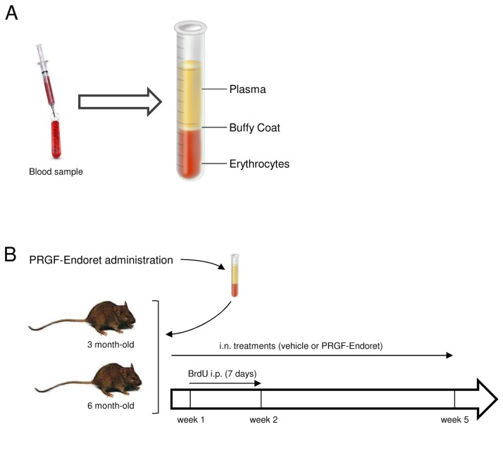
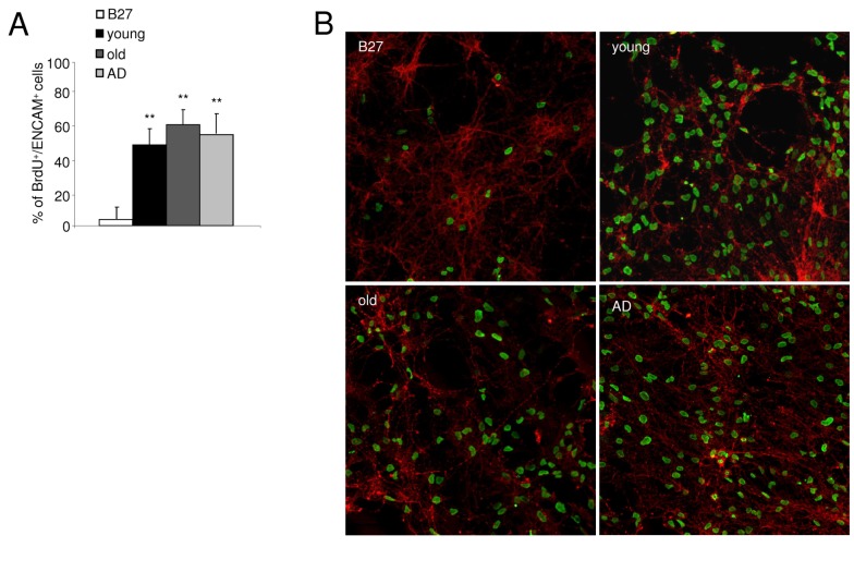
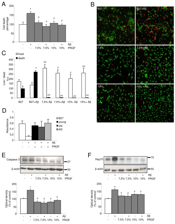
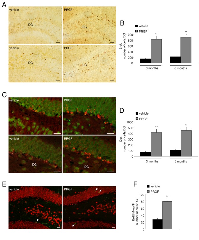
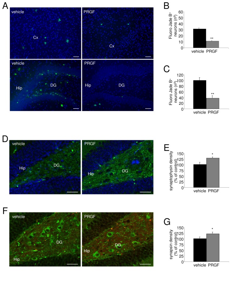
Neurodegeneration together with a reduction in neurogenesis are cardinal features of Alzheimer's disease (AD) induced by a combination of toxic amyloid-β peptide (Aβ) and a loss of trophic factor support. Amelioration of these was assessed with diverse neurotrophins in experimental therapeutic approaches. The aim of this study was to investigate whether intranasal delivery of plasma rich in growth factors (PRGF-Endoret), an autologous pool of morphogens and proteins, could enhance hippocampal neurogenesis and reduce neurodegeneration in an amyloid precursor protein/presenilin-1 (APP/PS1) mouse model. Neurotrophic and neuroprotective actions were firstly evident in primary neuronal cultures, where cell proliferation and survival were augmented by Endoret treatment. Translation of these effects in vivo was assessed in wild type and APP/PS1 mice, where neurogenesis was evaluated using 5-bromodeoxyuridine (BdrU), doublecortin (DCX), and NeuN immunostaining 5 weeks after Endoret administration. The number of BrdU, DCX, and NeuN positive cell was increased after chronic treatment. The number of degenerating neurons, detected with fluoro Jade-B staining was reduced in Endoret-treated APP/PS1 mice at 5 week after intranasal administration. In conclusion, Endoret was able to activate neuronal progenitor cells, enhancing hippocampal neurogenesis, and to reduce Aβ-induced neurodegeneration in a mouse model of AD.


Conflict of interest statement

Competing Interests: The authors have declared that no competing interests exist. EA, SP and GO are members of a foundation that investigates the potential of plasma rich in growth factors in Neurology. This does not alter the authors' adherence to PlOS ONE policies on sharing data and materials.

Figures

Figure 1
Figure 1. Use of Endoret treatment in a mouse model of AD.
A. Scheme of the different plasma fractions obtained with the Endoret technology from blood samples. B. Experimental design for the Endoret treatment in APP/PS1 mice.
Figure 2
Figure 2. Effects of Endoret on proliferation of primary cultured neurons.
A. Fluorescence microscopy images showing BrdU-labeling cells in B27- and Endoret-treated primary cultured neurons. B. Quantitative analysis of BrdU-labeling cells. C Calbindin immunocytochemistry shows an increase in the presence of calbindin-positive neurons in PRGF-treated primary cultured neurons than in B27-treated control group. D. The histogram shows quantitation of calbindin absorbance in each experimental group. E. Representative confocal microscopy images showing co-localization of ENCAM (green) with BrdU (red) in B27- and Endoret-treated primary cultured neurons. F. Quantitative analysis of double ENCAM (green) and BrdU (red)-labeling cells. One representative experiment is shown (n = 3 experiments). Data are mean ± SEM; *p<0.05, **p<0.01 vs control culture.
Figure 3
Figure 3. In vitro effects of Endoret from young, old and AD patient groups.
A. Endoret obtained from different donors (healthy young and old donors and old patients with AD) protects against Ab1-42 (10µM)-induced cell death in neuronal cell culture. B. Effects of Endoret on proliferation of primary cultured neurons. Representative confocal microscopy images showing co-localization of ENCAM (green) with BrdU (red) after exposure to 7.5% Endoret from young, elderly, and AD groups. One representative experiment is shown (n = 3 experiments). Data show mean ± SEM; **p<0.01 vs control culture.
Figure 4
Figure 4. Endoret inhibits apoptotic signaling pathway in Ab-stimulated neuronal cultures.
A. 7.5 and 10% Endoret treatment protects against Ab1-42 (10 µM)-induced cell death in neuronal cell culture assayed with a cell death ELISA. B. Fluorescent images of living cells (green) and dead cells (red) in neuronal cultures in control condition (B27), and after addition of Ab1-42 (10 µM), and 7.5 and 10% Endoret. C. Quantification of Ab1-42-induced cell death after 48 h in vitro. Representative blots and quantitative analysis of (D) caspase-3, and (E) HSP-70 expression in neuronal cultures treated with Aβ42 (10 µM) and 7.5 and 10% Endoret. One representative experiment is shown (n = 3 experiments). Data show mean ± SEM; *p<0.05 and **p<0.01 vs control culture. #p<0.05 and # #p<0.01 vs Aβ42 -treated culture.
Figure 5
Figure 5. Endoret treatment modulates neurogenesis in APP/PS1 mice.
A. Representative fields of BrdU immunostaining in the hippocampal dentate gyrus (DG) of 3 (upper) and 6 months of age (bottom) APP/PS1 mice treated with vehicle or Endoret. Scale bar = 20µm. B. Quantification of BrdU-positive cells after vehicle or Endoret intranasal administration in 3 and 6 month-old APP/PS1 mice. C. Representative confocal microscopy images DG of brain sections immunostained for DCX (red) in combination with NeuN (green) shown in 3 (upper) and 6-month old (bottom) APP/PS1 mice treated with vehicle or Endoret. Scale bar = 20µm. D. Quantification of neurogenesis in 3 and 6 month-old APP/PS1 mice. E. Representative confocal microscopy images DG of brain sections immunostained for BrdU (green) and NeuN (red) in 6 month-old APP/PS1 mice. Arrows point to merged signals. Scale bar = 20µm. F. Quantification of the relative number of BrdU and NeuN double positive cells. n = 9 mice per group. Data show mean ± SEM; **p<0.01 vs APP/PS1 + vehicle.
Figure 6
Figure 6. Endoret treatment decreased neurodegeneration in the brain of APP/PS1 mice.
A. Representative photomicrographs showing degenerating neurons by Fluoro-Jade B staining in the cerebral cortex (Cx), and hippocampus (Hip) of 6 month-old APP/PS1 mice treated with vehicle and Endoret. Scale bar=20µm. Densitometric analysis of the number (left histogram) and size (in % right histogram) of Fluoro-Jade B-positive plaques in the cerebral frontal cortex (B) and the hippocampus (C) of APP/PS1 mice treated with vehicle and Endoret. D. Representative immunoblots of caspase-3 and synaptic proteins extracted from hippocampal synaptosome preparations. Densitometric quantification of changes in gray values expressed as mean ± SEM (vehicle-treated group, or control, is indicated as 100%). (D) Synaptophysin (green) in combination with DAPI-staining nuclei (blue), and (F) synapsin (red) in combination with bIII-Tubulin (green) labeling in hippocampus of APP/PS1 mice treated with vehicle and PRGF. Quantification of the effect of Endoret administration on synaptophysin (E) and synapin (G) immunoreactivity in hippocampus of APP/PS1 mice. n= 9 mice per group. Data show mean ± SEM; *p<0.05, **p<0.01 vs APP/PS1 + vehicle. Scale bar = 20µm.

References

    1. Querfurth HW, LaFerla FM (2010) Alzheimer’s disease. N Engl J Med 362: 329-344. doi:10.1056/NEJMra0909142. PubMed: 20107219. - DOI - PubMed
    1. Zhang C, McNeil E, Dressler L, Siman R (2007) Long-lasting impairment in hippocampal neurogenesis associated with amyloid deposition in a knock-in mouse model of familial Alzheimer’s disease. Exp Neurol 204: 77-87. doi:10.1016/j.expneurol.2006.09.018. PubMed: 17070803. - DOI - PMC - PubMed
    1. Verret L, Jankowsky JL, Xu GM, Borchelt DR, Rampon C (2007) Alzheimer’s-type amyloidosis in transgenic mice impairs survival of newborn neurons derived from adult hippocampal neurogenesis. J Neurosci 27: 6771-6780. doi:10.1523/JNEUROSCI.5564-06.2007. PubMed: 17581964. - DOI - PMC - PubMed
    1. Ermini FV, Grathwohl S, Radde R, Yamaguchi M, Staufenbiel M et al. (2008) Neurogenesis and alterations of neural stem cells in mouse models of cerebral amyloidosis. Am J Pathol 172: 1520-1528. doi:10.2353/ajpath.2008.060520. PubMed: 18467698. - DOI - PMC - PubMed
    1. Rodríguez JJ, Jones VC, Tabuchi M, Allan SM, Knight EM et al. (2008) Impaired adult neurogenesis in the dentate gyrus of a triple transgenic mouse model of Alzheimer’s disease. PLOS ONE 3: e2935. doi:10.1371/journal.pone.0002935. PubMed: 18698410. - DOI - PMC - PubMed

MeSH terms