. 2013 Sep 12;8(9):e74706.
doi: 10.1371/journal.pone.0074706. eCollection 2013.

Mu opioid receptors on primary afferent nav1.8 neurons contribute to opiate-induced analgesia: insight from conditional knockout mice

Affiliations

Mu opioid receptors on primary afferent nav1.8 neurons contribute to opiate-induced analgesia: insight from conditional knockout mice

Raphaël Weibel et al. PLoS One. .

Abstract

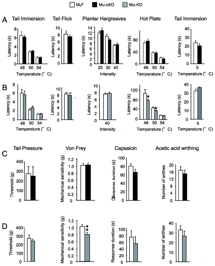
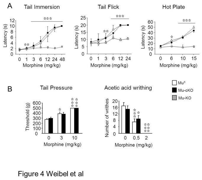
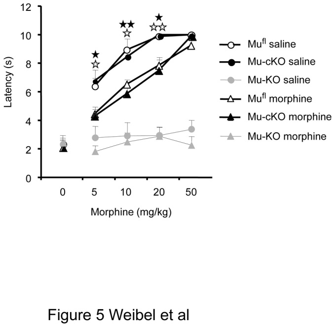
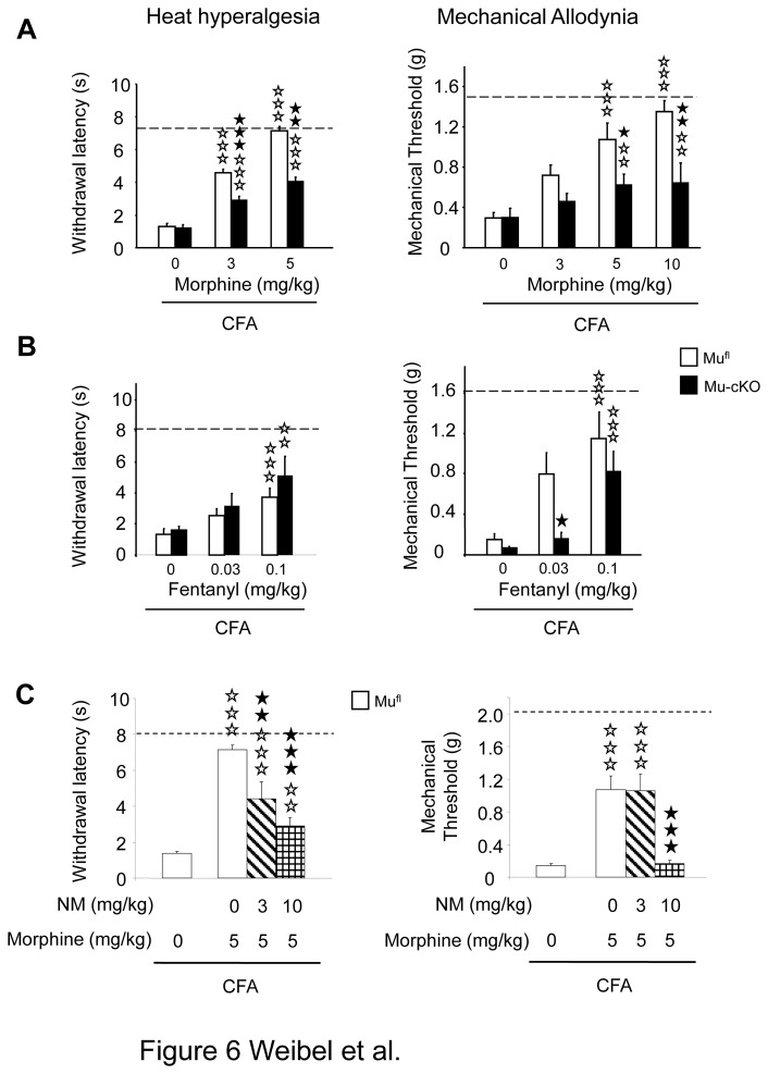
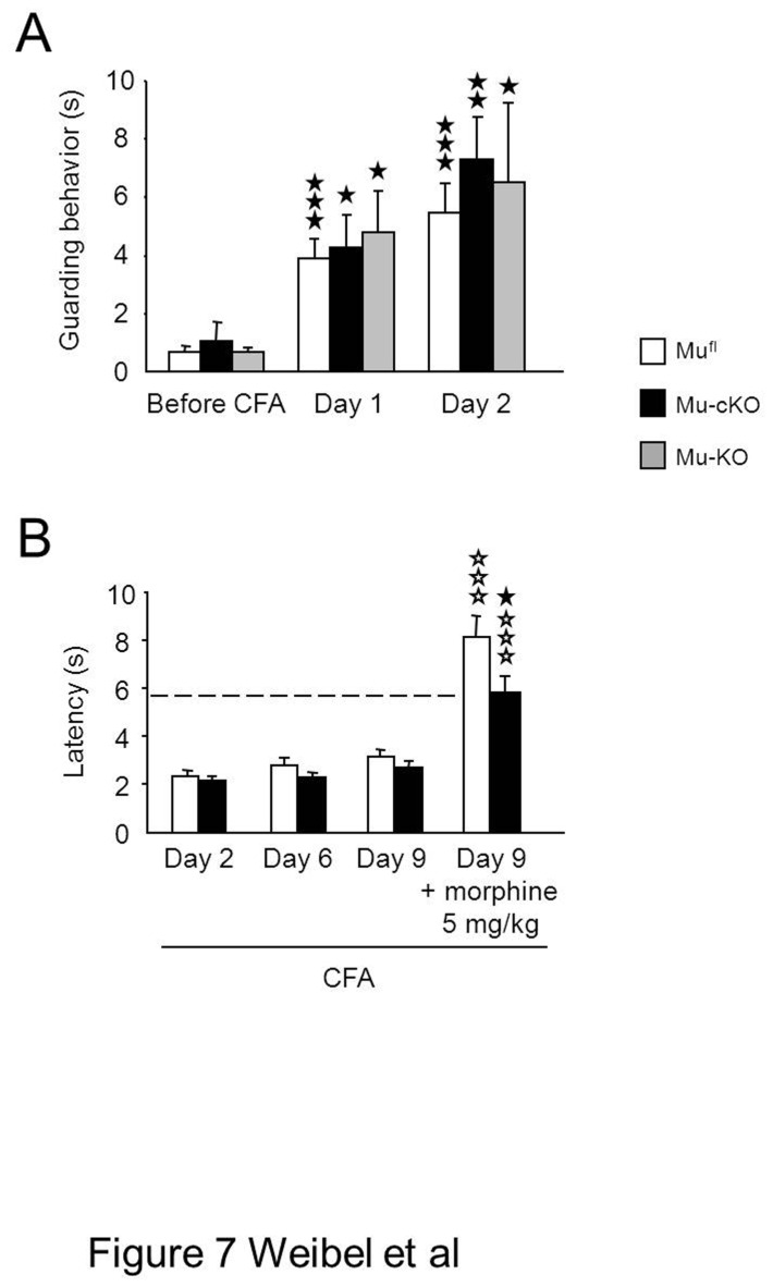
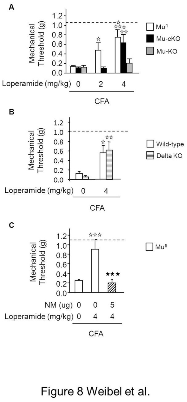
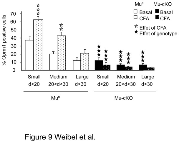
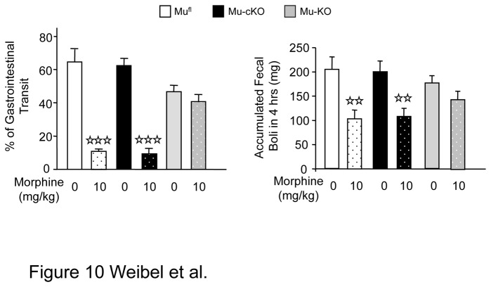
Opiates are powerful drugs to treat severe pain, and act via mu opioid receptors distributed throughout the nervous system. Their clinical use is hampered by centrally-mediated adverse effects, including nausea or respiratory depression. Here we used a genetic approach to investigate the potential of peripheral mu opioid receptors as targets for pain treatment. We generated conditional knockout (cKO) mice in which mu opioid receptors are deleted specifically in primary afferent Nav1.8-positive neurons. Mutant animals were compared to controls for acute nociception, inflammatory pain, opiate-induced analgesia and constipation. There was a 76% decrease of mu receptor-positive neurons and a 60% reduction of mu-receptor mRNA in dorsal root ganglia of cKO mice. Mutant mice showed normal responses to heat, mechanical, visceral and chemical stimuli, as well as unchanged morphine antinociception and tolerance to antinociception in models of acute pain. Inflammatory pain developed similarly in cKO and controls mice after Complete Freund's Adjuvant. In the inflammation model, however, opiate-induced (morphine, fentanyl and loperamide) analgesia was reduced in mutant mice as compared to controls, and abolished at low doses. Morphine-induced constipation remained intact in cKO mice. We therefore genetically demonstrate for the first time that mu opioid receptors partly mediate opiate analgesia at the level of Nav1.8-positive sensory neurons. In our study, this mechanism operates under conditions of inflammatory pain, but not nociception. Previous pharmacology suggests that peripheral opiates may be clinically useful, and our data further demonstrate that Nav1.8 neuron-associated mu opioid receptors are feasible targets to alleviate some forms of persistent pain.


Conflict of interest statement

Competing Interests: The authors have declared that no competing interests exist.

Figures

Figure 1
Figure 1. Generation of mu opioid receptor conditional knockout mice.
(A) The conditional Oprm1 (Oprm1 fl) allele was created by homologous recombination. The scheme shows the wild-type Oprm1 allele, the targeting vector, targeted allele and conditional allele obtained after excision of Hygror by a Cre recombinase treatment of ES cells. The Oprm1 fl conditional allele - or “floxed” allele - harbors two loxP sites flanking the Oprm1 exon 2 and 3. Black boxes, exons; Mf, Mfe1; Sa, SalI, Sp, Spe1 restriction sites; white triangles, loxP sites; Hygro box, floxed hygromycin-resistance cassette, grey box, probe for Southern blot analysis. Dash lines indicate expected labeled DNA fragments in Southern blot analysis. (B) Southern blot analysis of wild-type and targeted alleles in ES cells. Genomic DNA was digested using Mfe1 and hybridized to a 3’ external probe, shown in 1A. The expected bands at 8.5 and 15.7 kb were obtained. (C) Conditional mutant mice. Right part shows the Oprm1 fl conditional allele and excised allele (deletion of exons 2 & 3) after intercrossing Oprm1 fl/fl mice with Nav1.8-Cre mice. A and B indicate PCR primers used to detect gene excision, and C & D PCR primers for the floxed allele. PCR shows exon 2-3 deletion in DRGs but not brain of mu-cKO mice. In DRGs, the two bands result from gene excision in Nav1.8+ neurons but not in other Nav1.8-negative cells. Mu-KO mice show full deletion in both DRGs and brain. (D) Conditional knockout of mu opioid receptor gene in DRGs but not brain. Quantitative RT-PCR was used to measure Oprm1 mRNA levels from mufl, mu-cKO and mu-KO mice. Oprm1 mRNA expression was normalized to mufl control samples, and is decreased in mu-cKO animals. Oprm1 transcripts were undetectable in DRG and brain from mu-KO animals. ★★ P<0.01, mu-cKO vs mufl controls. (E) Conditional KO of the mu opioid receptor gene occurs in small/medium DRG cells. Left, representative in situ hybridization on DRG sections from mufl, mu-cKO and mu-KO mice. Thin, medium and large arrows point to small, medium and large cells, respectively. Scale bar = 100 µm. Right, cell size distribution of Oprm1-positive neurons in DRGs. The % of Oprm1-positive neurons in control and mu-cKO DRGs are shown in white and black, respectively. The % of Oprm1-positive cells is significantly reduced in small and medium, but not large diameter (>700 µm2) neurons from mu-cKO mice. ★★★ P<0.001 mu-cKO vs mufl controls, Student t-test.
Figure 2
Figure 2. Mu opioid agonist-induced [35S]-GTPγS binding is comparable on spinal cord membrane preparation from mu-cKO and mufl mice.
Spinal cord membranes were incubated in the absence or presence of the mu opioid agonist DAMGO (10-9-10-4 M) in assay buffer containing [35S] GTPγS. Basal level (100%) represents the specific [35S]-GTPγS binding in the absence of agonist. DAMGO significantly increases [35S]-GTPγS binding, in a comparable manner for mu-cKO and mufl mice. [35S]-GTPγS binding was absent on spinal cord membranes from mu-KO mice, and was decreased by half with a 50%-50% mix of membranes from mufl and mu-KO animals, containing half mu receptors as compared to mufl membranes, indicating that this assay allows to detect reduced receptor expression. Results are presented as means ± sem of 5-6 experiments on 5 distinct membrane preparations. ★★★ P<0.001, ★ P<0.05 50%-50% mu-cKO, mufl or 50%-50% mix vs mu-KO, Student t-test.
Figure 3
Figure 3. Acute pain responses are unchanged in conditional mu-cKO mice.
(A) Acute thermal responses were similar in mu-cKO mice and mufl controls in the tail immersion test at 48, 50 and 54°C (n=15/genotype), tail flick test (n=10/genotype), Hargreaves test at three different intensities (n=10/genotype), hot plate test (48, 50 and 54°C, n=15 /genotype) and cold tail immersion test at 5°C (n=19/genotype). (B) In the experimental conditions of (A), mu receptor total knockout mice displayed higher sensitivity in the hot plate assay only (conventional KO vs WT, n =11-14/genotype, ★ P<0.05) whereas they behaved as controls for heat tail immersion (n=11-14/genotype), tail flick (n=12/genotype), Hargreaves (n=20/genotype) and cold (5°C) tail immersion (n=12-13/genotype) tests. (C) Nociceptive responses to mechanical and chemical stimuli were unchanged in the conditional mutant mice when assessed in the tail pressure (n = 8/genotype) or von Frey filaments (n = 10-13/genotype) test, nocifensive responses to capsaicin (n=10/genotype) and abdominal writhing induced by acetic acid (n=13-14/genotype). (D) In the same experimental conditions as in (C), mu receptor total knockout mice were more sensitive than control mice in the von Frey filaments test for touch perception (conventional KO vs WT, n = 33-57/genotype, ★★ P<0.01, Student t-test). Total knockout mice behaved as controls in the tail pressure test (n = 8/genotype), nocifensive responses to capsaicin (n=6/genotype) and abdominal writhing induced by acetic acid (n=4/genotype).
Figure 4
Figure 4. Conditional mu-cKO and control mice show comparable systemic morphine analgesia in nociceptive assays.
Top. Morphine induced dose-dependent antinociception in both mu-cKO and mufl mice in the three heat assays, (A) tail immersion, (B) tail flick and (C) hot plate. Morphine-induced analgesia was abolished in conventional mu-KO animals (tail immersion, n=10/genotype; tail flick, n=10-14/genotype; hot plate, n=6-17/ genotype), confirming the selective effect of morphine on mu receptor. Bottom. Mu-cKO and mufl control mice show similar systemic morphine analgesia in the tail pressure (n=9-13/genotype) and acetic acid-induced visceral nociceptive (n=13-14/genotype) assays. Two-way ANOVA, post-hoc Fisher test for individual time points, ✰ P <0.05, ✰P<0.01, ✰✰✰ P<0.001, morphine vs saline.
Figure 5
Figure 5. The conditional deletion of mu receptor in Nav1.8 primary neurons does not abrogate tolerance to morphine-induced antinociception.
Morphine dose-dependent antinociception was measured following repeated 4-day i.p. injections of morphine or saline in mufl controls, mu-cKO and mu-KO animals. The shift to right for both mufl and mu-cKO chronic-morphine animals indicates the development of a comparable tolerance to analgesia. Total mu-KO animals show no antinociception. n=6-7/genotype/treatment, two-way ANOVA (genotype x treatment F(1,30) = 19.919, P <0.001 for treatment; F(2,30) = 97.039, P <0.001 for genotype); post-hoc Fisher test for individual morphine doses, ✰ P <0.05, ✰✰ P<0.01, chronic morphine (tolerance) vs chronic saline in mufl mice; ★ P<0.05, ★★ P<0.00 chronic morphine (tolerance) vs chronic saline in mu-cKO mice.
Figure 6
Figure 6. Conditional mu-cKO mice show decreased opiate-induced analgesia in the CFA-induced inflammatory pain model.
(A) Two days after CFA, morphine (i.p.) dose-dependently reduced heat and mechanical hypersensitivities in mufl control mice. This analgesia was diminished in mu-cKO mice. Dashed lines represent baseline (pre-CFA) sensitivities. White bars, mufl mice; black bars, mu-cKO mice. For plantar test, n=13-20/genotype. two-way ANOVA (genotype x treatment F(1,90) = 75.336, P <0.001 for treatment; F(1,90) = 23.313, P <0.001 for genotype. Post-hoc Bonferroni test, ✰✰✰ P<0.001, morphine vs saline; ★★ P<0.01, ★★★ P<0.001 cKO vs flox controls. For Von Frey filaments test, n=7-18/genotype. two-way ANOVA (genotype x treatment F(1,91) = 41.573, P <0.001 for treatment; F(1,91) = 27.378, P <0.001 for genotype. Post-hoc Bonferroni test, ✰✰ P<0.01, ✰✰✰ P<0.001 morphine vs saline; ★ P<0.05, ★★ P<0.01 mu-cKO vs mufl controls. (B) Fentanyl produced a dose-dependent analgesia in mufl control mice 2 days after CFA. Mu-cKO mice displayed a decreased analgesic response in the mechanical sensitivity test for the 0.03 mg/kg fentanyl dose. n=5-10/genotype. For plantar test, two-way ANOVA (genotype X treatment F(1,54) = 8.979, P <0.001 for treatment, P= 0.19 for genotype), post-hoc Bonferroni test; ✰✰ P <0.01, ✰✰✰ P<0.001, fentanyl 0.1 mg/kg vs saline; for fentanyl 0.03 mg/kg, P = 0.0675 in mufl animals, P = 0.21 in mu-cKO animals. For Von Frey test, two-way ANOVA (genotype X treatment F(1,45) = 22.802, P <0.001 for treatment, F(1,45) = 9.316, P <0.01 for genotype). Post-hoc Bonferroni test for treatment, ✰✰✰ P<0.001, fentanyl 0.1 mg/kg vs saline; for fentanyl 0.03 mg/kg, P = 0.0575 mufl animals, P = 0.28 mu-cKO animals. Post-hoc Fisher test for genotype, ★ P<0.05, mu-cKO vs mufl mice. (C) Morphine-induced analgesia is reduced by systemic administration of the peripheral antagonist naloxone methiodide (NM). Morphine (5 mg/kg) induced an antihyperalgesic effect in mufl mice for both heat and mechanical responses. Systemic NM diminished morphine-induced analgesia. Heat hypersensitivity, one-way ANOVA for treatment F(3,42) = 29.778, P<0.001 ; post-hoc Bonferroni test, ✰✰ P <0.01, ✰✰✰ P <0.001 morphine vs saline; ★★ P<0.01 ★★★ P<0.001 NM + morphine vs morphine. Mechanical hypersensitivity, one-way ANOVA for treatment F(3,52) = 23.467, P<0.001 ; post-hoc Bonferroni test, ✰✰✰ P <0.001 morphine vs saline; ★★★ P<0.001 NM + morphine vs morphine.
Figure 7
Figure 7. Spontaneous guarding pain behavior after paw-CFA and CFA-inflammatory pain at day 9 in conditional mu-cKO mice.
(A) The effect the conditional mutation on ongoing pain behavior was evaluated by quantifying the duration of guarding behavior over 6 min in mufl, mu-cKO and mu-KO mice before CFA-induced inflammation and at days 1 and 2 post-CFA. All mouse lines showed the same behavior (mufl, n=14; mu-cKO, n=6; mu-KO, n=12). Results are expressed as means ± sem. ★ P<0.05, ★★ P<0.01, ★★★ P<0.001 post-CFA vs naïve. (B) Following CFA injection into tail, mu-cKO and mufl mice showed similar heat hyperalgesia at days 2, 6 and 9. The dashed line represents baseline (pre-CFA) sensitivity in the tail immersion tests at 48°C. Morphine (i.p.) produced anti-hyperalgesia in both genotypes, and that was reduced in mu-cKO mice as compared to controls. n=19/genotype., two-way ANOVA (genotype x treatment F(1,71) = 48.812, P <0.001 for treatment; F(1,71) = 5.999, P <0.05 for genotype. Post-hoc Bonferroni test, ✰✰✰ P<0.001, morphine vs saline; ★ P<0.05, cKO vs flox controls.
Figure 8
Figure 8. Conditional mu-cKO mice show decreased analgesia to the peripheral opiate loperamide in the paw inflammatory pain model.
(A) Two-days after CFA, loperamide (s.c.) dose-dependently reduced mechanical hypersensitivity of mufl control mice. Analgesia produced by 2mg/kg loperamide was diminished in the conditional mutant mice and 4mg/kg loperamide induced no analgesia in the full mu-KO mice. Dashed lines represent baseline (pre-CFA) sensitivity. White bars, mufl mice; black bars, mu-cKO mice; grey bars mu-KO mice; n=4-10/genotype. Two-way ANOVA for mufl and mu-cKO mice (genotype X treatment F(1,78) = 17.508, P <0.001 for treatment; P = 0.0657 for genotype), post-hoc Bonferroni test, ✰ P <0.05, ✰✰✰ P <0.001, loperamide vs saline. (B) Loperamide-induced antihyperalgesia was comparable in control and delta opioid receptor total KO mice; n=9-13/genotype. White bars, control mice; stripped bars, delta receptor KO mice, two-way ANOVA (genotype X treatment F(1,38) = 16.363, P <0.001 for treatment; P = 0.959 for genotype), post-hoc Fisher test, ✰ P <0.05, ✰✰ P <0.01, loperamide vs saline. (C) Analgesia produced by 4 mg/kg loperamide in mufl mice was abolished by pretreatment with the peripheral antagonist naloxone methiodide (NM); n=11/group, white bars, mufl mice; hatched bars, NM-pretreated mufl mice; one-way ANOVA for treatment F(2,42) = 16.925, P <0.001 for treatment; post-hoc Bonferroni test , ✰✰✰ P <0.001, loperamide vs saline; ★★★ P<0.001, loperamide vs NM + loperamide.
Figure 9
Figure 9. Inflammation increased the number of small/medium Oprm1-positive neurons in DRGs of mufl but not of mu-cKO mice.
Inflammation was induced by intra-paw CFA as in previous figures. The cell size distribution of Oprm1-positive neurons in DRGs was evaluated by In Situ Hybridization. The % of Oprm1-positive neurons in naïve mufl and mu-cKO DRGs are shown in white and black, respectively. The % of Oprm1-positive neurons in ipsilateral DRGs of CFA mufl and mu-cKO DRGs are shown in dotted white and black bars. ✰✰ P <0.01, ✰✰✰ P <0.001, CFA vs naïve; ★★★ P<0.001 mu-cKO vs mufl, Student t-test.
Figure 10
Figure 10. Morphine-induced constipation is maintained in conditional mu-cKO mice.
Left: Morphine-induced inhibition of small intestinal transit. Mice were treated with saline (0 mg/kg morphine bars) or morphine (10 mg/kg) and 20 min. later given a charcoal gavage. For determination of small intestine transit, the distance travelled by charcoal was measured relative to the total length of the small intestine. White bars, mufl mice; black bars, mu-cKO mice; grey bars, mu-KO mice, n=5/group, two-way ANOVA (genotype X treatment F(2,24) = 18.206, P <0.001 for treatment; P = 0.184 for genotype), post-hoc Fisher test, ✰✰✰ P <0.001, morphine vs saline. Right: Morphine-induced inhibition of fecal boli accumulation. Mice were administered morphine or saline and fecal boli were collected after 4 hrs. n=10-12/group, two-way ANOVA (genotype X treatment F(2,62) = 21.138, P <0.001 for treatment; P = 0.934 for genotype), post-hoc Fisher test, ✰✰ P <0.01, morphine vs saline.

Similar articles

Cited by

References

    1. Le Merrer J, Becker JA, Befort K, Kieffer BL (2009) Reward processing by the opioid system in the brain. Physiol Rev 89: 1379-1412. doi:10.1152/physrev.00005.2009. PubMed: 19789384. - DOI - PMC - PubMed
    1. Mansour A, Fox CA, Akil H, Watson SJ (1995) Opioid-receptor mRNA expression in the rat CNS: anatomical and functional implications. Trends Neurosci 18: 22-29. doi:10.1016/0166-2236(95)93946-U. PubMed: 7535487. - DOI - PubMed
    1. Lutz PE, Kieffer BL (2013) The multiple facets of opioid receptor function: implications for addiction. Curr Opin Neurobiol, 23: 473–9. PubMed: 23453713. - PMC - PubMed
    1. Stein C, Machelska H (2011) Modulation of peripheral sensory neurons by the immune system: implications for pain therapy. Pharmacol Rev 63: 860-881. doi:10.1124/pr.110.003145. PubMed: 21969325. - DOI - PubMed
    1. Ossipov MH, Dussor GO, Porreca F (2010) Central modulation of pain. J Clin Invest 120: 3779-3787. doi:10.1172/JCI43766. PubMed: 21041960. - DOI - PMC - PubMed

Publication types

MeSH terms

Substances

LinkOut - more resources