. 2013 Sep 17;8(9):e74712.
doi: 10.1371/journal.pone.0074712. eCollection 2013.

A degenerative retinal process in HIV-associated non-infectious retinopathy

Affiliations

A degenerative retinal process in HIV-associated non-infectious retinopathy

Igor Kozak et al. PLoS One. .

Abstract

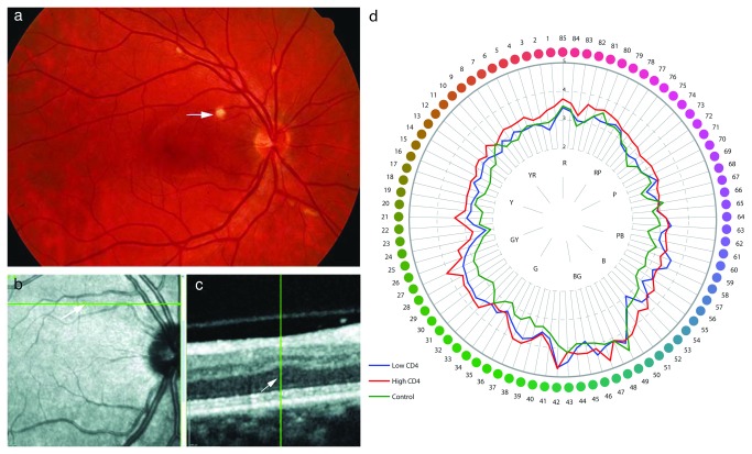
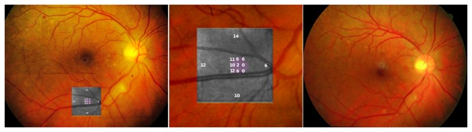
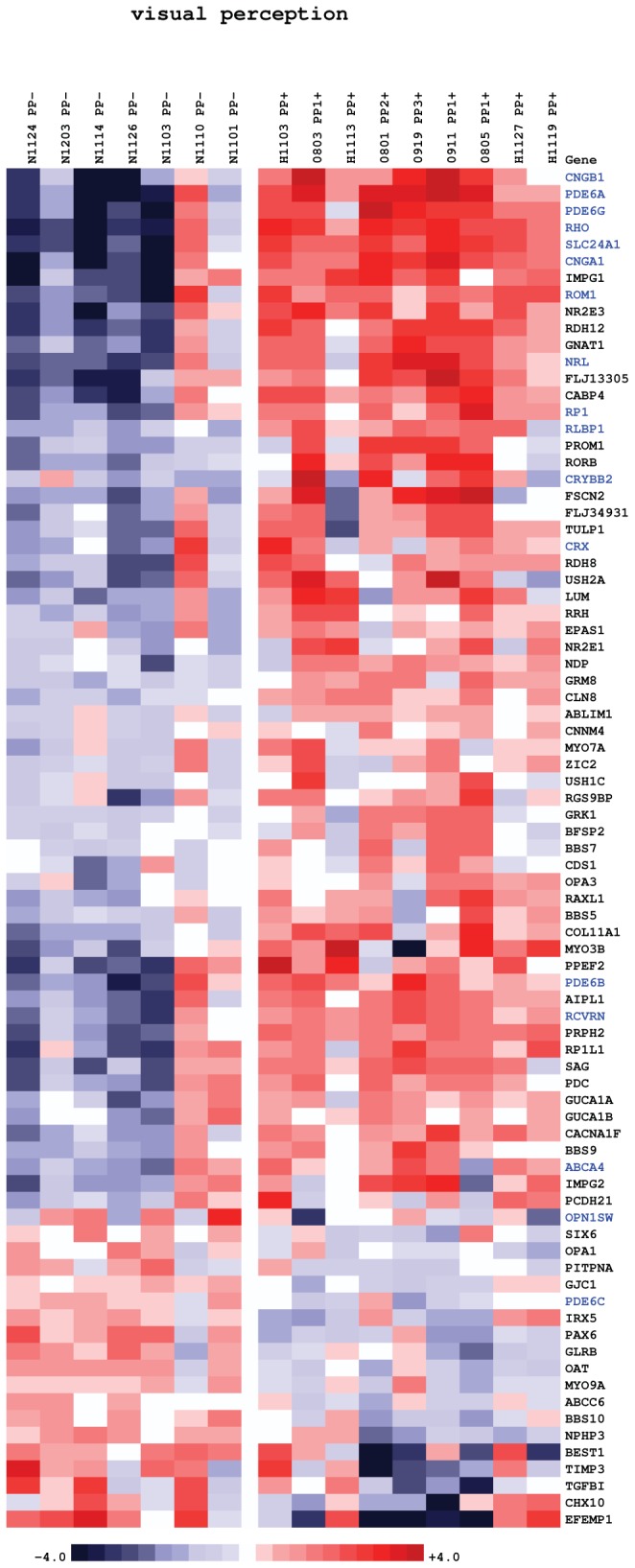
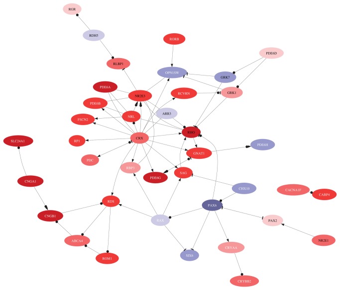
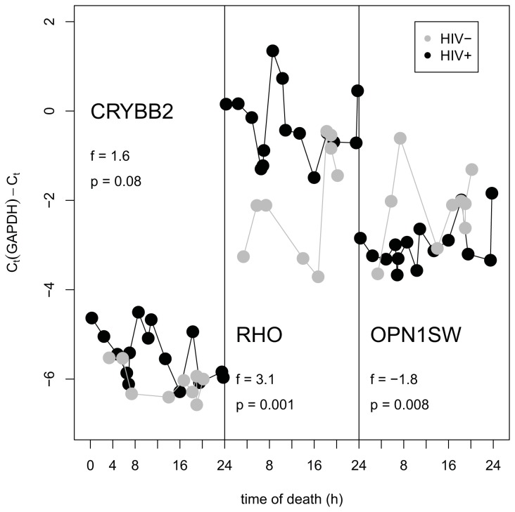
HIV retinopathy is the most common non-infectious complication in the eyes of HIV-positive individuals. Oncotic lesions in the retinal nerve fiber layer, referred to as cotton wool spots (CWS), and intraretinal (IR) hemorrhages are frequently observed but are not unique to this pathology. HIV-positive patients have impaired color vision and contrast sensitivity, which worsens with age. Evidence of inner-retinal lesions and damage have been documented ophthalmoscopically, however their long term structural effect has not been investigated. It has been hypothesized that they may be partially responsible for loss of visual function and visual field. In this study we utilized clinical data, retinal imaging and transcriptomics approaches to comprehensively interrogate non-infectious HIV retinopathy. The methods employed encompassed clinical examinations, fundus photography, indirect ophthalmoscopy, Farmsworth-Munsell 100 hue discrimination testing and Illumina BeadChip analyses. Here we show that changes in the outer retina, specifically in the retinal pigment epithelium (RPE) and photoreceptor outer segments (POS) contribute to vision changes in non-infectious HIV retinopathy. We find that in HIV-positive retinae there is an induction of rhodopsin and other transcripts (including PDE6A, PDE6B, PDE6G, CNGA1, CNGB1, CRX, NRL) involved in visual transduction, as well as structural components of the rod photoreceptors (ABCA4 and ROM1). This is consistent with an increased rate of renewal of rod outer segments induced via increased phagocytosis by HIV-infected RPE previously reported in culture. Cone-specific transcripts (OPN1SW, OPN1LW, PDE6C, PDE6H and GRK7) are uniformly downregulated in HIV positive retina, likely due to a partial loss of cone photoreceptors. Active cotton wool spots and intraretinal hemorrhages (IRH) may not affect photoreceptors directly and the interaction of photoreceptors with the aging RPE may be the key to the progressive vision changes in HIV-positive patients.


Conflict of interest statement

Competing Interests: The authors have declared that no competing interests exist.

Figures

Figure 1
Figure 1. Oncotic lesions (CWS) in the retinal nerve fiber layer were investigated.
After resolution permanent damage remained to the inner retinal layer. A: Color photograph of the retina of an HIV-positive patient with non-infectious retinopathy. Cotton wool spots (arrow) are present along the retinal blood vessels. B: Infrared image of the same retina 18 years after resolution of the cotton wool spot with the arrow pointing to its previous location. The green line represents high-resolution spectral-domain optical coherence tomography (SD-OCT) sectioning through the ischemic spot co-localizing it to an adjacent B-scan. C: Adjacent B-scan showing all retinal layers. The hyper-reflective layer on the top is the nerve fiber layer, original location of the cotton wool spot, which shows thinning. There is a compensatory shift of outer retinal layers including the outer nuclear layer towards the inner retina (arrowhead). D: Farnsworth-Munsell diagram showing averaged data for all three groups of eyes. Each group - the group with high CD4, the group with low CD4, and the control group – was significantly different from every other group (p < 0.0001 in each comparison).
Figure 2
Figure 2. Example of the CWS microperimetric abnormality demonstrating long term-damage.
Left Panel: Microperimetry examination overlaid on color fundus photograph, at the area of the previous cotton wool spot. An intraretinal hemorrhage is present superotemporal to the macula and a CWS is inferior to the optic nerve. Middle Panel: Enlarged image of microperimetry overlay showing sensitivity values in dB over the area of CWS shows decreased sensitivity (lower numbers in violet) over the area of previous cotton wool spot demonstrating long-term damage. Right Panel: Color photograph of the retina of the same eye with a dot hemorrhage superotemporal to the macula and fading cotton wool spot.
Figure 3
Figure 3. Heatmap of gene expression changes between posterior pole retinal punches of HIV-positive (10 samples on the right) and HIV-negative donor eyes (six samples on the left).
Red and blue boxes indicate relative over- and under-expression with respect to a mean level between the two groups. Only genes with regularized t-statistic > 1 have been included. Symbols in blue indicate genes discussed in text. The samples are ordered by the donors’ time of death from left to right in each group.
Figure 4
Figure 4. Interaction network of genes involved in visual perception using MetaCore™ knowledge base [48].
The color reflects direction and size of regulation in HIV-positive retinae compared to HIV-negative controls. A link signifies interaction, a bullet is binding of protein products, an arrow means regulation of transcription or activation, and a hammer means inhibition or deactivation.
Figure 5
Figure 5. Abundance of transcript measured by qPCR (relative to a reference gene GAPDH) in retinae of HIV-negative and HIV-positive donors as function of time of death.

References

    1. Lansky A, Brooks JT, DiNenno E, Heffelfinger J, Hall HI et al. (2010) Epidemiology of HIV in the United States. JAIDS. J Acquir Immune Defic Syndr, 53: 55–61: S64-S68 doi:10.1097/QAI.1090b1013e3181fbbe1015. PubMed: 19927003. - DOI - PubMed
    1. Campsmith ML, Rhodes PH, Hall HI, Green TA (2010) Undiagnosed HIV prevalence among adults and adolescents in the United States at the end of 2006. J Acquir Immune Defic Syndr 53: 619-624. PubMed: 19838124. - PubMed
    1. Jaffe HW, Valdiserri RO, De Cock KM (2007) The reemerging HIV/AIDS epidemic in men who have sex with men. JAMA 298: 2412-2414. doi:10.1001/jama.298.20.2412. PubMed: 18042919. - DOI - PubMed
    1. Prejean J, Song R, Hernandez A, Ziebell R, Green T et al. (2011) Estimated HIV incidence in the United States, 2006-2009. PLOS ONE 6: e17502 PubMed: 21826193. - PMC - PubMed
    1. Mayer KH, Hamilton CD (2010) Synergistic Pandemics: Confronting the Global HIV and Tuberculosis Epidemics. Clin Infect Dis 50: S67-S70. PubMed: 20397958. - PubMed

Publication types

MeSH terms