

Noise-induced tinnitus using individualized gap detection analysis and its relationship with hyperacusis, anxiety, and spatial cognition
- PMID: 24069375
- PMCID: PMC3771890
- DOI: 10.1371/journal.pone.0075011
Noise-induced tinnitus using individualized gap detection analysis and its relationship with hyperacusis, anxiety, and spatial cognition
Abstract
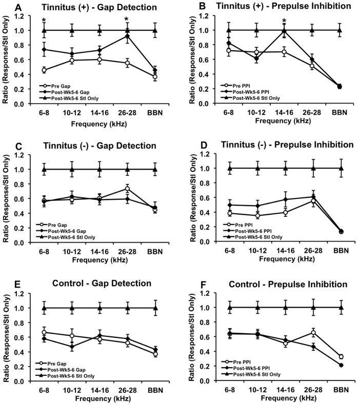
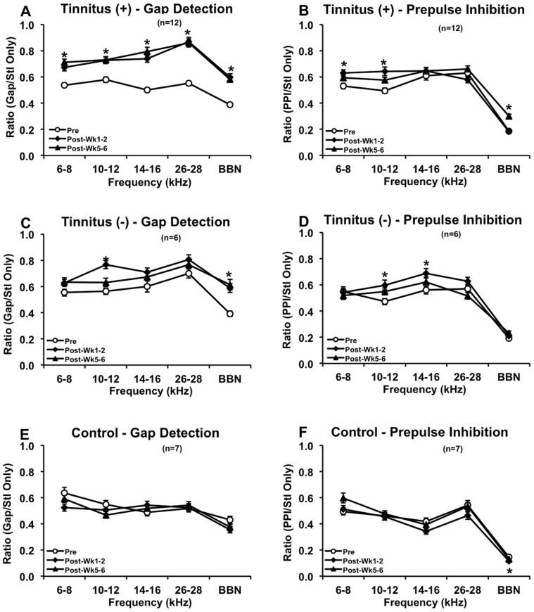
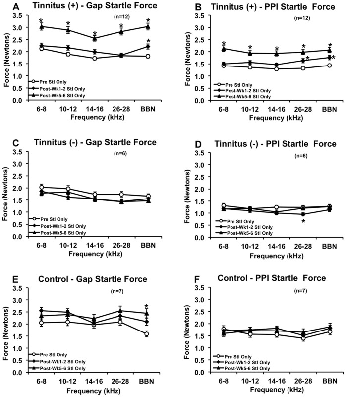
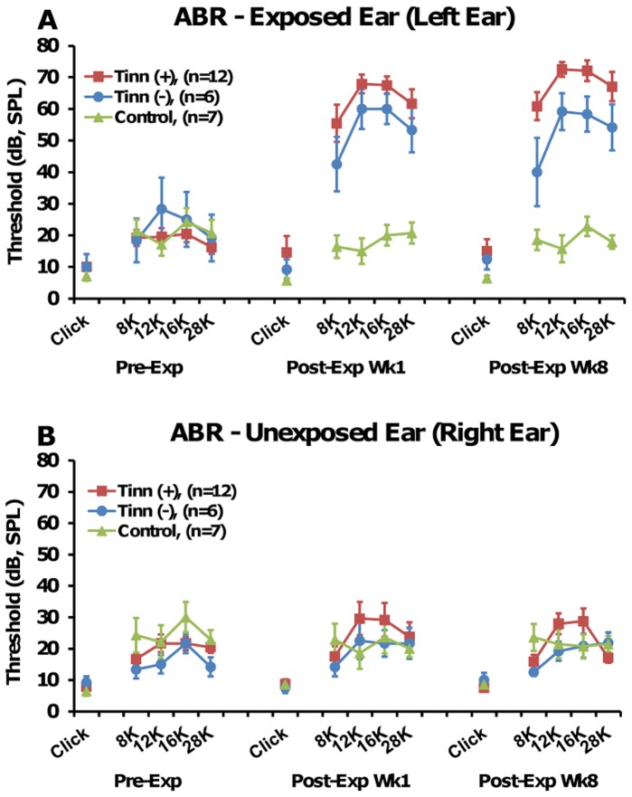
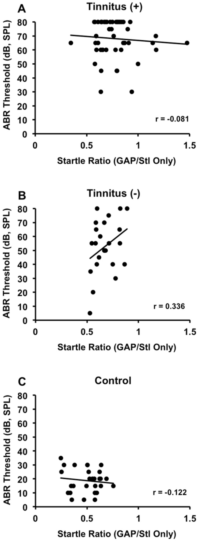
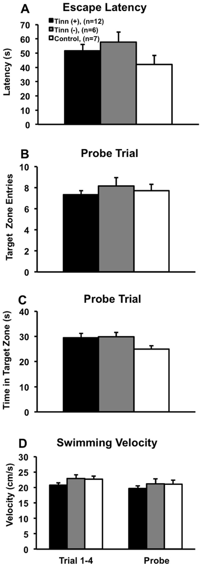
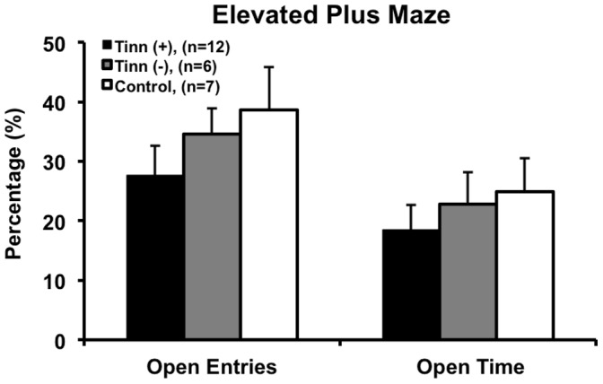
Tinnitus has a complex etiology that involves auditory and non-auditory factors and may be accompanied by hyperacusis, anxiety and cognitive changes. Thus far, investigations of the interrelationship between tinnitus and auditory and non-auditory impairment have yielded conflicting results. To further address this issue, we noise exposed rats and assessed them for tinnitus using a gap detection behavioral paradigm combined with statistically-driven analysis to diagnose tinnitus in individual rats. We also tested rats for hearing detection, responsivity, and loss using prepulse inhibition and auditory brainstem response, and for spatial cognition and anxiety using Morris water maze and elevated plus maze. We found that our tinnitus diagnosis method reliably separated noise-exposed rats into tinnitus((+)) and tinnitus((-)) groups and detected no evidence of tinnitus in tinnitus((-)) and control rats. In addition, the tinnitus((+)) group demonstrated enhanced startle amplitude, indicating hyperacusis-like behavior. Despite these results, neither tinnitus, hyperacusis nor hearing loss yielded any significant effects on spatial learning and memory or anxiety, though a majority of rats with the highest anxiety levels had tinnitus. These findings showed that we were able to develop a clinically relevant tinnitus((+)) group and that our diagnosis method is sound. At the same time, like clinical studies, we found that tinnitus does not always result in cognitive-emotional dysfunction, although tinnitus may predispose subjects to certain impairment like anxiety. Other behavioral assessments may be needed to further define the relationship between tinnitus and anxiety, cognitive deficits, and other impairments.
Conflict of interest statement
Figures
Similar articles
-
Effects of noise exposure on development of tinnitus and hyperacusis: Prevalence rates 12 months after exposure in middle-aged rats.Hear Res. 2016 Apr;334:30-6. doi: 10.1016/j.heares.2015.11.004. Epub 2015 Nov 14. Hear Res. 2016. PMID: 26584761
-
Prolonged Exposure of CBA/Ca Mice to Moderately Loud Noise Can Cause Cochlear Synaptopathy but Not Tinnitus or Hyperacusis as Assessed With the Acoustic Startle Reflex.Trends Hear. 2018 Jan-Dec;22:2331216518758109. doi: 10.1177/2331216518758109. Trends Hear. 2018. PMID: 29532738 Free PMC article.
-
Behavioral evaluation of auditory function abnormalities in adult rats with normal hearing thresholds that were exposed to noise during early development.Physiol Behav. 2019 Oct 15;210:112620. doi: 10.1016/j.physbeh.2019.112620. Epub 2019 Jul 17. Physiol Behav. 2019. PMID: 31325509
-
Psychophysical and neural correlates of noised-induced tinnitus in animals: Intra- and inter-auditory and non-auditory brain structure studies.Hear Res. 2016 Apr;334:7-19. doi: 10.1016/j.heares.2015.08.006. Epub 2015 Aug 20. Hear Res. 2016. PMID: 26299842 Review.
-
Noise: Acoustic Trauma to the Inner Ear.Otolaryngol Clin North Am. 2020 Aug;53(4):531-542. doi: 10.1016/j.otc.2020.03.008. Epub 2020 Apr 30. Otolaryngol Clin North Am. 2020. PMID: 32362563 Review.
Cited by
-
Tinnitus Correlates with Downregulation of Cortical Glutamate Decarboxylase 65 Expression But Not Auditory Cortical Map Reorganization.J Neurosci. 2019 Dec 11;39(50):9989-10001. doi: 10.1523/JNEUROSCI.1117-19.2019. Epub 2019 Nov 8. J Neurosci. 2019. PMID: 31704784 Free PMC article.
-
A Conditioned Behavioral Paradigm for Assessing Onset and Lasting Tinnitus in Rats.PLoS One. 2016 Nov 11;11(11):e0166346. doi: 10.1371/journal.pone.0166346. eCollection 2016. PLoS One. 2016. PMID: 27835697 Free PMC article.
-
Insult-induced adaptive plasticity of the auditory system.Front Neurosci. 2014 May 23;8:110. doi: 10.3389/fnins.2014.00110. eCollection 2014. Front Neurosci. 2014. PMID: 24904256 Free PMC article. Review.
-
Behavioral Animal Model of the Emotional Response to Tinnitus and Hearing Loss.J Assoc Res Otolaryngol. 2018 Feb;19(1):67-81. doi: 10.1007/s10162-017-0642-8. Epub 2017 Oct 18. J Assoc Res Otolaryngol. 2018. PMID: 29047013 Free PMC article.
-
Salicylate-induced hearing loss and gap detection deficits in rats.Front Neurol. 2015 Feb 20;6:31. doi: 10.3389/fneur.2015.00031. eCollection 2015. Front Neurol. 2015. PMID: 25750635 Free PMC article.
References
-
- Adams PF, Marano MA (1995) Current estimates from the National Health Interview Survey, 1994. Vital Health Stat10: 1–260. - PubMed
-
- Shargorodsky J, Curhan GC, Farwell WR (2010) Prevalence and characteristics of tinnitus among US adults. AmJMed 123: 711–718. - PubMed
-
- Hebert S, Fullum S, Carrier J (2011) Polysomnographic and quantitative electroencephalographic correlates of subjective sleep complaints in chronic tinnitus. JSleep Res 20: 38–44. - PubMed
Publication types
MeSH terms
LinkOut - more resources
Full Text Sources
Other Literature Sources
Medical
Miscellaneous
