. 2013 Sep 17;8(9):e75311.
doi: 10.1371/journal.pone.0075311. eCollection 2013.

Role for Krüppel-like transcription factor 11 in mesenchymal cell function and fibrosis

Affiliations

Role for Krüppel-like transcription factor 11 in mesenchymal cell function and fibrosis

Angela Mathison et al. PLoS One. .

Abstract

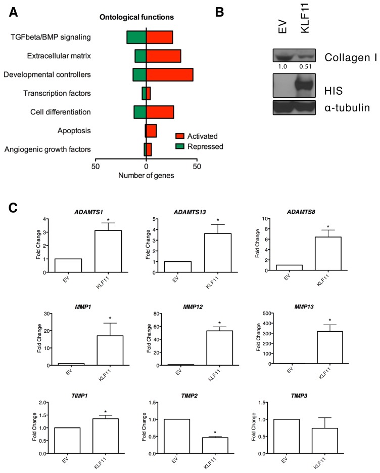
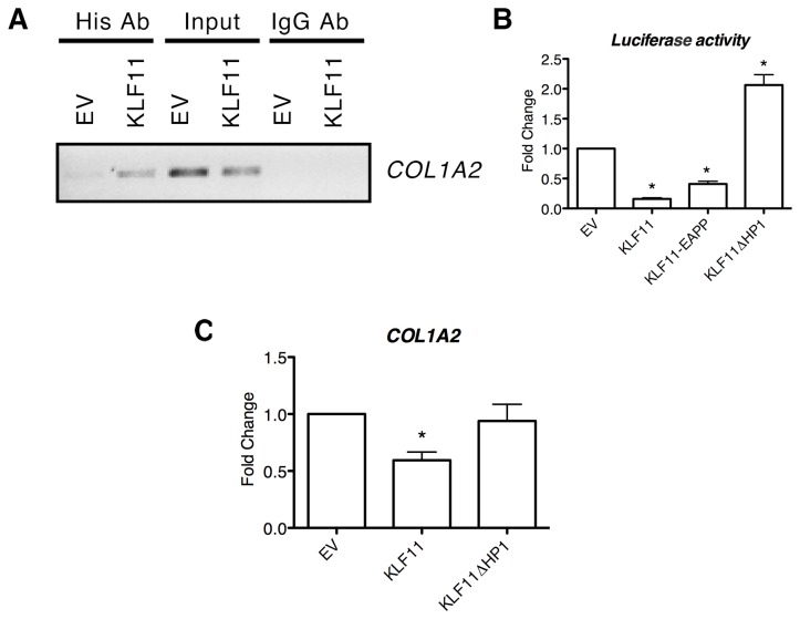
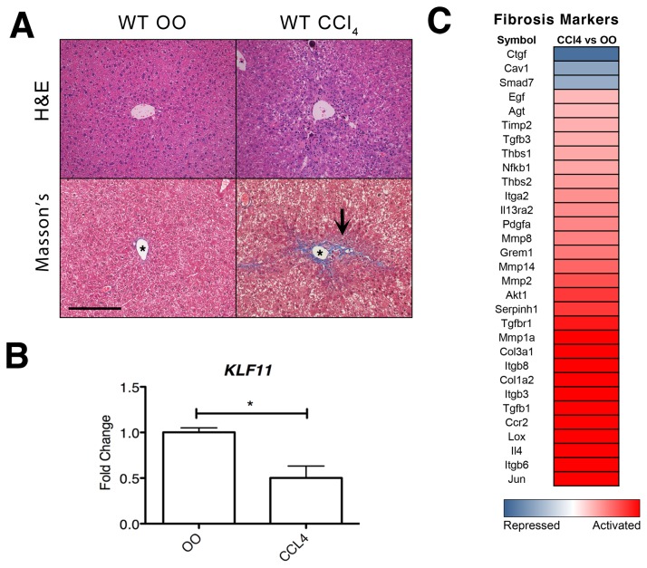
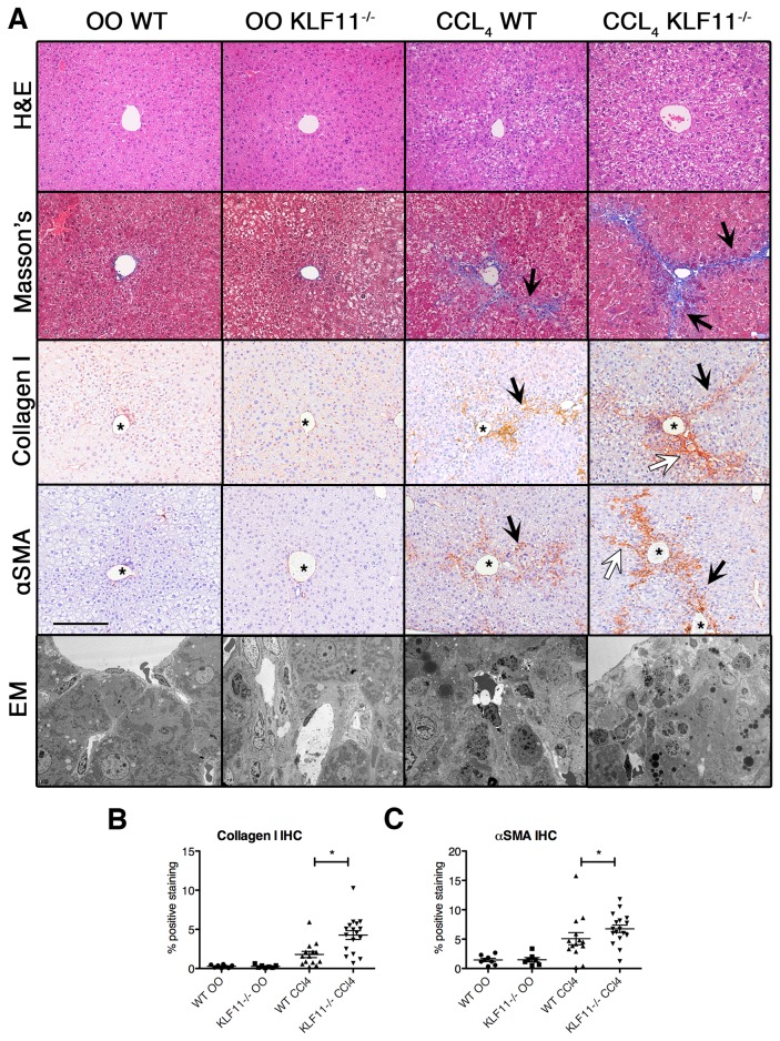
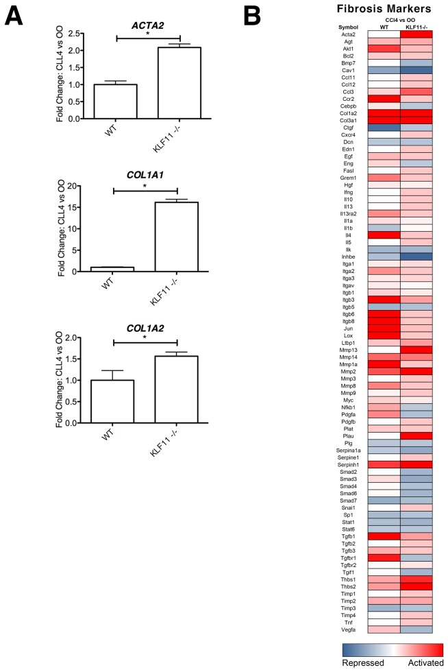
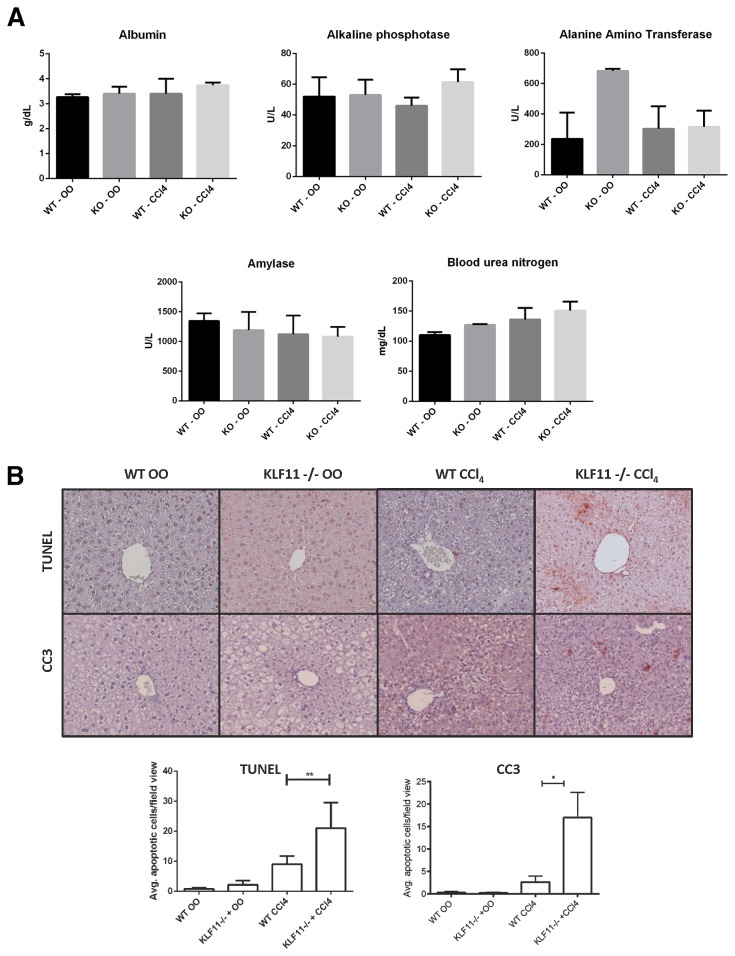
Krüppel-like factor 11 (KLF11) and the highly homologous KLF10 proteins are transcription factors originating from duplication of the Drosophila melanogaster ancestor cabut. The function of these proteins in epithelial cells has been previously characterized. In the current study, we report a functional role for KLF11 in mesenchymal cells and in mesenchymal cell dysfunction, namely, fibrosis, and subsequently perform a detailed cellular, molecular, and in vivo characterization of this phenomenon. We find that, in cultured mesenchymal cells, enhanced expression of KLF11 results in activated extracellular matrix pathways, including collagen gene silencing and matrix metalloproteinases activation without changes in tissue inhibitors of metalloproteinases. Combined, reporter and chromatin immunoprecipitation assays demonstrate that KLF11 interacts directly with the collagen 1a2 (COL1A2) promoter in mesenchymal cells to repress its activity. Mechanistically, KLF11 regulates collagen gene expression through the heterochromatin protein 1 gene-silencing pathway as mutants defective for coupling to this epigenetic modifier lose the ability to repress COL1A2. Expression studies reveal decreased levels of KLF11 during liver fibrogenesis after chemically induced injury in vivo. Congruently, KLF11(-/-) mice, which should be deficient in the hypothesized anti-fibrogenic brake imposed by this transcription factor, display an enhanced response to liver injury with increased collagen fibril deposition. Thus, KLFs expands the repertoire of transcription factors involved in the regulation of extracellular matrix proteins in mesenchymal cells and define a novel pathway that modulates the fibrogenic response during liver injury.


Conflict of interest statement

Competing Interests: The authors have declared that no competing interests exist.

Figures

Figure 1
Figure 1. KLF11 is critical to a variety of biological processes in liver mesenchymal cells.
(A) LX2 cells overexpressing KLF11 elicit a variety of changes in extracellular matrix, TGFβ response, and growth factor genes. The biosynthetic response of 232 unique genes was measured by qPCR arrays (SA Biosciences) in LX2 hepatic stellate cells comparing empty vector (EV) and KLF11 transduced cells after 48 hours. The full list of altered genes may be found in Table S1. The number of genes significantly regulated by KLF11 compared to empty vector control across a variety of ontological categories is represented here. (B) Protein levels of collagen are decreased in response to KLF11 overexpression in LX2 cells. Collagen I levels were measured by western blot in LX2 hepatic stellate cells comparing EV and KLF11 transduced cells after 48 hours of infection. In the same samples, overexpression of His-KLF11 was confirmed by Omni-D8 western blot and α-tubulin confirms equal loading of lysates. Relative densitometry of collagen I levels is shown, normalized to α-tubulin control for each sample. (C) Transcript levels of matrix metalloproteinases are greatly induced while their regulators, TIMPs, are generally unchanged or downregulated. For KLF11 overexpression compared to empty vector: ADAMTS1, 3.12 ± 0.57; ADAMST13, 3.62 ± 0.85; ADAMTS8, 6.41 ±1.35; MMP1, 17.07 ± 7.30; MMP12, 53.08 ± 6.24; MMP13, 318.10 ± 65.98; TIMP1, 1.35 ± 0.14; TIMP2, 0.46 ± 0.04; TIMP3, 0.74 ± 0.31. * p-value <0.05.
Figure 2
Figure 2. Collagen 1 is directly repressed by KLF11 in mesenchymal cells coupled to the HP1-HMT pathway.
(A) To illustrate that KLF11 was a direct regulator of the collagen I promoter, chromatin immunoprecipitation was performed on LX2 cells transduced with empty vector or KLF11. Immunoprecipitation of KLF11 displayed amplification of the collagen 1a2 promoter by PCR, thus verifying the direct binding of KLF11 to the promoter of collagen1a2. Input controls for empty vector and KLF11 are included to ensure equal volume of precipitated DNA as well as the results of amplification from immunoprecipitates using a non-specific IgG antibody. (B) Further evidence that collagen is a direct target of the sequence specific transcription factor KLF11 was observed in luciferase assays. Sections of the collagen 1a2 promoter containing GC consensus sequences were cloned upstream of a luciferase reporter. The reporter gene was significantly repressed upon overexpression of KLF11 in LX2 cells (84 ± 1.94% compared to empty vector, p<0.05). Examination of the effect of overexpression of KLF11-EAPP mutant reveals only a slight change in the repression of the collagen I promoter (41 ± 4.5% repression compared to empty vector, p<0.05). However, overexpression of the KLF11ΔHP1 mutant lead to completion de-repression of the collagen I promoter (206 ± 17% compared to empty vector, p<0.05), indicating that the repression of the gene occurs through the HP1-HMT chromatin remodeling pathway. (C) PCR of collagen 1a2 expression in LX2 cells transduced with empty vector, KLF11, or KLF11ΔHP1 demonstrate that expression of the gene is repressed in the presence of KLF11 (0.59 ± 0.11 fold compared to empty vector) but depressed in the presence of the inactivating KLF11ΔHP1 mutant (0.92 ± 0.14 fold compared to empty vector).
Figure 3
Figure 3. KLF11 is repressed during liver injury caused by chronic CCl4 exposure.
(A) C57Bl/6 wild type mice were treated with CCl4 or olive oil (OO) for a period of six weeks. Liver sections with hematoxylin and eosin (H and E) labeling are provided to demonstrate overall tissue architecture. In CCl4-treated mice, Masson’s trichrome staining indicates increase extracellular matrix accumulation (bright blue) around the central vein. All images are X200 magnification with scale bar equaling 200µm. Asterisk in image denotes central vein. Black arrows denote C-C septa. (B) In wild type mice, levels of KLF11 expression were measured by qPCR. Data are expressed as fold change over olive oil control after normalization to housekeeping genes beta-actin (ACTB) and glyceraldehyde-3-phosphate dehydrogenase (GAPDH). The data are means ± SEM, n=4 to 6 per experimental condition. Mice treated with CCl4 had significantly downregulated (0.5 ± 0.13) levels of KLF11 compared to OO-treated control animals (* = p<0.05). (C) qPCR measured the changes in 31 fibrosis markers in pooled liver samples from wild type mice treated with CCl4 for six weeks normalized to OO control. All genes with a fold change <-2 and >2.0 are considered differentially regulated between the CCl4 and OO treatments. Scaling is from -5 fold change (repressed) to 1 (unchanged) to +5 fold change (activated).
Figure 4
Figure 4. Inactivation of KLF11 correlates with an increased severity of fibrosis.
(A) C57Bl/6 wild type mice were treated with CCl4 or olive oil (OO) control for a period of six weeks. The extent of fibrosis was observed with Masson’s trichrome staining with no fibrosis observed under olive oil treatment in either KLF11-/- or wild type mice. In contrast, fibrosis was observed in CCl4-treated wild type mice as indicated by an increase in extracellular matrix deposition. Collagen I immunohistochemistry reveals similar increases in fibrous septa formation. Similarly, the stellate cell marker α-smooth muscle actin (αSMA) appears increased in CCl4-treated wild type mice compared to OO-treated wild type mice. CCl4-treated KLF11-/- mice display significant increase in αSMA and collagen I labeling as compared to CCl4-treated wild type. There is an increase in the extent of C-C septa formation as well as the presence of C-P septa in CCl4-treated KLF11-/- mice compared to wild type, indicating a more severe fibrotic response to the chemical insult. Electronic microscopy also revealed disrupted cellular architecture in the CCl4-treated KLF11-/- and wild type mice compared to OO-treated controls. All images are X200 magnification with scale bar equaling 200µm. Black arrows indicated C-C septa and white arrows denote C-P septa. Asterisks label the central vein. (B-D) Masson’s trichrome, collagen I, and α-smooth muscle actin (αSMA) staining were quantified using Imaging System KS400 as detailed in the materials and methods. The levels of all markers are significantly increased in the CCl4-treated treated animals and the CCl4-treated KLF11-/- mice have significantly high percentage of αSMA and collagen I staining than the similarly treated wild type animals (* = p<0.05). Each datapoint represents the average of the intensity values of 10 random fieldviews.
Figure 5
Figure 5. Mesenchymal cell activation and fibril deposition are increased in response to chronic CCl4 treatment in the absence of KLF11.
(A) In a subset of CCl4-treated KLF11-/- mice and CCl4-treated wild type mice, ACTA2, COL1A1, and COL1A2 transcript levels were measured by PCR, validating the values obtained by the fibrotic marker array depicted in part B and the data for which appear in Table S2. Each of these genes was found to be upregulated in CCl4-treated KLF11-/- mice as compared to wild type control. Data are expressed as fold change over olive oil control for each genotype after normalization to housekeeping genes beta-actin (ACTB) and glyceraldehyde-3-phosphate dehydrogenase (GAPDH). ACTA2 (KLF11-/-, 2.09 ± 0.10 compared to WT). COL1A1 (KLF11-/-: 16.16 ± 0.72 compared to WT). COL1A2 (KLF11-/-: 1.56 ± 0.10 compared to WT). * = p<0.05. (B) qPCR array measured the changes in 84 fibrosis makers in pooled liver samples from CCl4-treated KLF11-/- mice and CCl4-treated wild type mice normalized to the their respective olive oil (OO) controls. Regardless of the genetic background, CCl4 treatment results in significant upregulation of collagen I. However, a number of TGFβ pathway markers are repressed only in the absence of KLF11, indicating that the fibrogenic response possesses KLF11-dependent and independent mechanisms. Levels of ACTA2 are increased in the absence of KLF11, suggesting that the HSC response is mediated by this transcription factor. Numerical data may be found in Table S2.
Figure 6
Figure 6. KLF11 inactivation does not affect liver function but increases hepatocyte apoptosis.
(A) In a subset of olive oil and CCl4-treated KLF11-/- and wild type mice, sera were screened for a panel of liver injury markers (as detailed in results). No differences were found between wild type and knockout animals under CCl4 treatment for the displayed markers. A significant difference was found between wild type and knockout animals under olive oil treatment owing to a previously characterized defect in fatty acid oxidation in the absence of KLF11. * = p<0.05. (B) Immunohistochemistry for cleaved caspase 3 and TUNEL quantification reveals that CCl4-treated KLF11-/- experienced increased apoptosis, exclusive to hepatocytes, compared to CCl4-treated wild type animals. * = p<0.05. ** = p<0.01. All images appear at X200 magnification with scale bar equaling 200µm.

References

    1. Botella LM, Sánchez-Elsner T, Sanz-Rodriguez F, Kojima S, Shimada J et al. (2002) Transcriptional activation of endoglin and transforming growth factor-beta signaling components by cooperative interaction between Sp1 and KLF6: their potential role in the response to vascular injury. Blood 100: 4001-4010. PubMed: 12433697. - PubMed
    1. Botella LM, Sanz-Rodriguez F, Komi Y, Fernandez LA, Varela E et al. (2009) TGF-beta regulates the expression of transcription factor KLF6 and its splice variants and promotes co-operative transactivation of common target genes through a Smad3-Sp1-KLF6 interaction. Biochem J 419: 485-495. PubMed: 19076057. - PMC - PubMed
    1. Miele L, Beale G, Patman G, Nobili V, Leathart J et al. (2008) The Kruppel-like factor 6 genotype is associated with fibrosis in nonalcoholic fatty liver disease. Gastroenterology 135: 282-291 . PubMed: 18515091. - PMC - PubMed
    1. Ratziu V, Lalazar A, Wong L, Dang Q, Collins C et al. (1998) Zf9, a Kruppel-like transcription factor up-regulated in vivo during early hepatic fibrosis. Proc Natl Acad Sci U S A 95: 9500-9505. PubMed: 9689109. - PMC - PubMed
    1. Stärkel P, Sempoux C, Leclercq I, Herin M, Deby C et al. (2003) Oxidative stress, KLF6 and transforming growth factor-beta up-regulation differentiate non-alcoholic steatohepatitis progressing to fibrosis from uncomplicated steatosis in rats. J Hepatol 39: 538-546. PubMed: 12971963. - PubMed

Publication types

MeSH terms