. 2013 Sep 19;8(9):e76282.
doi: 10.1371/journal.pone.0076282. eCollection 2013.

Transcriptome sequencing reveals differences between primary and secondary hair follicle-derived dermal papilla cells of the Cashmere goat (Capra hircus)

Affiliations

Transcriptome sequencing reveals differences between primary and secondary hair follicle-derived dermal papilla cells of the Cashmere goat (Capra hircus)

Bing Zhu et al. PLoS One. .

Abstract

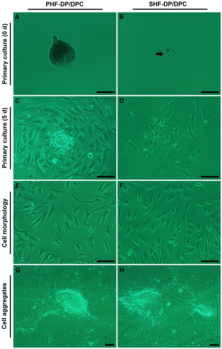
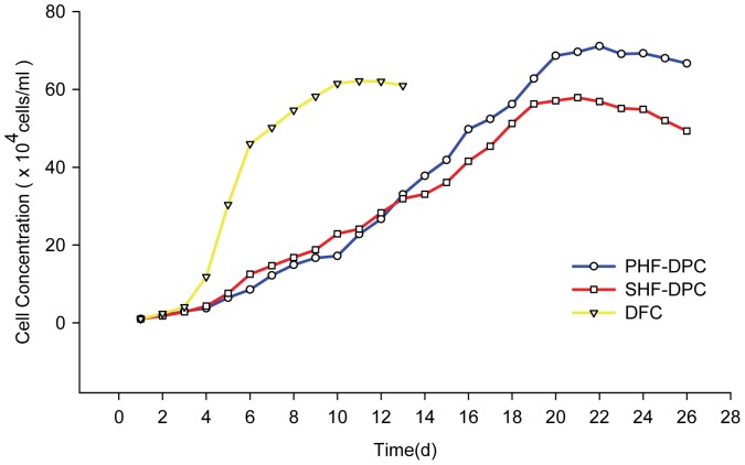
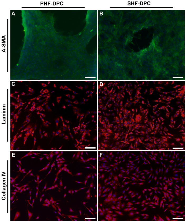
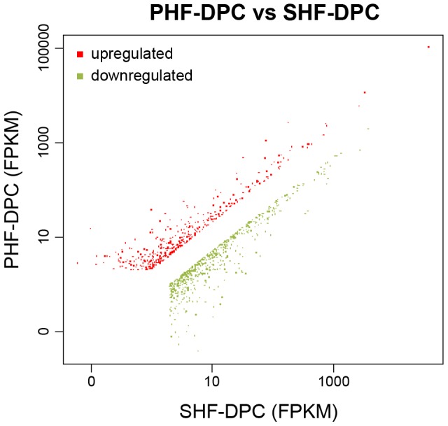
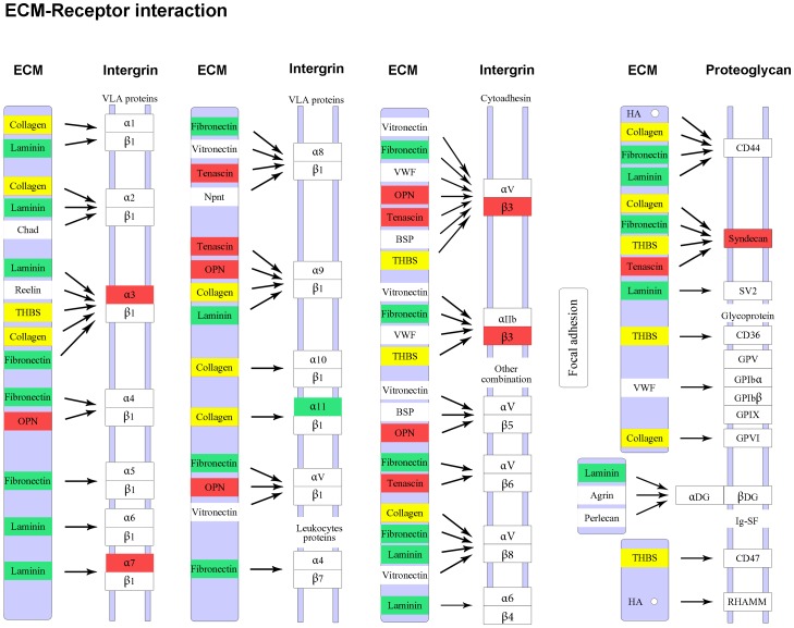
The dermal papilla is thought to establish the character and control the size of hair follicles. Inner Mongolia Cashmere goats (Capra hircus) have a double coat comprising the primary and secondary hair follicles, which have dramatically different sizes and textures. The Cashmere goat is rapidly becoming a potent model for hair follicle morphogenesis research. In this study, we established two dermal papilla cell lines during the anagen phase of the hair growth cycle from the primary and secondary hair follicles and clarified the similarities and differences in their morphology and growth characteristics. High-throughput transcriptome sequencing was used to identify gene expression differences between the two dermal papilla cell lines. Many of the differentially expressed genes are involved in vascularization, ECM-receptor interaction and Wnt/β-catenin/Lef1 signaling pathways, which intimately associated with hair follicle morphogenesis. These findings provide valuable information for research on postnatal morphogenesis of hair follicles.


Conflict of interest statement

Competing Interests: The authors have declared that no competing interests exist.

Figures

Figure 1
Figure 1. Culture of dermal papilla cells (DPCs) in the DMEM/F12 Medium plus 10% newborn calf serum.
Primary culture of the primary hair follicle-dermal papilla cells (PHF-DPCs) (A, C), and secondary hair follicle-dermal papilla cells (SHF-DPCs) (B, D). The DPCs exhibited a triangular or polygon shape (Figure 1C–F) at primary and subsequent passage. At the second passage both the PHF-DPCs (E) and SHF-DPCs (F) formed cell aggregates during further culture for approximately 20 days. Black arrow in (B) shows the tiny DP isolated from SHF. Scale bars  = 100 µm.
Figure 2
Figure 2. Growth curves of primary hair follicle-dermal papilla cells (PHF-DPCs), secondary hair follicle-dermal papilla cells (SHF-DPCs) and dermal fibroblast cells (DFCs).
The inoculum was 1.0×10⁴ cells/ml. Cell numbers were counted daily and recorded. The population doubling time was approximately 3.6days for PHF-DPC, 3.7days for SHF-DPC and 0.86days for DFC.
Figure 3
Figure 3. Immunocytochemical analysis of cultured dermal papilla cells (DPCs).
Immunocytochemistry of DPCs using anti-α-SMA antibody (green) was performed (A&B) when cells started to exhibit an aggregative growth behavior, while the other two antibodies were performed on monolayer cultured DPCs. Both primary hair follicle-dermal papilla cells (PHF-DPCs) and secondary hair follicle-dermal papilla cells (SHF-DPCs) were positive for α-SMA (green, A & B), Laminin (red, C & D), and Collagen IV (red, E & F). Nuclei in A–F were marked by DAPI staining (blue). Scale bars  = 100 µm.
Figure 4
Figure 4. Schematic representation of the differentially expressed genes between primary hair follicle-dermal papilla cells (PHF-DPCs) and secondary hair follicle-dermal papilla cells (SHF-DPCs).
Of the 1044 differentially expressed genes, 620 were upregulated (top-left, red) and 424 were downregulated (bottom-right, green) in PHF-DPCs compared with SHF-DPCs.
Figure 5
Figure 5. Pearson correlation coefficient* of differential expression ratios between RNA-Seq and qRT-PCR.
*Correlation is significant at the 0.01 level.
Figure 6
Figure 6. Differentially expressed genes between primary hair follicle-dermal papilla cells (PHF-DPCs) and secondary hair follicle-dermal papilla cells (SHF-DPCs) involved in extracellular matrix (ECM)-receptor interaction pathway.
The red color labels genes upregulated in the PHF-DPCs compared with the SHF-DPCs. The green color labels genes downregulated in the PHF-DPCs. The yellow color labels genes that some were upregulated and others were downregulated in the PHF-DPCs.

References

    1. Driskell RR, Clavel C, Rendl M, Watt FM (2011) Hair follicle dermal papilla cells at a glance. J Cell Sci 124: 1179–1182. - PMC - PubMed
    1. Driskell RR, Giangreco A, Jensen KB, Mulder KW, Watt FM (2009) Sox2-positive dermal papilla cells specify hair follicle type in mammalian epidermis. Development 136: 2815–2823. - PMC - PubMed
    1. Bitgood MJ, McMahon AP (1995) Hedgehog and Bmp genes are coexpressed at many diverse sites of cell-cell interaction in the mouse embryo. Dev Biol 172: 126–138. - PubMed
    1. Oro AE, Scott MP (1998) Splitting hairs: dissecting roles of signaling systems in epidermal development. Cell 95: 575–578. - PubMed
    1. Millar SE (2002) Molecular mechanisms regulating hair follicle development. J Invest Dermatol 118: 216–225. - PubMed

Publication types