Review
. 2013 Jul 1;2(3):e24137.
doi: 10.4161/jkst.24137. Epub 2013 Aug 15.

The involvement of the JAK-STAT signaling pathway in chronic inflammatory skin disease atopic dermatitis

Affiliations
Review

The involvement of the JAK-STAT signaling pathway in chronic inflammatory skin disease atopic dermatitis

Lei Bao et al. JAKSTAT. .

Abstract

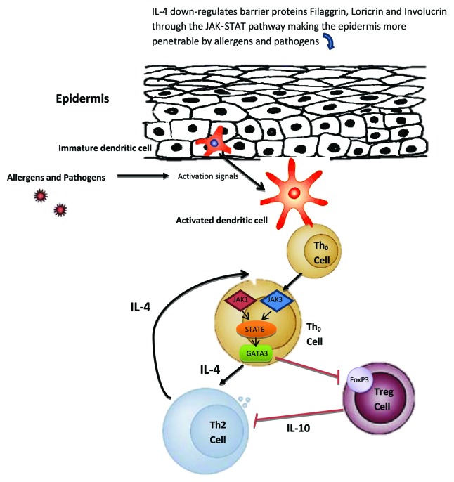
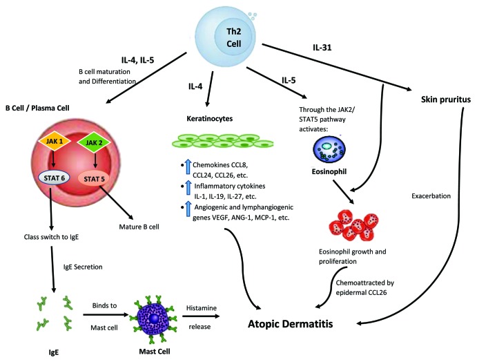
Atopic dermatitis (AD), a common chronic inflammatory skin disease, is characterized by inflammatory cell skin infiltration. The JAK-STAT pathway has been shown to play an essential role in the dysregulation of immune responses in AD, including the exaggeration of Th2 cell response, the activation of eosinophils, the maturation of B cells, and the suppression of regulatory T cells (Tregs). In addition, the JAK-STAT pathway, activated by IL-4, also plays a critical role in the pathogenesis of AD by upregulating epidermal chemokines, pro-inflammatroy cytokines, and pro-angiogenic factors as well as by downregulating antimicrobial peptides (AMPs) and factors responsible for skin barrier function. In this review, we will highlight the recent advances in our understanding of the JAK-STAT pathway in the pathogenesis of AD.

Keywords: JAK-STAT; Th2; atopic dermatitis; inflammation; skin.


Figures

None
Figure 1. Proposed mechanism of JAK-STAT involvement in atopic dermatitis (AD) development, part I. Skin barrier function defects are critical for the development of AD. In addition to the filaggrin gene mutation defects in some AD patients, IL-4 is also able to downregulate barrier proteins filaggrin, loricrin and involucrin through the JAK-STAT pathway making the epidermis more penetrable by allergens and pathogens. Once penetrated through the epidermis, allergens/pathogens are detected by dendritic cells, which become subsequently activated to present these antigens to naïve Th0 cells. The Th0 cell can then differentiate into the Th2 cell through the JAK1,3-STAT6 pathway under the influence of IL-4. In the Th0 cells, the STAT6 pathway can also upregulate GATA3, a master regulator of Th2 cells. GATA3 in turn suppresses Foxp3, the master regulator in Treg cells, thus allowing more T cells to be activated. Black arrows indicate activation pathway. Red lines indicate inhibition pathway.
None
Figure 2. Proposed mechanism of JAK-STAT involvement in atopic dermatitis (AD) development, part II. Here, Th2 cells play a significant role. By their abilities to provide IL-4 and IL-5 stimulation via the JAK-STAT pathway, immature B cells could be differentiated into mature B cell and plasma cells would undergo antibody heavy chain switching to IgE class. The subsequent binding of IgE to skin mast cells could lead to release of histamine, which is known to exacerbate AD. Similarly, this hyper Th2 immune milieu, particularly IL-4, could trigger epidermal cells to produce and release various chemokines (such as CCL26), pro-inflammatory cytokines, and angiogenic factors, leading to AD pathophysiology. Moreover, IL-5 released from this hyper Th2 milieu could, through JAK-STAT pathway, activate eosinophils, and attract them to the skin via CCL26, further worsening the AD condition. In addition, by way of IL-31, an inducer of pruritus, AD symptoms could become increasingly intensified. Black arrows indicate activation pathway. Red lines indicate inhibition pathway.

Similar articles

Cited by

References

    1. Saito H. Much atopy about the skin: genome-wide molecular analysis of atopic eczema. Int Arch Allergy Immunol. 2005;137:319–25. doi: 10.1159/000086464. - DOI - PubMed
    1. Kapoor R, Menon C, Hoffstad O, Bilker W, Leclerc P, Margolis DJ. The prevalence of atopic triad in children with physician-confirmed atopic dermatitis. J Am Acad Dermatol. 2008;58:68–73. doi: 10.1016/j.jaad.2007.06.041. - DOI - PubMed
    1. Guttman-Yassky E, Nograles KE, Krueger JG. Contrasting pathogenesis of atopic dermatitis and psoriasis--part II: immune cell subsets and therapeutic concepts. J Allergy Clin Immunol. 2011;127:1420–32. doi: 10.1016/j.jaci.2011.01.054. - DOI - PubMed
    1. Guttman-Yassky E, Nograles KE, Krueger JG. Contrasting pathogenesis of atopic dermatitis and psoriasis--part I: clinical and pathologic concepts. J Allergy Clin Immunol. 2011;127:1110–8. doi: 10.1016/j.jaci.2011.01.053. - DOI - PubMed
    1. Chen L, Martinez O, Overbergh L, Mathieu C, Prabhakar BS, Chan LS. Early up-regulation of Th2 cytokines and late surge of Th1 cytokines in an atopic dermatitis model. Clin Exp Immunol. 2004;138:375–87. doi: 10.1111/j.1365-2249.2004.02649.x. - DOI - PMC - PubMed