Review
. 2013:2013:284873.
doi: 10.1155/2013/284873. Epub 2013 Aug 28.

The role of changes in extracellular matrix of cartilage in the presence of inflammation on the pathology of osteoarthritis

Affiliations
Review

The role of changes in extracellular matrix of cartilage in the presence of inflammation on the pathology of osteoarthritis

Maricela Maldonado et al. Biomed Res Int. 2013.

Abstract

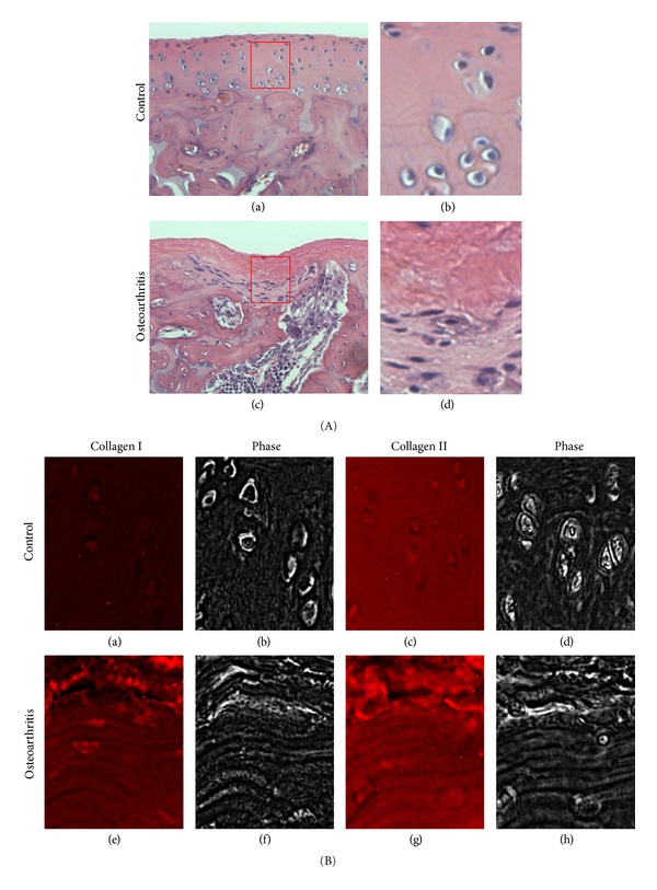
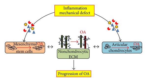
Osteoarthritis (OA) is a degenerative disease that affects various tissues surrounding joints such as articular cartilage, subchondral bone, synovial membrane, and ligaments. No therapy is currently available to completely prevent the initiation or progression of the disease partly due to poor understanding of the mechanisms of the disease pathology. Cartilage is the main tissue afflicted by OA, and chondrocytes, the sole cellular component in the tissue, actively participate in the degeneration process. Multiple factors affect the development and progression of OA including inflammation that is sustained during the progression of the disease and alteration in biomechanical conditions due to wear and tear or trauma in cartilage. During the progression of OA, extracellular matrix (ECM) of cartilage is actively remodeled by chondrocytes under inflammatory conditions. This alteration of ECM, in turn, changes the biomechanical environment of chondrocytes, which further drives the progression of the disease in the presence of inflammation. The changes in ECM composition and structure also prevent participation of mesenchymal stem cells in the repair process by inhibiting their chondrogenic differentiation. This review focuses on how inflammation-induced ECM remodeling disturbs cellular activities to prevent self-regeneration of cartilage in the pathology of OA.


Figures

Figure 1
Figure 1
Changes in the extracellular matrix structure and composition of cartilage afflicted by osteoarthritis (OA). Experimental OA was induced by intra-articular injection of monoiodoacetate (MIA) similar to the previously described protocol using a rat model [4]. OA induced rats were sacrificed at day 11, and the medial condyles of the arthritic knees (A (c-d); B (e–h)) were histologically (H&E staining (A)) and immunohistologically (collagen type I (B (a) and (e)) and type II (Figure B (c) and (g)) compared to that of the saline-injected sham control ((A (a-b); B (a–d)). (A) Microscopic features of OA cartilage (grade 2-3) show cartilage lesion formation, articular surface fissurization, subchondral bone advancement, and bone marrow edema/cyst. In addition, cell clustering and fibrocartilage formation is apparent in OA samples. (A) (b and d) are magnified images of the area indicated in (A) (a and c), respectively, to reveal the changes in cellular morphology. (B) Consecutive sections of the healthy and OA cartilages were stained using monoclonal antibodies for collagen type I or type II. An increase in intensity for collagen type I is observed in the OA cartilage, while it is not present in the control cartilage. Collagen type II is readily observed for both the healthy and OA cartilage. (B) (b, d, f, and h) are phase-contrast images of (B) (a, c, e, and g), respectively, to reveal tissue morphologies.
Figure 2
Figure 2
Schematic of the interplay between the extracellular matrix and cellular activities under inflammation during the progression of osteoarthritis (OA). Wear and tear or trauma induces inflammation and mechanical defects in cartilage, which initiate OA. These altered microenvironments affect the residential chondrocytes to produce nonchondrocytic extracellular matrix (ECM) that, in turn, further drives the dedifferentiation of the chondrocytes. The changes in microenvironments also negatively affect the chondrogenic differentiation of mesenchymal stem cells that originate from subchondral bone marrow, preventing the self-regeneration of cartilage. The positive feedback loop between mal-formed ECM and cellular activities drives the progression of OA.

References

    1. Nam J, Perera P, Liu J, et al. Sequential alterations in catabolic and anabolic gene expression parallel pathological changes during progression of monoiodoacetate-induced arthritis. PLoS ONE. 2011;6(9)e24320 - PMC - PubMed
    1. Pearle AD, Warren RF, Rodeo SA. Basic science of articular cartilage and osteoarthritis. Clinics in Sports Medicine. 2005;24(1):1–12. - PubMed
    1. Martel-Pelletier J, Boileau C, Pelletier J, Roughley PJ. Cartilage in normal and osteoarthritis conditions. Best Practice and Research. 2008;22(2):351–384. - PubMed
    1. Nam J, Perera P, Liu J, et al. Transcriptome-wide gene regulation by gentle treadmill walking during the progression of monoiodoacetate-induced arthritis. Arthritis & Rheumatism. 2011;63(6):1613–1625. - PMC - PubMed
    1. Lark MW, Bayne EK, Flanagan J, et al. Aggrecan degradation in human cartilage: evidence for both matrix metalloproteinase and aggrecanase activity in normal, osteoarthritic, and rheumatoid joints. The Journal of Clinical Investigation. 1997;100(1):93–106. - PMC - PubMed

MeSH terms