. 2013:2013:361906.
doi: 10.1155/2013/361906. Epub 2013 Aug 28.

Activation of the ERK1/2 signaling pathway during the osteogenic differentiation of mesenchymal stem cells cultured on substrates modified with various chemical groups

Affiliations

Activation of the ERK1/2 signaling pathway during the osteogenic differentiation of mesenchymal stem cells cultured on substrates modified with various chemical groups

Bing Bai et al. Biomed Res Int. 2013.

Abstract

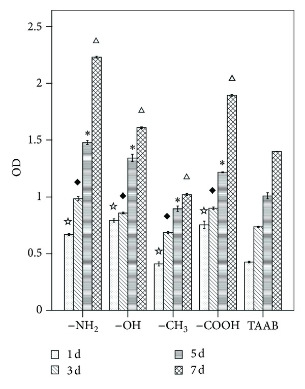
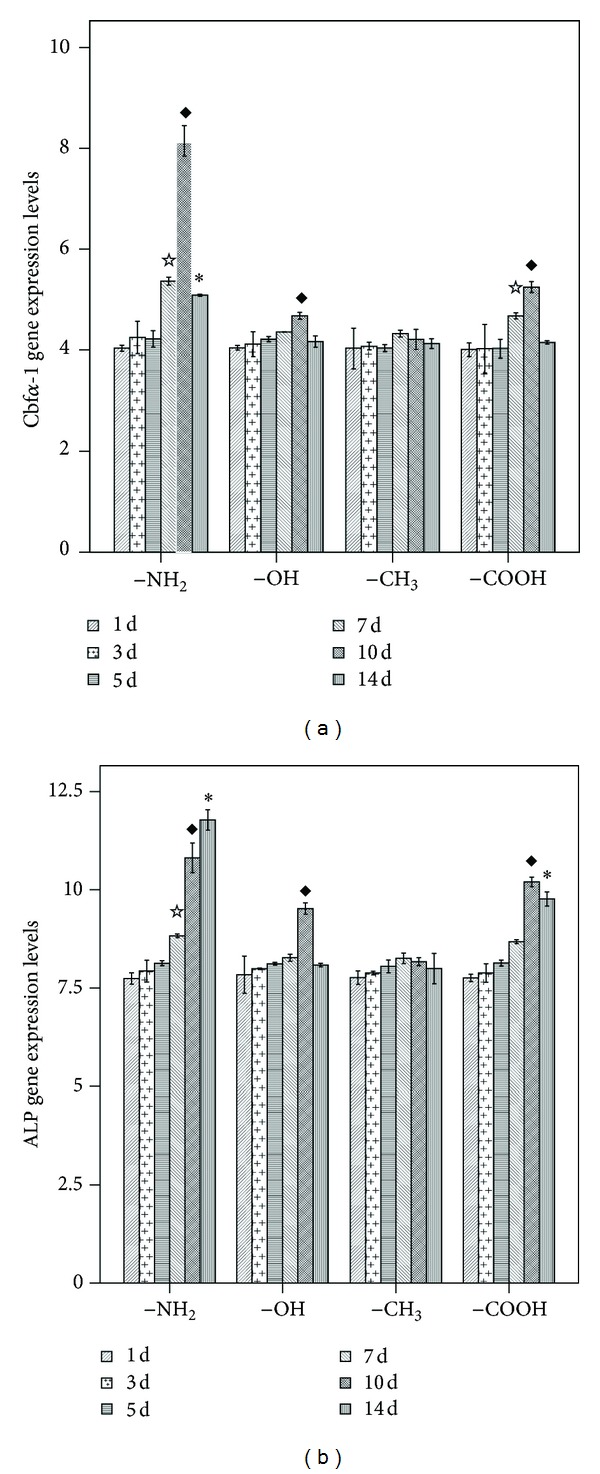
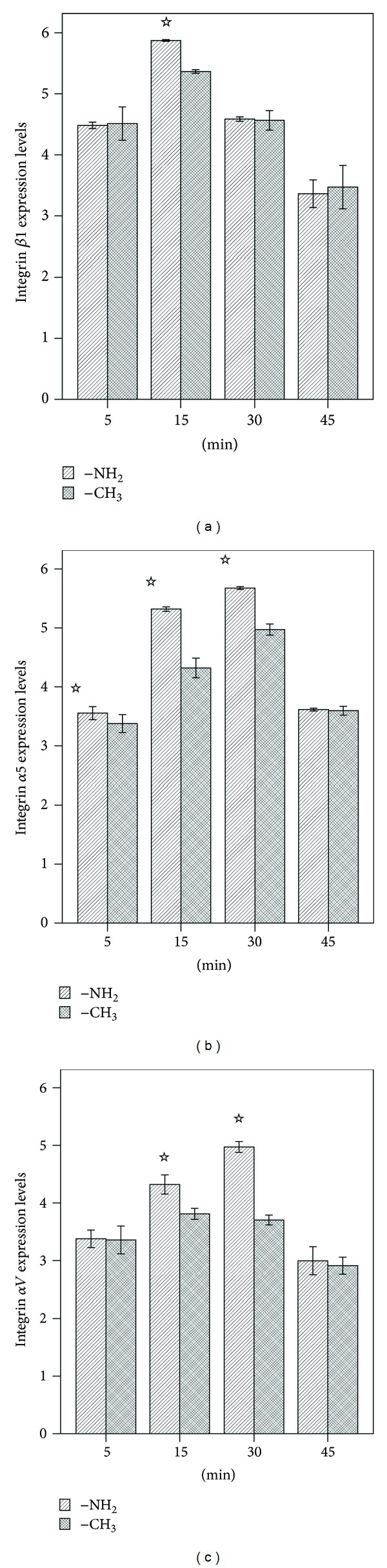
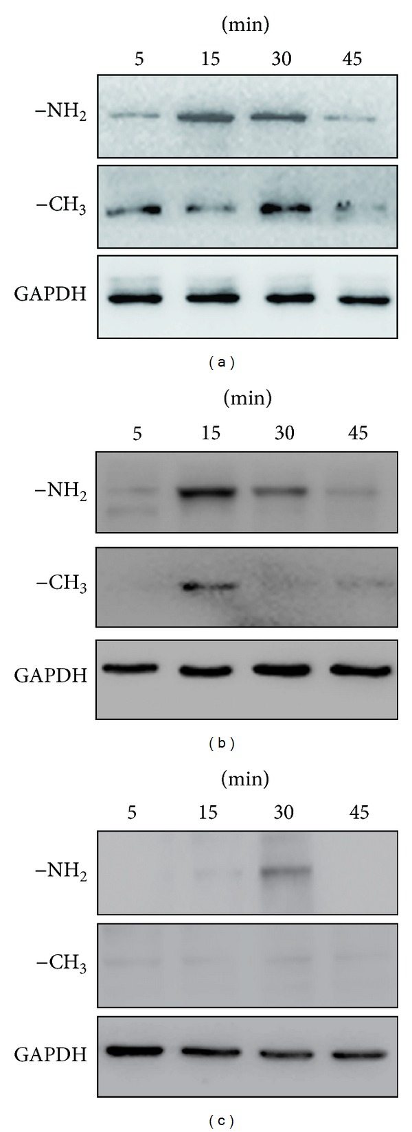
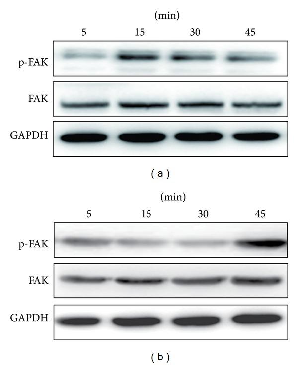
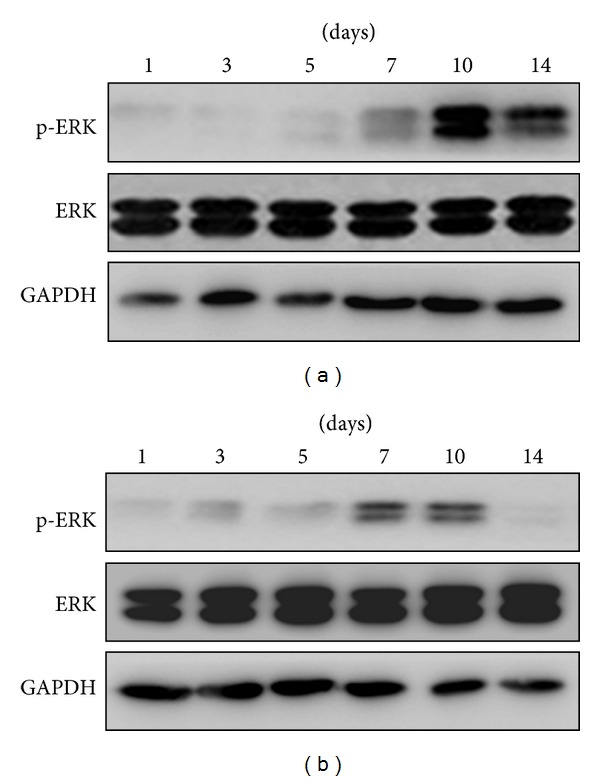
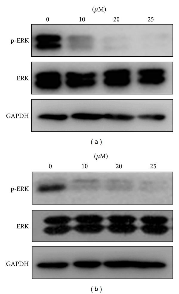
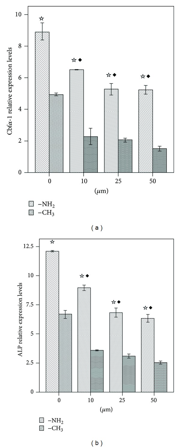
The current study examined the influence of culture substrates modified with the functional groups -OH, -COOH, -NH2, and -CH3 using SAMs technology, in conjunction with TAAB control, on the osteogenic differentiation of rabbit BMSCs. The CCK-8 assay revealed that BMSCs exhibited substrate-dependent cell viability. The cells plated on -NH2- and -OH-modified substrates were well spread and homogeneous, but those on the -COOH- and -CH3-modified substrates showed more rounded phenotype. The mRNA expression of BMSCs revealed that -NH2-modified substrate promoted the mRNA expression and osteogenic differentiation of the BMSCs. The contribution of ERK1/2 signaling pathway to the osteogenic differentiation of BMSCs cultured on the -NH2-modified substrate was investigated in vitro. The -NH2-modified substrate promoted the expression of integrins; the activation of FAK and ERK1/2. Inhibition of ERK1/2 activation by PD98059, a specific inhibitor of the ERK signaling pathway, blocked ERK1/2 activation in a dose-dependent manner, as revealed for expression of Cbf α -1 and ALP. Blockade of ERK1/2 phosphorylation in BMSCs by PD98059 suppressed osteogenic differentiation on chemical surfaces. These findings indicate a potential role for ERK in the osteogenic differentiation of BMSCs on surfaces modified by specific chemical functional groups, indicating that the microenvironment affects the differentiation of BMSCs. This observation has important implications for bone tissue engineering.


Figures

Figure 1
Figure 1
CCK-8 analysis of BMSCs cultured on different chemical functional groups after 1, 3, 5, and 7 days. P < 0.05 versus TAAB at day 1; P < 0.05 versus TAAB at day 3; *P < 0.05 versus TAAB at day 5; P < 0.05 versus TAAB at day 7.
Figure 2
Figure 2
Confocal fluorescence microscopy of the cytoskeleton demonstrating the differentiated cell phenotypes from BMSCs cultured on the various test surfaces after 1 and 3 d of culture. Immunofluorescence staining of anti-F-actin (Green: (a), (e), (i), (m)), DAPI nuclear staining (Blue: (b), (f), (j), (n)), antivinculin (Red: (c), (g), (k), (o)) and merged images ((d), (h), (l), (p)). Scale bar = 50 μm. –NH2-modified substrate for 1 day ((a), (b), (c), (d)) and 3 days ((e), (f), (g), (h)), –OH-modified substrate for 1 day ((i), (j), (k), (l)) and 3 days ((m), (n), (o), (p)).
Figure 3
Figure 3
Confocal fluorescence microscopy of the cytoskeleton demonstrating the differentiated cell phenotypes from BMSCs cultured on the various test surfaces after 1 and 3 d of culture. Immunofluorescence staining of anti-F-actin (Green: (a), (e), (i), (m)), DAPI nuclear staining (Blue: (b), (f), (j), (n)), antivinculin (Red: (c), (g), (k), (o)), and merged images ((d), (h), (l), (p)). Scale bar = 50 μm. –CH3-modified substrate for 1 day ((a), (b), (c), (d)) and 3 days ((e), (f), (g), (h)), –COOH-modified substrate for 1 day ((i), (j), (k), (l)) and 3 days ((m), (n), (o), (p)).
Figure 4
Figure 4
Real-time PCR analysis of the expression of osteogenesis-related genes in BMSCs cultured on surfaces modified with different chemical functional groups. (a) CBFA-1; (b) ALP. P < 0.05 versus the respective –CH3-modified substrate at 7 d; P < 0.05 versus the respective –CH3-modified substrate at 10 d; *P < 0.05 versus the respective –CH3-modified substrate at 14 d.
Figure 5
Figure 5
Real-time PCR analysis of the expression of osteogenesis-related genes in BMSCs cultured on the –NH2- and –CH3-modified substrates. (a) Integrin β1, (b) integrin α5, (c) integrin αV. P < 0.05 versus the respective –CH3-modified substrate.
Figure 6
Figure 6
The –NH2-modified substrate induces the activation of osteogenesis-related genes. (a) Integrin β1, (b) integrin α5 and (c) integrin αV  in BMSCs. Cells were cultured on the –NH2- and –CH3-modified substrates, and lysates were prepared at the indicated times. Lysates were subjected to immunoblot analysis using integrin antibodies. An aliquot of each lysate was subjected to a kinase detection assay using SDS-PAGE. GAPDH was used as a control.
Figure 7
Figure 7
The –NH2-modified substrate induces the activation of FAK in BMSCs. Cells were cultured on the –NH2-modified substrate (a) and the –CH3-modified substrate (b), and lysates were prepared at the indicated times. Lysates were subjected to immunoblot analysis using phosphospecific and nonactivated FAK antibodies. An aliquot of each lysate was subjected to a kinase detection assay using SDS-PAGE. GAPDH was used as a control.
Figure 8
Figure 8
The –NH2-modified substrate induces the activation of ERK1/2 in BMSCs. Cells were cultured on the –NH2-modified substrate (a) and the –CH3-modified substrate (b), and lysates were prepared at the indicated times. Lysates were subjected to immunoblot analysis using phosphospecific and nonactivated ERK1/2 kinase antibodies. An aliquot of each lysate was subjected to a kinase detection assay using SDS-PAGE. GAPDH was used as a control.
Figure 9
Figure 9
PD98059 inhibits ERK1/2 activation in a dose-dependent manner. Cells were cultured on the –NH2- and –CH3-modified substrates containing 0, 10, 25, and 50 μM of PD98059 for 10 days. (a) –NH2-modified substrate and (b) –CH3-modified substrate. Cell lysates were analyzed by immunoblots using phospho-ERK-specific and nonactivated ERK1/2 antibodies; GAPDH was used as a control.
Figure 10
Figure 10
Real-time RT-PCR analysis of the expression of osteogenesis-related genes in BMSCs cultured on the –NH2- and –CH3-modified substrates in the presence of 0, 10, 25, and 50 μM of PD98059 for 10 days. (a) Cbfα-1 and (b) ALP. P < 0.05 versus the respective –CH3-modified substrate; P < 0.05 versus the 0 μM dose group.

Similar articles

Cited by

References

    1. Buma P, Schreurs W, Verdonschot N. Skeletal tissue engineering—from in vitro studies to large animal models. Biomaterials. 2004;25(9):1487–1495. - PubMed
    1. Pittenger MF, Mackay AM, Beck SC, et al. Multilineage potential of adult human mesenchy-mal stem cells. Science. 1999;284(5411):143–147. - PubMed
    1. Dallari D, Fini M, Stagni C, et al. In vivo study on the healing of bone defects treated with bone marrow stromal cells, platelet-rich plasma, and freeze-dried bone allografts, alone and in combination. Journal of Orthopaedic Research. 2006;24(5):877–888. - PubMed
    1. Lutolf MP, Gilbert PM, Blau HM. Designing materials to direct stem-cell fate. Nature. 2009;462(7272):433–441. - PMC - PubMed
    1. Chai C, Leong KW. Biomaterials approach to expand and direct differentiation of stem cells. Molecular Therapy. 2007;15(3):467–480. - PMC - PubMed

Publication types

MeSH terms

Substances