

A structural comparison of lipopolysaccharide biosynthesis loci of Legionella pneumophila serogroup 1 strains
- PMID: 24069939
- PMCID: PMC3766260
- DOI: 10.1186/1471-2180-13-198
A structural comparison of lipopolysaccharide biosynthesis loci of Legionella pneumophila serogroup 1 strains
Abstract
Background: The lipopolysaccharide (LPS) is the major immuno-dominant antigen of all Legionella species including L. pneumophila. Its diversity is the basis for the classification of L. pneumophila into serogroups and monoclonal subgroups and is thought to be involved in strain specific virulence. The understanding of the genetic basis of the LPS-antigen is incomplete. Thus, we analyzed the genetic locus involved in LPS-biosynthesis of L. pneumophila serogroup 1 (Sg1) strains with the focus on strain specific gene composition.
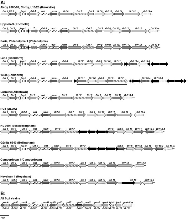
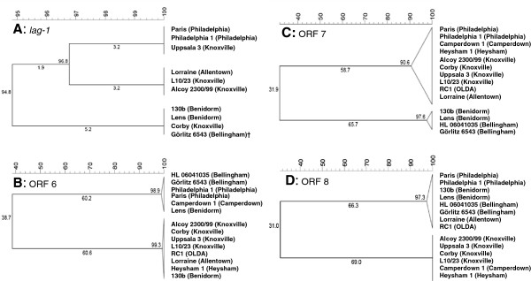
Results: The LPS-biosynthesis loci of 14 L. pneumophila Sg1 strains comprise two distinct regions: A 15 kb region containing LPS-biosynthesis genes that can be found in all L. pneumophila strains and a Sg1-specific 18 kb region. The 15 kb region is highly conserved among Sg1 strains as reflected by high homologies of single ORFs and by a consistent ORF arrangement. In contrast, the Sg1 specific 18 kb region is variable and partially disrupted by phage related genes. We propose that the region spanning from ORF 6 to ORF 11 of the Sg1-specific region is likely involved in late LPS-modification. Due to the high variability of this small region and various combinations of single ORFs within this region a strain specific LPS-structure could be synthesized including modifications of legionaminic acid derivates.
Conclusions: Our data clearly demonstrate that the gene structure of the LPS-biosynthesis locus of L. pneumophila Sg1 strains show significant interstrain variability. These data can be used for further functional analysis of the LPS synthesis to understand pathogenesis and reactivity with monoclonal antibodies. Moreover, variable but strain specific regions can serve as basis for the development of novel genotyping assays.
Figures
Similar articles
-
Cloning and functional characterization of a 30 kb gene locus required for lipopolysaccharide biosynthesis in Legionella pneumophila.Int J Med Microbiol. 2000 Mar;290(1):37-49. doi: 10.1016/S1438-4221(00)80104-6. Int J Med Microbiol. 2000. PMID: 11043980
-
Specific real-time PCR for simultaneous detection and identification of Legionella pneumophila serogroup 1 in water and clinical samples.Appl Environ Microbiol. 2011 Mar;77(5):1708-17. doi: 10.1128/AEM.02261-10. Epub 2010 Dec 30. Appl Environ Microbiol. 2011. PMID: 21193672 Free PMC article.
-
Multigenome analysis identifies a worldwide distributed epidemic Legionella pneumophila clone that emerged within a highly diverse species.Genome Res. 2008 Mar;18(3):431-41. doi: 10.1101/gr.7229808. Epub 2008 Feb 6. Genome Res. 2008. PMID: 18256241 Free PMC article.
-
Prevalence of Infection-Competent Serogroup 6 Legionella pneumophila within Premise Plumbing in Southeast Michigan.mBio. 2018 Feb 6;9(1):e00016-18. doi: 10.1128/mBio.00016-18. mBio. 2018. PMID: 29437918 Free PMC article.
-
The lipopolysaccharide of Legionella pneumophila serogroup 1 (strain Philadelphia 1): chemical structure and biological significance.Prog Clin Biol Res. 1995;392:113-39. Prog Clin Biol Res. 1995. PMID: 8524918 Review.
Cited by
-
Legionella pneumophila type II secretome reveals a polysaccharide deacetylase that impacts intracellular infection, biofilm formation, and resistance to polymyxin- and serum-mediated killing.mBio. 2025 Jul 9;16(7):e0139325. doi: 10.1128/mbio.01393-25. Epub 2025 Jun 20. mBio. 2025. PMID: 40539790 Free PMC article.
-
Population analysis of Legionella pneumophila reveals a basis for resistance to complement-mediated killing.Nat Commun. 2021 Dec 9;12(1):7165. doi: 10.1038/s41467-021-27478-z. Nat Commun. 2021. PMID: 34887398 Free PMC article.
-
Dynamics and impact of homologous recombination on the evolution of Legionella pneumophila.PLoS Genet. 2017 Jun 26;13(6):e1006855. doi: 10.1371/journal.pgen.1006855. eCollection 2017 Jun. PLoS Genet. 2017. PMID: 28650958 Free PMC article.
-
Legionella pneumophila strain associated with the first evidence of person-to-person transmission of Legionnaires' disease: a unique mosaic genetic backbone.Sci Rep. 2016 May 19;6:26261. doi: 10.1038/srep26261. Sci Rep. 2016. PMID: 27196677 Free PMC article.
-
Host-Specific Adaptation of Legionella pneumophila to Single and Multiple Hosts.Mol Biol Evol. 2025 Jul 30;42(8):msaf161. doi: 10.1093/molbev/msaf161. Mol Biol Evol. 2025. PMID: 40609045 Free PMC article.
References
Publication types
MeSH terms
Substances
Associated data
- Actions
- Actions
- Actions
- Actions
- Actions
LinkOut - more resources
Full Text Sources
Other Literature Sources
Molecular Biology Databases
