. 2013 Sep 27:14:106.
doi: 10.1186/1471-2202-14-106.

Tissue sparing, behavioral recovery, supraspinal axonal sparing/regeneration following sub-acute glial transplantation in a model of spinal cord contusion

Affiliations

Tissue sparing, behavioral recovery, supraspinal axonal sparing/regeneration following sub-acute glial transplantation in a model of spinal cord contusion

Helen R Barbour et al. BMC Neurosci. .

Abstract

Background: It has been shown that olfactory ensheathing glia (OEG) and Schwann cell (SCs) transplantation are beneficial as cellular treatments for spinal cord injury (SCI), especially acute and sub-acute time points. In this study, we transplanted DsRED transduced adult OEG and SCs sub-acutely (14 days) following a T10 moderate spinal cord contusion injury in the rat. Behaviour was measured by open field (BBB) and horizontal ladder walking tests to ascertain improvements in locomotor function. Fluorogold staining was injected into the distal spinal cord to determine the extent of supraspinal and propriospinal axonal sparing/regeneration at 4 months post injection time point. The purpose of this study was to investigate if OEG and SCs cells injected sub acutely (14 days after injury) could: (i) improve behavioral outcomes, (ii) induce sparing/regeneration of propriospinal and supraspinal projections, and (iii) reduce tissue loss.

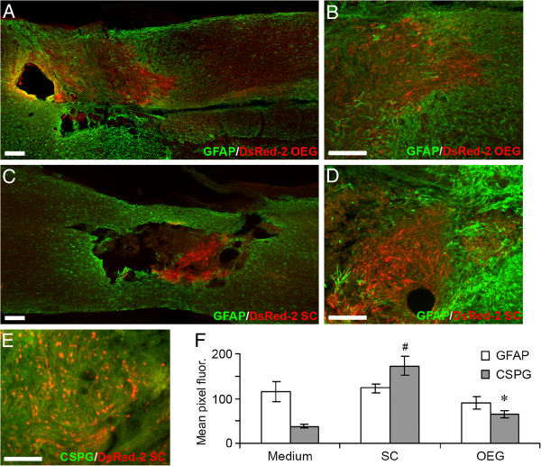
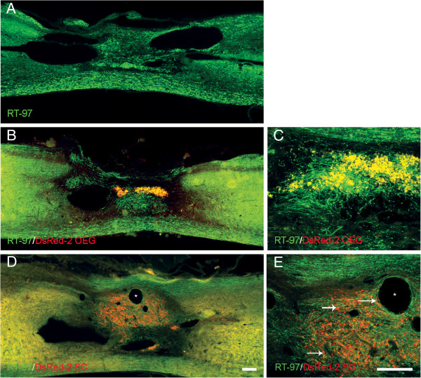
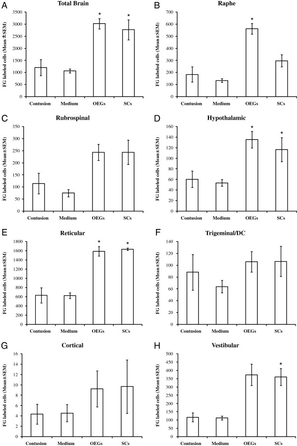
Results: OEG and SCs transplanted rats showed significant increased locomotion when compared to control injury only in the open field tests (BBB). However, the ladder walk test did not show statistically significant differences between treatment and control groups. Fluorogold retrograde tracing showed a statistically significant increase in the number of supraspinal nuclei projecting into the distal spinal cord in both OEG and SCs transplanted rats. These included the raphe, reticular and vestibular systems. Further pairwise multiple comparison tests also showed a statistically significant increase in raphe projecting neurons in OEG transplanted rats when compared to SCs transplanted animals. Immunohistochemistry of spinal cord sections short term (2 weeks) and long term (4 months) showed differences in host glial activity, migration and proteoglycan deposits between the two cell types. Histochemical staining revealed that the volume of tissue remaining at the lesion site had increased in all OEG and SCs treated groups. Significant tissue sparing was observed at both time points following glial SCs transplantation. In addition, OEG transplants showed significantly decreased chondroitin proteoglycan synthesis in the lesion site, suggesting a more CNS tolerant graft.

Conclusions: These results show that transplantation of OEG and SCs in a sub-acute phase can improve anatomical outcomes after a contusion injury to the spinal cord, by increasing the number of spared/regenerated supraspinal fibers, reducing cavitation and enhancing tissue integrity. This provides important information on the time window of glial transplantation for the repair of the spinal cord.


Figures

Figure 1
Figure 1
Graphic describing the amount of total tissue remaining and intact tissue at the site of the spinal cord contusion injury at day of transplant 2 weeks and 4 months. Quantification of gold chloride and Nissl stained sagittal sections to measure the amount of intact tissue and total tissue remaining within each of the experimental groups using Image Pro. (A): Total tissue remaining 2 weeks and 4 months after cell transplantation. (B): Intact tissue remaining 2 weeks and 4 months after cell transplantation. There was a significant increase (* p<0.05) in intact tissue found in the OEG and Schwann cell treated groups when compared to medium controls. (C, G): Appearance of spinal cord lesion site 2 weeks after injury (i.e. day of cell transplantation) (D) Total tissue remaining at 2 weeks after medium injection. (E) Appearance of spinal cord lesion site 2 weeks after OEG transplantation (4 weeks after injury). (F) Appearance of the spinal cord lesion epicenter 2 weeks after Schwann cell transplantation (4 weeks after initial injury). (H): Appearance of spinal cord lesion site 4 months after medium injection. (I) Demonstrates the spinal cord tissue 4 months after OEG transplantation. Note the considerable increased retention of intact tissue and a reduction in cystic volume when compared to image (H). (J): Injured spinal cord at 4 months after being injected with Schwann cells at the 14 day treatment time point. Note the presence of small microcysts (see black arrow) within the spinal cord parenchyma, but only where the Schwann cells are present. Scale bar: 200 μm.
Figure 2
Figure 2
Photomicrographs representing lentiviral transduced and transplanted OEG and Schwann cells. A: OEG in vitro labeled with lentiviral DsRed2. B: Schwann cells in vitro labeled with lentiviral DsRed2. Scale bar (A B): 50 μm. C D: Localization of transplanted OEG (C) and Schwann cells (D) within the spinal cord lesion site 2 weeks after transplantation. Schwann cells appear to have survived in greater numbers than the OEG. However, OEG are more dispersed through the spinal cord tissue while Schwann cells remain in a dense aggregate. Scale bar (C D): 100 μm.
Figure 3
Figure 3
Glial scar formation at the lesion site indicated by GFAP and CSPG immunoreactivity. A-D: GFAP in green DsRed2-labeled transplanted cells in red. A-B: Lesion site 2 weeks after transplantation of OEG. C-D: Lesion site 2 weeks after transplantation of Schwann cells. Dispersal of transplanted cells is greater for OEG than for Schwann cells. Schwann cells remain in a dense aggregate at the lesion site surrounded by strong GFAP immunoreactivity. E: Schwann cells are surrounded by dense proteoglycan network CSPG (green), DsRed2-labeled transplanted cells (red). Scale bars: A-D: 100 μm, E: 200 μm. F: Graph showing the quantitative fluorescence (measured by mean pixels) of GFAP and CSPG at the distal interface of the transplant zone (n=10 for each group). OEG had significantly less (p<0.05) CSPG expression when compared to Schwann cells (see *). Schwann cells had significantly greater (p< 0.05) CSPG expression when compared to medium injected controls (see #).
Figure 4
Figure 4
Schwann cell, OEG and medium treated spinal cords at 4 months. Photomicrographs depicting the distribution and immunoreactivity for laminin-1 (A, B, D-K) and collagen IV (C, L-S) at the site of spinal cord contusion lesion with or without the transplantation of SCs and OEG. Areas of strong immunoreactivity for laminin can be seen to surround the areas of the transplanted cells (see arrow in D depicting DsRed-2 OEG). Laminin immunostaining does not appear to be co-localized on the surface of the OEG cells (see arrows in G). Laminin is also found surrounding Schwann cell transplants (see H). Arrow depicts the central core of the transplant. Laminin is very strong in the region of the Schwann cells (see I-J) with the host spinal cord below the dashed line has reduced intensity of staining. Collagen IV immunostaining depicts a central core of staining close to the lesion site surrounding any surviving OEG (see L) but labeled cells (see arrows in O) are not co-labeled with collagen IV. Collagen IV immunostaining in Schwann cell transplanted spinal cords show intense staining surrounding and within the DsRed-2 SC transplant area (see arrow). In higher power the staining is mostly co-localized (see above dashed line) but some areas are not. Scale bars: 100 μm. Scale bar in A also serves C, D, H, L and P. Scale bar in B also serves E, F, I, J, M, N, Q and R.
Figure 5
Figure 5
Axonal staining at the lesion site of Schwann cell, OEG and medium groups. Immunoreactivity for RT-97 at the site of spinal cord contusion 4 months after medium (A) OEG (B-C) and Schwann cells (D-E) transplantation. RT97 positive axons are observed within the lesion site in all groups but number and distribution varied. Axons in the OEG group (B) were arranged in fascicular bundles but did not have rostral to caudal orientation unlike axons beneath the graft area which constitute the ventral white matter. See (C) for higher power image of (B). RT97 staining after Schwann cell transplantation (see D). These axons showed a close association with DsRed-2 positive cells but followed a non linear path. Arrows in (E) depict DsRed-2 labeled Schwann cells deep within the transplants or close to microcysts (*). Scale bars: 100 μm. Scale bar in D also serves A and B scale bar in E also serves C.
Figure 6
Figure 6
Photomicrographs depicting p75 immunoreactivity of spinal cord sections in the lesion site after SCI. Endogenous p75 positive cells Schwann cells are present within the lesion site 2 weeks (A) and 4 months post injury (B). p75 positive profiles are present in large numbers in OEG (C-D) and Schwann cell (E-F) transplanted groups. The arrows in C and D represent DsRED-2 positive transplanted OEG within the lesion site co-mingled with host p75 positive Schwann cells. In the Schwann cell transplanted group the p75 positive endogenous Schwann cells (see arrows) also surround the transplanted DsRed-2 positive cells The Schwann cell transplants are tightly clustered unlike the dispersed OEG (see C-D). Scale bar in C (100 μm) also serves A and B.
Figure 7
Figure 7
Number of neurons in brain/brainstem nuclei retrogradely labeled with Fluorogold injected 8 mm caudal to the thoracic spinal cord lesion site. OEG transplantation into the contused spinal cord 14 days after injury promotes significant axonal sparing/regeneration of Raphe-, Vestibular- and Reticulo-spinal axons. Graphs depict the mean numbers of total FG-labeled neurons in the brains and brainstems of the four groups (A). Mean neuron numbers of the Raphe (B), Rubrospinal (C), Hypothalamic (D), Reticular (E), Trigeminal/DC (F), Cortical layer V (G) and Vestibular (H). Experimental groups were compared using a one-way analysis of variance (ANOVA), followed by the Dunnett’s method of multiple comparisons versus a single control group (injury only) and also the Tukey test for multiple comparison procedures between all experimental and control groups.
Figure 8
Figure 8
Propriospinal neurons rostral to the lesion site were retrogradely labeled with Fluorogold injected caudal (8 mm) to the lesion site (A). Significantly higher numbers of propriospinal neurons (n=10, p<0.05) were observed in the Schwann cell-transplanted group (#) and OEG-transplanted groups (*) relative to the medium-injected group (B).
Figure 9
Figure 9
Mean BBB scores over 4 months after injection of medium, Schwann cells and OEG plotted against contusion injury only. The red dotted line depicts the transplant day. A statistically significant increase in BBB scores was seen in the Schwann cell group when compared to contusion only (#, p<0.05). A significant increase in BBB scores was also seen in the OEG group when compared to control injury only (*, p<0.05).
Figure 10
Figure 10
Ladder walking behavioral testing. A: representative photo of a rat traversing the horizontal ladder. B: ladder scores of treatment groups on horizontal ladder walking test 6 weeks after cell transplantation date. C: as for B at 4 months after cell transplantation date. At no stage were statistical differences between groups observed on the horizontal ladder test.

References

    1. Doucette R. PNS-CNS transitional zone of the first cranial nerve. J Comp Neurol. 1991;312(3):451–466. doi: 10.1002/cne.903120311. - DOI - PubMed
    1. Ramer LM, Richter MW, Roskams AJ, Tetzlaff W, Ramer MS. Peripherally-derived olfactory ensheathing cells do not promote primary afferent regeneration following dorsal root injury. Glia. 2004;47(2):189–206. doi: 10.1002/glia.20054. - DOI - PubMed
    1. Richter MW, Roskams AJ. Olfactory ensheathing cell transplantation following spinal cord injury: Hype or hope? Exp Neurol. 2008;209(2):353–367. doi: 10.1016/j.expneurol.2007.06.011. - DOI - PubMed
    1. Ruitenberg MJ, Vukovic J, Sarich J, Busfield SJ, Plant GW. Olfactory ensheathing cells: characteristics genetic engineering and therapeutic potential. J Neurotrauma. 2006;23(3–4):468–478. - PubMed
    1. Graziadei PP, Monti Graziadei GA. Neurogenesis and neuron regeneration in the olfactory system of mammals III. Deafferentation and reinnervation of the olfactory bulb following section of the fila olfactoria in rat. J Neurocytol. 1980;9(2):145–162. doi: 10.1007/BF01205155. - DOI - PubMed

Publication types