. 2013 Nov 19;46(11):2725-39.
doi: 10.1021/ar400149m. Epub 2013 Sep 26.

Geometric and electronic structure contributions to function in non-heme iron enzymes

Affiliations

Geometric and electronic structure contributions to function in non-heme iron enzymes

Edward I Solomon et al. Acc Chem Res. .

Abstract

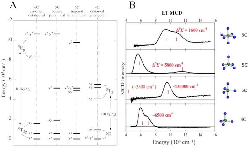
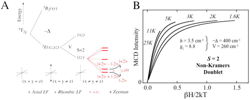
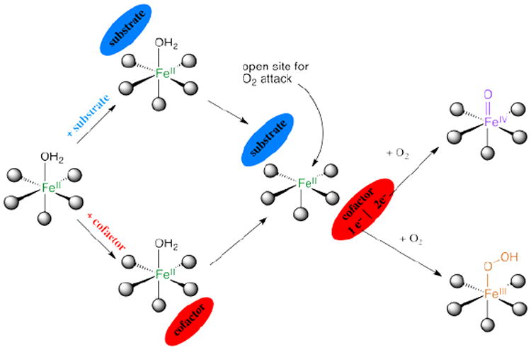
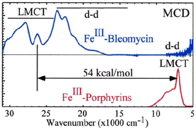
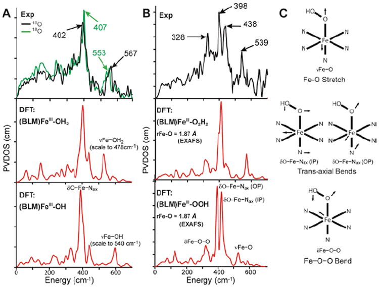
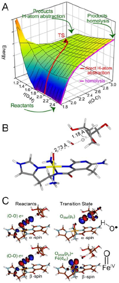
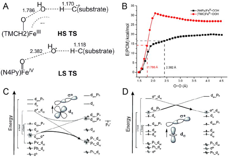
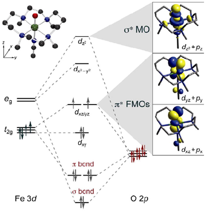
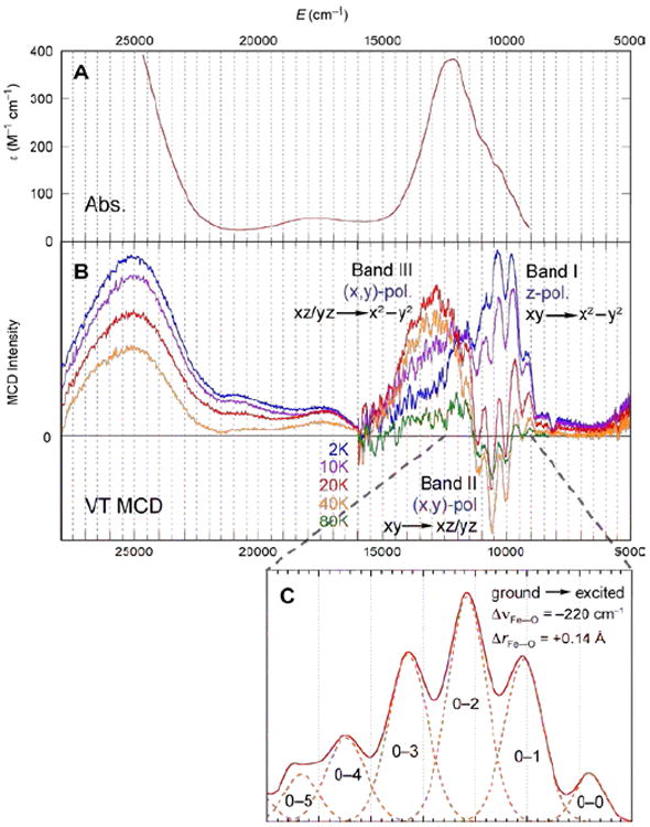
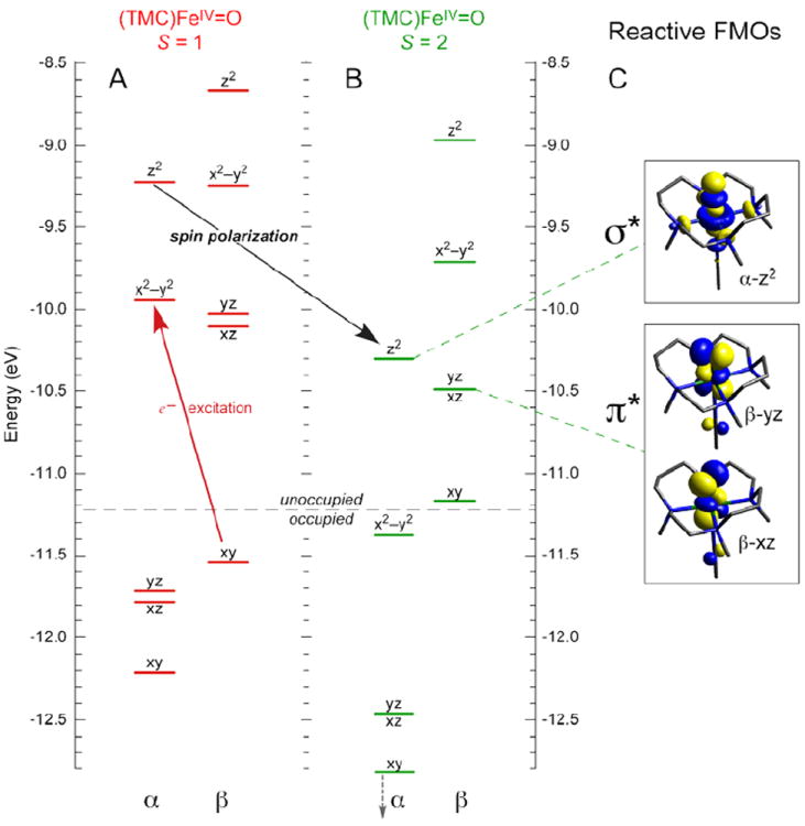
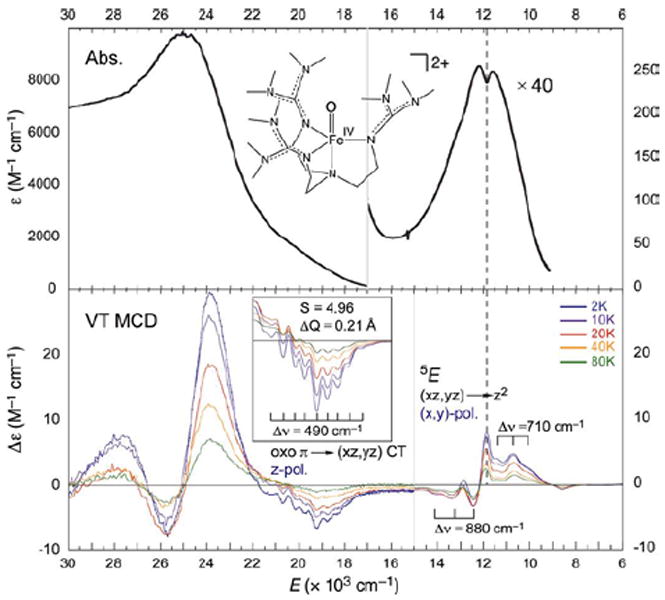
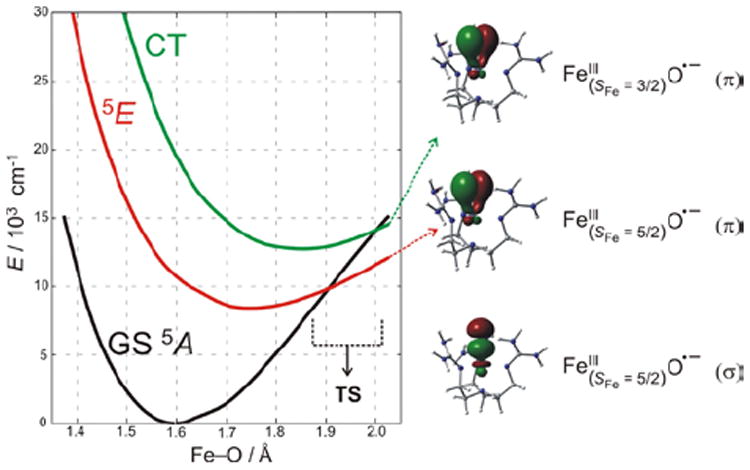
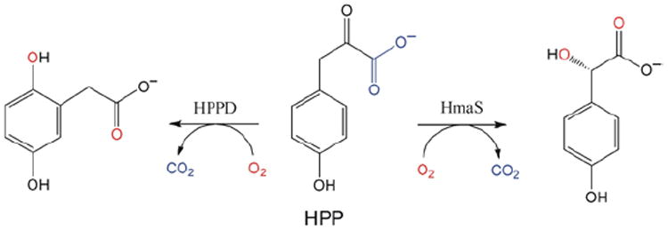
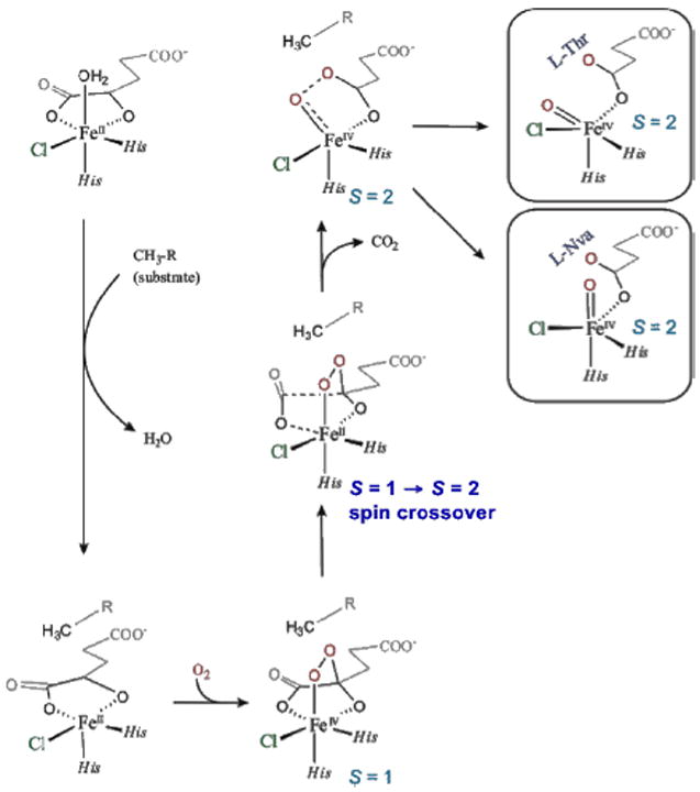
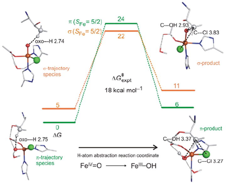
Mononuclear non-heme Fe (NHFe) enzymes play key roles in DNA repair, the biosynthesis of antibiotics, the response to hypoxia, cancer therapy, and many other biological processes. These enzymes catalyze a diverse range of oxidation reactions, including hydroxylation, halogenation, ring closure, desaturation, and electrophilic aromatic substitution (EAS). Most of these enzymes use an Fe(II) site to activate dioxygen, but traditional spectroscopic methods have not allowed researchers to insightfully probe these ferrous active sites. We have developed a methodology that provides detailed geometric and electronic structure insights into these NHFe(II) active sites. Using these data, we have defined a general mechanistic strategy that many of these enzymes use: they control O2 activation (and limit autoxidation and self-hydroxylation) by allowing Fe(II) coordination unsaturation only in the presence of cosubstrates. Depending on the type of enzyme, O2 activation either involves a 2e(-) reduced Fe(III)-OOH intermediate or a 4e(-) reduced Fe(IV)═O intermediate. Nuclear resonance vibrational spectroscopy (NRVS) has provided the geometric structure of these intermediates, and magnetic circular dichroism (MCD) has defined the frontier molecular orbitals (FMOs), the electronic structure that controls reactivity. This Account emphasizes that experimental spectroscopy is critical in evaluating the results of electronic structure calculations. Therefore these data are a key mechanistic bridge between structure and reactivity. For the Fe(III)-OOH intermediates, the anticancer drug activated bleomycin (BLM) acts as the non-heme Fe analog of compound 0 in heme (e.g., P450) chemistry. However BLM shows different reactivity: the low-spin (LS) Fe(III)-OOH can directly abstract a H atom from DNA. The LS and high-spin (HS) Fe(III)-OOHs have fundamentally different transition states. The LS transition state goes through a hydroxyl radical, but the HS transition state is activated for EAS without O-O cleavage. This activation is important in one class of NHFe enzymes that utilizes a HS Fe(III)-OOH intermediate in dioxygenation. For Fe(IV)═O intermediates, the LS form has a π-type FMO activated for attack perpendicular to the Fe-O bond. However, the HS form (present in the NHFe enzymes) has a π FMO activated perpendicular to the Fe-O bond and a σ FMO positioned along the Fe-O bond. For the NHFe enzymes, the presence of π and σ FMOs enables enzymatic control in determining the type of reactivity: EAS or H-atom extraction for one substrate with different enzymes and halogenation or hydroxylation for one enzyme with different substrates.


Figures

FIGURE 1
FIGURE 1
NIR MCD transitions for NHFeII sites. (A) Theoretical d orbital splittings for various geometries. (B) Experimentally-observed transitions.
FIGURE 2
FIGURE 2
VTVH MCD for FeII sites. (A) Splitting of the 5T2g ground state upon axial and rhombic perturbations. S.O. Refers to the effect of spin-orbit coupling. (B) Nested MCD isotherms for an S = 2 non-Kramers doublet.
FIGURE 3
FIGURE 3
MCD studies of PAH. (A) MCD spectra of resting PAH (black) with substrate-bound (blue), pterin-bound (green) and substrate/pterin-bound (red). All spectra were collected at 5 K. (B) VTVH MCD data for resting (left) and substrate/pterin-bound (right) PAH. (C) Derived d-orbital energy level diagrams.
FIGURE 4
FIGURE 4
General mechanistic strategy for O2-activation by NHFeII enzymes.
FIGURE 5
FIGURE 5
(A) Steric and H-bonding contributions to loss of coordinated H2O. CAD stands for C-terminal activation domain of the substrate HIF-1α. (B) Effect of a second-sphere H-bonding partner on the facial triad carboxylate.
FIGURE 6
FIGURE 6
Optimized structure of ABLM.
FIGURE 7
FIGURE 7
MCD spectral comparison of LMCT energies of (BLM)FeIII (deprotonated amide to FeIII) and a prototypical FeIII-heme (porphyrin to FeIII).
FIGURE 8
FIGURE 8
(A) NRVS spectra (PVDOS) of 16O and 18O (BLM)FeIII (top) and DFT-simulated PVDOS spectra of two models with H2O (middle) and OH (bottom) as axial ligands. (B) NRVS spectrum of ABLM (top) and DFT-simulated spectra with H2O2 (middle) and OOH (bottom) as axial ligands. (C) Major NRVS-active vibrational modes of ABLM.
FIGURE 9
FIGURE 9
(A) PES for H-atom abstraction by ABLM. (B) TS of direct H-atom abstraction. (C) FMOs of ABLM before (left) and at the TS (right).
FIGURE 10
FIGURE 10
(A) TSs of HS (TMC)FeIII–OOH and LS (N4Py)FeIII–OOH in H-atom abstraction. (B) PESs of HS (TMC)FeIII–OOH and LS (N4Py)FeIII–OOH O–O homolysis. Calculations were performed using the Polarizable Continuum Model (PCM) with a dielectric constant of acetone. (C) and (D) Forbidden and allowed orbital crossings for O–O bond homolysis of LS and HS FeIII–alkylperoxo complexes. (See Ref for details)
FIGURE 11
FIGURE 11
MO diagram for S = 1 (TMC)FeIV=O (inset, top left) producing π* FMOs and σ* MO (right).
FIGURE 12
FIGURE 12
(A) 233 K Abs. and (B) VT MCD spectra of (TMC)FeIV=O. (C) Vibronic progression of band II (40 K; plotted positive).
FIGURE 13
FIGURE 13
MO diagrams of (TMC)FeIV=O in (A) S = 1 (ground) and (B) S = 2 states, showing that excitation of β-dxy e into α-dx2–y2 orbital leads to spin-polarization of the α-manifold and a low-energy α-dz2 FMO for reactivity. (C) Isosurface plots of S = 2 FMOs.
FIGURE 14
FIGURE 14
233 K Abs. (top) and VT MCD (7T, bottom) spectra of (TMG3tren)FeIV=O showing structured features corresponding to FMOs.
FIGURE 15
FIGURE 15
(left) PES of S = 2 states along the Fe—O coordinate for (TMG3tren)FeIV=O, which lead to one σ and two π FMOs of FeIII—oxyl character (right) at the TS.
FIGURE 16
FIGURE 16
Reactions of HPP with HPPD (left, EAS) and HmaS (right, H-atom abstraction) to produce homogentisate and (4-hydroxy)mandelate respectively.
FIGURE 17
FIGURE 17
CD spectra of HPP binding (red lines) to HmaS (top) and HPPD (bottom) FeII sites. Resting FeII d-d transitions (blue), and pyruvate-bound-FeII (green) as controls.
FIGURE 18
FIGURE 18
Reactivities of FeIV=O intermediates in HPPD (left) and HmaS (right), with different substrate (HPP) conformations leading to a σ TS for EAS in HPPD and a π TS for H-atom abstraction in HmaS. Formation of FeIII—O•− species at TS provides FMOs primed for reactivity.
FIGURE 19
FIGURE 19
SyrB2 catalyzes both chlorination of native substrate l-Thr (top) and hydroxylation of alternative substrate l-Nva (bottom).
FIGURE 20
FIGURE 20
Experimental (top) and simulated (middle) NRVS spectra of FeIV=O intermediate of SyrB2, ligated at FeIV=O active site by either Cl (green) or Br (red). Simulated spectra based on TBP structure with inert substrate l-Cpg (bottom).
FIGURE 21
FIGURE 21
O2 activation in SyrB2 leading to two FeIV=O intermediate orientations relative to substrate (right): perpendicular (native l-Thr) and parallel (non-native l-Nva).
FIGURE 22
FIGURE 22
SyrB2 H-atom abstraction reaction coordinates for π-trajectory (green) and σ-trajectory (orange).

References

    1. Solomon EI, Brunold TC, Davis MI, Kemsley JN, Lee S-K, Lehnert N, Neese F, Skulan AJ, Yang Y-S, Zhou J. Geometric and electronic structure/function correlations in non-heme iron enzymes. Chem Rev. 2000;100:235–349. - PubMed
    1. Que L, Jr, Ho RYN. Dioxygen Activation by Enzymes with Mononuclear Non-Heme Iron Active Sites. Chem Rev. 1996;96:2607–2624. - PubMed
    1. Krebs C, Galonić Fujimori D, Walsh CT, Bollinger JM. Non-heme Fe(IV)-oxo intermediates. Acc Chem Res. 2007;40:484–492. - PMC - PubMed
    1. Kovaleva EG, Niebergall MB, Chakrabarty S, Lipscomb JD. Finding Intermediates in the O2 Activation Pathways of Non-Heme Iron Oxygenases. Acc Chem Res. 2007;40:475–483. - PMC - PubMed
    1. Brown CD, Neidig ML, Lipscomb JD, Solomon EI. VTVH-MCD and DFT Studies of Thiolate Bonding to {FeNO}7/{FeO2}8 Complexes of Isopenicillin N Synthase: Substrate Determination of Oxidase versus Oxygenase Activity in Nonheme Fe Enzymes. J Am Chem Soc. 2007;129:7427–7438. - PMC - PubMed

Publication types