. 2014 Feb;20(3-4):611-21.
doi: 10.1089/ten.TEA.2013.0229. Epub 2013 Nov 6.

Bone regeneration potential of stem cells derived from periodontal ligament or gingival tissue sources encapsulated in RGD-modified alginate scaffold

Affiliations

Bone regeneration potential of stem cells derived from periodontal ligament or gingival tissue sources encapsulated in RGD-modified alginate scaffold

Alireza Moshaverinia et al. Tissue Eng Part A. 2014 Feb.

Abstract

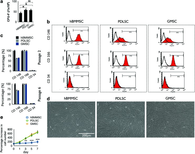
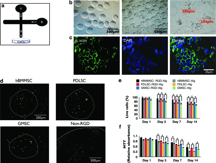
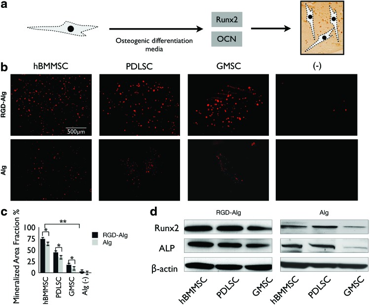
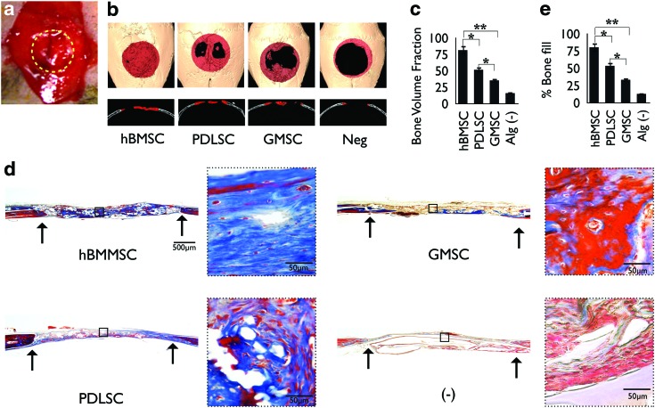
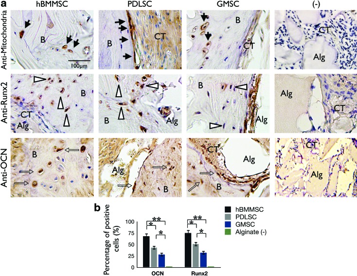
Mesenchymal stem cells (MSCs) provide an advantageous alternative therapeutic option for bone regeneration in comparison to current treatment modalities. However, delivering MSCs to the defect site while maintaining a high MSC survival rate is still a critical challenge in MSC-mediated bone regeneration. Here, we tested the bone regeneration capacity of periodontal ligament stem cells (PDLSCs) and gingival mesenchymal stem cells (GMSCs) encapsulated in a novel RGD- (arginine-glycine-aspartic acid tripeptide) coupled alginate microencapsulation system in vitro and in vivo. Five-millimeter-diameter critical-size calvarial defects were created in immunocompromised mice and PDLSCs and GMSCs encapsulated in RGD-modified alginate microspheres were transplanted into the defect sites. New bone formation was assessed using microcomputed tomography and histological analyses 8 weeks after transplantation. Results confirmed that our microencapsulation system significantly enhanced MSC viability and osteogenic differentiation in vitro compared with non-RGD-containing alginate hydrogel microspheres with larger diameters. Results confirmed that PDLSCs were able to repair the calvarial defects by promoting the formation of mineralized tissue, while GMSCs showed significantly lower osteogenic differentiation capability. Further, results revealed that RGD-coupled alginate scaffold facilitated the differentiation of oral MSCs toward an osteoblast lineage in vitro and in vivo, as assessed by expression of osteogenic markers Runx2, ALP, and osteocalcin. In conclusion, these results for the first time demonstrated that MSCs derived from orofacial tissue encapsulated in RGD-modified alginate scaffold show promise for craniofacial bone regeneration. This treatment modality has many potential dental and orthopedic applications.


Figures

<b>FIG. 1.</b>
FIG. 1.
(a) Generation of colony-forming units in cultures seeded with 1×10⁶ bone marrow mesenchymal stem cells (BMMSCs), periodontal ligament stem cells (PDLSCs), and gingival mesenchymal stem cells (GMSCs) at a low density for 10 days. (b) Expression of cell surface markers on stem cells (passage 2) as determined by flow cytometric analysis. (c) Quantification of percentage of cells that express stem cell markers determined by flow cytometry (mean±standard deviation). The results are representative of at least five independent experiments from passages 2–6. (d) Comparison of the morphology and growth properties of PDLSCs, GMSCs, and human BMMSCs (hBMMSCs). (e) Proliferation and cell counts of PDLSCs, GMSCs, and hBMMSCs (n=5). NS, not significant. *p<0.05, **p<0.01, ***p<0.001. Color images available online at www.liebertpub.com/tea
<b>FIG. 2.</b>
FIG. 2.
(a) Schematic representation of microfluidic device. Channel 1: alginate injection channel; Channel 2: soybean oil injection channel. Alginate droplets were sheared off by the soybean oil and flowed out of the device through Channel 3. (b) Microspheres produced by the microfluidic device. The diameter of the microspheres was between 196 and 581 μm (scale bar=500 μm). (c) Cytoskeleton organization of MSCs encapsulated in alginate microspheres stained with phalloidin Alexa Fluor 568 for F-actin (green) and 4′,6-diamidino-2-phenylindole (DAPI, blue) for nucleus. (d) Live/dead staining of the stem cell microspheres after 1 week of culturing (scale bar=200 μm). White dots show the peripheries of each microcapsule. Note the larger diameter of the non-RGD-containing alginate microsphere (with PDLSCs) with average diameter of 1 mm, fabricated via traditional methods.18,19 Larger microspheres show more dead positive cells after 1 week than RGD-coupled microspheres fabricated using microfluidics. (e) Viability of the encapsulated PDLSCs, GMSCs, and hBMMSCs: live/dead staining, percentage of live cells in either RGD-coupled alginate microspheres or in alginate microspheres without RGD. (f ) 3-(4,5-Dimethylthiazol-2-yl)-2, 5-diphenyltetrazolium bromide (MTT) assay of metabolic activity of cells. No significant difference was observed between the stem cell groups at each time interval. *p<0.05. Color images available online at www.liebertpub.com/tea
<b>FIG. 3.</b>
FIG. 3.
(a) Simple schematic representation of the transcription factors regulating the differentiation of MSCs and osteogenesis. (b) Osteo-differentiation and mineralization of encapsulated PDLSCs, GMSCs, and hBMMSCs in alginate in vitro after 4 weeks of culturing in osteogenic differentiation media. The specimens were stained by xylenol orange and emitted red fluorescence. (c) Mineralization area fraction was defined as the area of stained mineralization divided by the total area of the field of view of the image. (d) Western blot analysis of the expression of ALP and Runx2. *p<0.05, **p<0.01. Color images available online at www.liebertpub.com/tea
<b>FIG. 4.</b>
FIG. 4.
In vivo calvarial defect model in mice. (a) Calvarial defects (5 mm) were generated along the yellow dots in nude mice and encapsulated PDLSCs, GMSCs, or hBMMSCs were transplanted in the defect sites. (b) Microcomputed tomography (CT) three-dimensional reconstruction of bone repair in mouse calvarial defects implanted with PDLSCs, GMSCs, or hBMMSCs encapsulated in RGD-modified alginate. Red dots represent the periphery of the defect site. (c) Semiquantitative analysis of bone formation via micro-CT images. (d) Histomicrographs (trichrome staining) of mouse calvarial bone defects after 8 weeks of transplantation. Arrows indicate the boundaries of defects. (e) Histomorphometric analysis of calvarial defects showing the relative amount of bone formation. *p<0.05, **p<0.01, n=4 for each group. Color images available online at www.liebertpub.com/tea
<b>FIG. 5.</b>
FIG. 5.
Characterization of the origin and fate of PDLSCs and GMSCs after transplantation. (a) Upper panel: The cells of human origin were confirmed by immunohistochemical staining with a specific antibody for human mitochondria (black arrows). After 8 weeks of transplantation in immunocompromised mice, both PDLSCs and GMSCs were able to form bone. (b) Osteogenic cells were positive for anti-osteocalcin (OCN; middle panel, open arrows in black) and Runx2 (lower panel, white arrows) antibody staining, while negative control (−) immunohistochemical staining results failed to express any of these osteogenic markers. (b) Semiquantitative analysis of percent of positive cells for anti-OCN and anti-Runx2 antibodies via immunohistochemical staining images. *p<0.05, **p<0.01. Scale bar=100 μm. Color images available online at www.liebertpub.com/tea

Similar articles

Cited by

References

    1. Sachlos E., and Czernuszka J.T.Making tissue engineering scaffolds work. Review on the application of solid freeform fabrication technology to the production of tissue engineering scaffold. Eur Cell Mater 5,29, 2003 - PubMed
    1. Chen F.M., Zhang J., Zhang M., An Y., Chen F., and Wu Z.F.A review on endogenous regenerative technology in periodontal regenerative medicine. Biomaterials 31,7892, 2010 - PubMed
    1. Monaco E., Bionaz M., Hollister S.J., and Wheeler M.B.Strategies for regeneration of the bone using porcine adult adipose-derived mesenchymal stem cells. Theriogenology 75,1381, 2011 - PubMed
    1. Gronthos S., Mankani M., Brahim J., Robey P.G., and Shi S.Postnatal human dental pulp stem cells (DPSCs) in vitro and in vivo. Proc Natl Acad Sci U S A 97,13625, 2000 - PMC - PubMed
    1. Seo B.M., Miura M., Gronthos S., Bartold P.M., Batouli S., Brahim J., et al. . Investigation of multipotent postnatal stem cells from human periodontal ligament. Lancet 364,149, 2004 - PubMed

Publication types

MeSH terms

LinkOut - more resources